|
Exhibition Hall 13:30 - 14:30 |
|
|
|
Computer # |
|
4413.
 |
74 |
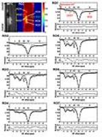
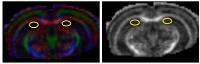
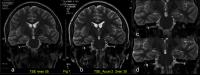
HASTE imaging with EPI volumetric navigators for real-time fetal
head motion detection 
Borjan Gagoski1,2, Patrick McDaniel3,
André J. W. van der Kouwe2,4, Himanshu Bhat5,
Lawrence L. Wald2,4,6, Elfar Adalsteinsson3,6,
P. Ellen Grant1,2, and M. Dylan Tisdall2,4
1Fetal Neonatal Neuroimaging and Developmental
Science Center, Boston Children's Hospital, Boston, MA,
United States, 2Radiology,
Harvard Medical School, Boston, MA, United States, 3Electrical
Engineering and Computer Science, Massachusetts Institute of
Technology, Cambridge, MA, United States, 4Athinoula
A. Martinos Center for Biomedical Imaging, Massachusetts
General Hospital, Charlestown, MA, United States, 5Siemens
Medical Solutions USA Inc, Charlestown, MA, United States, 6Harvard-MIT
Health Sciences and Technology, Institute of Medical
Engineering and Science, Massachusetts Institute of
Technology, Cambridge, MA, United States
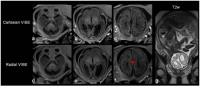
Although heavily used in clinical fetal imaging due to its
encoding efficiency, the image quality of T2-weighted
singe-shot fast-spin-echo (ss-FSE, or HASTE) acquisitions is
often compromised by fetal head motion. We have implemented
and tested an enhanced version of the HASTE acquisition
scheme that includes EPI-based volumetric navigators (EPI-vNavs)
played each TR, enabling detection and estimation of fetal
head motion along six degrees of freedom in real time, while
maintaining equivalent T2 contrast
in the fetal head compared to the original HASTE
acquisition.
|
|
4426.
 |
87 |
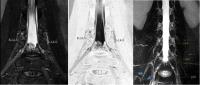
Distortion correction of fetal EPI using registration of
orthogonal stacks with Laplacian constraint
Maria Kuklisova Murgasova1, Georgia Lockwood
Estrin1, Rita G. Nunes1,2, Mary
Rutherford1, and Jo Hajnal1
1King's College London, London, United Kingdom, 2Instituto
de Biofisica e Engenharia Biomedica, Faculdade de Ciencias,
Universidade de Lisboa, Lisbon, Portugal
We present a novel method for correction of geometric
distortions induced by static B0 field in fetal EPI. The
method estimates distortion by including a
distortion-correction step in the slice to volume
reconstruction of orthogonal EPI stacks with orthogonal
phase encoding directions, in the form of non-rigid
registration with a Laplacian constraint. We show that the
proposed method achieves better consistency with
reconstructed ssFSE volumes than EPI volumes constructed
from data corrected by B0 field map. The registration-based
distortion correction is thus a viable alternative to
acquisition of B0 field map.
|
|
4431.
 |
92 |
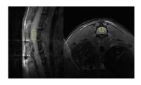
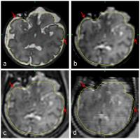
Optimal Slice Planning of the Fetal Brain Using Interactive
Real-Time MRI
Lau Brix1,2, Steffen Ringgaard1, Puk
Sandager3, Olav Bjørn Petersen3,
Thomas Sangild Sørensen4,5, Erik Lundorf1,
and Brian Stausbøl-Grøn1
1MR Research Centre, Aarhus University Hospital,
Skejby, Aarhus N, Denmark, 2Department
of Procurement & Clinical Engineering, Region Midt, Aarhus
N, Denmark, 3Department
of Obstetrics and Gynecology, Aarhus University Hospital,
Skejby, Aarhus N, Denmark, 4Department
of Clinical Medicine, Aarhus University, Aarhus N, Denmark, 5Department
of Computer Science, Aarhus University, Aarhus N, Denmark
Diagnostic image quality of MRI can be hampered by fetal
movements during data acquisition which may limit its
diagnostic use (1;2). We propose an interactive real-time
MRI technique which may serve as an alternative to
traditional fetal MRI for anthropometrics or as a supplement
for representation of fetal brain structures in cases in
which fetal motion causes challenges in relation to
obtaining optimal slice planes using conventional MRI
techniques.
|
|
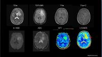
4414.
 |
75 |
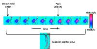
Free-breathing T1-weigthed gradient-echo imaging for fetus brain
bin zhang1
1Department of Radiology, Xijing Hospital, xi'an,
China, People's Republic of
Magnetic resonance (MR) imaging appears to be increasing
used for the diagnosis of abnormalities in fetuses because
of the absence of ionizing radiation and superior contrast
of soft tissues. However, the T1-weighted 3D MR imaging for
fetus remains very challenging due to the respiratory motion
of the mother and the movement of the fetus. In this study,
we evaluated the feasibility of a free-breathing 3D
T1-weighted gradient-echo imaging with radial data sampling
for fetus imaging, and compared with a standard breath-hold
imaging with Cartesian k-space acquisition
|
|
4415.
 |
76 |
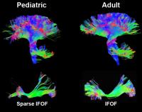
HARDI Acquisition in Neonates and Children using Modular
Multiband Multi-shell Sequence
Vincent Kyu Lee1, Meredith Monsour2,
Sudhir Pathak2, Vincent Schmithorst3,
Catherine Fissell2, Ashok Panigrahy1,3,
and Walt Schneider2
1Radiology, University of Pittsburgh, Pittsburgh,
PA, United States, 2University
of Pittsburgh, Pittsburgh, PA, United States, 3Children's
Hospital of Pittsburgh, Pittsburgh, PA, United States
High angular-resolution diffusion imaging (HARDI) is the
best imaging technique to distinguish crossing fibers and
high turning angle neuronal tracts, which is critical for
identifying and characterizing the microstructural changes
in neuro pathology and traumatic brain injury. Its main
disadvantage has been the lengthy scan time needed to
acquire analyzable images – a challenge especially in
children and neonates who do not tolerate long scanning
sessions. This study presents the preliminary fiber
tractography of healthy neonatal and pediatric subjects
acquired using multiband multi-shell HARDI sequence within a
practicable scan time without sacrificing image quality.
|
|
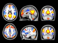
4421.
 |
82 |
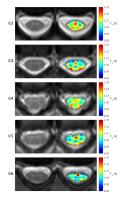
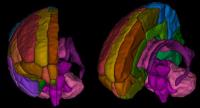
Parcellation of neonatal brain MRI into 107 regions using atlas
propagation through intermediate time points in childhood.
Manuel Blesa1, Ahmed Serag1, Alaistir
G Wilkinson2, Devasuda Anblagan1,3,
Emma J Telford1, Rozalia Pataki1,
Sarah A Sparrow1, Gillian Macnaught4,
Scott I Semple4, Mark E Bastin3, and
James P Boardman1,3
1MRC Centre for Reproductive Health, University
of Edinburgh, Edinburgh, United Kingdom, 2Department
of Radiology, Royal Hospital for Sick Children, Edinburgh,
United Kingdom, 3Centre
for Clinical Brain Sciences, University of Edinburgh,
Edinburgh, United Kingdom, 4Clinical
Research Imaging Centre, University of Edinburgh, Edinburgh,
United Kingdom
We created a neonatal brain atlas of healthy subjects that
can be applied to multi-modal MRI data. Structural and
diffusion 3T MRI scans were acquired after birth from 25
neonates born at term. The SRI24/TZO atlas was propagated to
the neonatal data using temporal registration via childhood
templates (NIHPD), with the final atlas (the Edinburgh
Neonatal Atlas, ENA25) constructed using iterative averaging
of T1-weighted volumes. The computed transformations were
applied to T2-weighted data, diffusion maps and tissue
probability maps to provide a multi-modal atlas with 107
anatomical regions; and we have generated a symmetric
version to facilitate studies of laterality.
|
|
4428.
 |
89 |
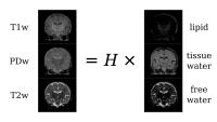
A simple method for myelin mapping using T1-weighted,
T2-weighted and PD-weighted images
J-Donald Tournier1,2, Rui Pedro A. G. Teixeira1,2,
Maria Murgasova1,2, A. David Edwards2,3,
Joseph V. Hajnal1,2, and Serena J. Counsell2,3
1Biomedical Engineering, King's College London,
London, United Kingdom, 2Centre
for the Developing Brain, King's College London, London,
United Kingdom, 3Perinatal
Imaging and Health, King's College London, London, United
Kingdom
Myelin mapping is of great interest, particularly to study
brain development. However, existing methods are time
consuming and/or noisy. We propose a simple method to obtain
semi-quantitative maps of myelin from routinely acquired
T1-, T2- and proton density weighted images, by modelling
the signal as a linear combination of non-exchanging tissue
types: lipid, tissue water and free water. The method is
calibrated empirically from the signal intensities in the
data themselves. We show promising results in neonatal
scans, showing the expected pattern of myelination in
infants at term-equivalent age.
|
|
4425.
 |
86 |
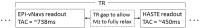
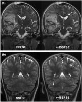
Variable Refocusing Flip Angle Single Shot Imaging For
Anesthesia-Free Brain MRI - Video
Not Available
Kristen W. Yeom1, Valentina Taviani1,
Andreas M. Loening1, Michael Iv1, and
Shreyas S. Vasanawala1
1Stanford University, Stanford, CA, United States
Conventional single shot fast spin echo (SSFSE) and variable
refocusing flip angle SSFSE (vrfSSFSE) were compared for
fast sedation-free pediatric brain MRI (N=33). Two
neuroradiologists independently and blindly evaluated SSFSE
and vrfSSFSE images for motion, perceived resolution
(sharpness), contrast and lesion conspicuity on a five-point
scale. vrfSSFSE gave less motion and misregistration
artefacts than conventional SSFSE, due to the shorter scan
duration. As for the other image quality metrics, vrfSSFSE
was found to be either comparable or superior to
conventional SSFSE.
|
|
4422.
 |
83 |
Assessing the effects of pediatric subject motion on T2
relaxation under spin tagging (TRUST) cerebral oxygenation
measurements using volume navigators (vNavs)
Jeffrey N Stout1, M. Dylan Tisdall2,
Patrick McDaniel3, Borjan Gagoski4,
Divya S Bolar2,5, Patricia Ellen Grant4,
and Elfar Adalsteinsson1,3,6
1Harvard-MIT Health Sciences and Technology,
Massachusetts Institute of Technology, Cambridge, MA, United
States, 2Martinos
Center for Biomedical Imaging, MGH/Harvard Medical School,
Boston, MA, United States, 3Department
of Electrical Engineering and Computer Science,
Massachusetts Institute of Technology, Cambridge, MA, United
States, 4Fetal-Neonatal
Neuroimaging and Developmental Science Center, Boston
Children’s Hospital, Boston, MA, United States, 5Department
of Radiology, Massachusetts General Hospital, Boston, MA,
United States, 6Institute
for Medical Engineering and Science, Cambridge, MA, United
States
When using the T2-relaxation under spin tagging
(TRUST) technique on non-compliant subjects, motion has an
unknown effect on estimations of cerebral oxygenation that
are derived from an empirical mapping between T2 and
blood oxygen saturation. Incorporating low resolution 3D-EPI
volume navigators into the TRUST pulse sequence permits
independent measurements of motion during scanning. We show
that for static scans vNav modules have only small effects
on resulting venous blood T2 estimates,
that poor exponential goodness of fit is not a sufficient
indicator of motion, and that T2 is
biased upwards with increasing motion.
|
|
4412.
 |
73 |
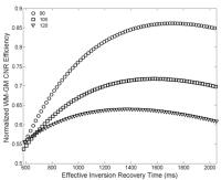
Improving the Quality of Neonatal Brain Structural MRI with
Shorter Acquisition Train Length
Lili He1, Jinghua Wang2, Mark Smith3,
Zhong-Lin Lu2, and Nehal A Parikh1,4
1Center for Perinatal Research, Nationwide
Children’s Hospital, Columbus, OH, United States, 2The
Ohio State Univeristy, Columbus, OH, United States, 3Radiology,
Nationwide Children’s Hospital, Columbus, OH, United States, 4Department
of Pediatrics, The Ohio State University College of
Medicine, Columbus, OH, United States
Three-dimensional (3D) T1-weighted sequences such
as MP-RAGE are invaluable for evaluation of neonatal and
infant brain injury/development. Sequence optimization for
neonates has been historically challenging because neonatal
brains exhibit reversed white matter–gray matter (WM-GM)
contrast on T1-weighted scans, and the contrast
is much lower than that of adult brains. In this study, we
show in preterm neonates that shortening the acquisition
train length of the MP-RAGE sequence significantly improved
SNR and CNR efficiencies. The proposed optimization
methodology can be easily extended to other populations
(e.g. term infants, adults and elders), and different
organs, field strengths and MR sequences.
|
|
4432.
 |
93 |
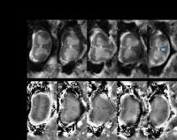
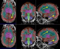
A hybrid premature neonatal segmentation pipeline for clinical
brain imaging acquired without dedicated neonatal coils.
Zachary Hill1, Mengyuan Liu1, Sandra
Juul2, and Colin Studholme3
1Bioengineering, University of Washington,
Seattle, WA, United States, 2Pediatrics,
University of Washington, Seattle, WA, United States, 3Pediatrics,
Bioengineering, Radiology, University of Washington,
Seattle, WA, United States
Due to the difference in individual cases, larger multi-site
studies of brain injury after premature birth may be needed,
but dedicated neonatal imaging technology isn't always
available. The use of older scanners with coils not
specifically aimed at imaging neonatal brains introduces a
severe intensity variation across the field of view, which
can cause conventional image analysis pipelines to fail. A
robust hybrid tissue segmentation pipeline was developed and
shown to improve tissue segmentations of four test subjects
with manual segmentations for reference. This enables
automated and consistent analysis to better quantitatively
study early human brain development.
|
|
4424.
 |
85 |
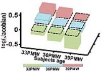
Age-specific gray and white matter DTI atlas for human brain at
33 and 36 postmenstrual weeks
Lei Feng1,2, Hang Li1,3, Kenichi Oishi4,
Virendra Mishra5, Minhui Ouyang1, Tina
Jeon1, Yun Peng3, Shuwei Liu2,
and Hao Huang1,6
1Department of Radiology, Children’s Hospital of
Philadelphia, Philadelphia, PA, United States, 2Research
Center for Sectional and Imaging Anatomy, Shandong
University School of Medicine, Jinan, China, People's
Republic of, 3Department
of Radiology, Beijing Children’s Hospital Affiliated to
Capital Medical University, Beijing, China, People's
Republic of, 4Department
of Radiology and Radiological Science, Johns Hopkins
University, Baltimore, MD, United States, 5Advanced
Imaging Research Center, University of Texas Southwestern
Medical Center, Dallas, TX, United States, 6Department
of Radiology, Perelman School of Medicine, University of
Pennsylvania, Philadelphia, PA, United States
The large brain morphological differences of the preterm
brain at 33 or 36 postmenstrual week (PMW) to that at 40 PMW
makes it necessary to establish age-specific atlases for
preterm brains. In this study, with diffusion MRI (dMRI)
data acquisition of 82 preterm and term normal neonates, we
aimed to establish a comprehensive digital atlas including
labeling of gray and white matter for preterm brains at 33
and 36 PMW. We demonstrated these atlases and showed the
differences of the major neural structures including
ganglionic eminence and uncinate fasciculus by comparison to
JHU-neonate-SS atlas for brains at around 40PMW.
|
|
4427.
 |
88 |
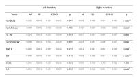
Age-related white matter changes on phase difference enhanced
imaging in children
Tetsu Niwa1, Tetsuya Yoneda2, Shuhei
Shibukawa1, Toshiki Kazama1, Taro
Takahara3, and Yutaka Imai1
1Radiology, Tokai University School of Medicine,
Isehara, Japan, 2Medical
Physics in Advanced Biomedical Sciences, Kumamoto
University, Kumamoto, Japan, 3Biomedical
Engineering, Tokai University School of Engineering,
Isehara, Japan
Recent reports suggest that phase shift in the white matter
may be related to myelin content. We assessed the
age-related phase changes of the white matter in small
children (age range, 0?6 years) on phase difference enhanced
imaging (PADRE). PADRE showed progression of the phase
changes in the white matter along with age, particularly in
the pyramidal tract and subcortical region in Rolandic are.
Whereas, less phase changes were noted in the subcortical
white matter in the temporal lobe. PADRE showed age-related
white matter phase shift, suggesting progression of
myelination and myelin content.
|
|
4430.
 |
91 |
Microstructural organization of the language connectome in
typically developing left-handed children: a DTI tractography
study
Marjolein Verly1, Robin Gerrits1,
Lieven Lagae2, Inge Zink1, Stefan
Sunaert3, and Nathalie Rommel1
1Dept. Neurosciences, KU Leuven, Leuven, Belgium, 2Dept.
Pediatrics, UZ Leuven, Leuven, Belgium, 3Dept.
Translational MRI, KU Leuven, Leuven, Belgium
The main objective of this study was to investigate the
relationship between the microstructural properties of
language-related white matter (WM) tracts and hand
preference in typically developing school-aged children. Our
DTI tractography results provide evidence for a different
structural connectivity pattern of the language connectome
in left-handed children. Whereas right-handed children show
a clear left-lateralized structural language network, our
group of left-handed children seems to have a more bilateral
organized language system. Those observed differences in WM
microstructure and lateralization might reflect an
interaction between handedness and the neural processing of
language in children.
|
|
4416.
 |
77 |
Gender-specific attention system subnetwork vulnerability in
prematurely born children
Elda Fischi-Gomez1,2, Lana Vasung1,
Sebastien Urben3,4, Cristina Borradori-Tolsa1,
François Lazeyras5, Jean-Philippe Thiran2,6,
and Petra Susan Hüppi1
1Division of Development and Growth. Department
of Pediatrics, University Hospital of Geneva, Geneva,
Switzerland, 2Signal
Processing Laboratory 5, École Polytechnique Fédérale de
Lausanne (EPFL), Lausanne, Switzerland, 3Child
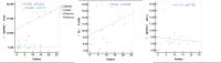
Clinical Neuropsychology Unit, Department of Psychology,
University of Geneva, Geneva, Switzerland, 4Research
Unit, University Service of Child and Adolescent Psychiatry,
Department of Psychiatry, University Hospital of Lausanne
(CHUV), Lausanne, Switzerland, 5Department
of Radiology and Medical Informatics, Faculty of Medicine,
University of Geneva, Geneva, Switzerland, 6Department
of Radiology, University Hospital Center (CHUV) and
University of Lausanne (UNIL), Lausanne, Switzerland
Within preterm-born children, being born male and at a lower
gestational age have both been associated with a heightened
risk for developmental difficulties. However, in this
population little is known about the combined effect and the
influence of these risk factors on the structural networks
subserving attention and executive. Using a diffusion-based
brain connectome approach, in this work we analyze the
effect of these two factors in the brain networks of
school-age preterm born children and provide evidence of a
gender-specific vulnerability in the executive attentional
subnetwork.
|
|
4433.
 |
94 |
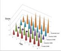
Utilization of Simultaneous Multi-slice Accelerated Turbo Spin
Echo in Pediatric Epilepsy
Michael Kean1,2, Lee Coleman2,3,
Simone Mandelstam3, Sonal Josan4,
Benjamin Schmitt4, and Dingxin Wang5,6
1Children MRI Centre, Royal Childrens Hospital,
Parkville, Australia, 2Murdoch
Childrens Research Institute, Parkville, Australia, 3Royal
Childrens Hospital, Parkville, Australia, 4Siemens
Healthcare, Bayswater, Australia, 5Centre
for Magnetic Resonance Research, University of Minnesota,
Minneapolis, MN, United States, 6Siemens
Medical Solutions, Malvern, PA, United States
The objective of our prospective study was to examine the
clinical utility of Simultaneous Multi-Slice(SMS)
Accelerated TSE at 3T imaging paediatric patients who
present with seizures.A randomly selected cohort of
patients were enrolled in the protocol covering a broad
spectrum of clinical entities. A direct comparison was
undertaken with anatomically matched conventional TSE and
SMS TSE acquisitions with matched in-plane and through plane
resolution, echo train lengths and echo spacing.
Analysis of the data confirmed that although there were
minimal variations in the quantitative measures recorded
both sequences provided images of consistent image quality
and diagnostic confidence with a significant scan time
reduction attributed to the SMS TSE acquisition.
|
|
4420.
 |
81 |
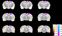
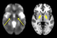
Iron deposition in the globus pallidus of healthy youth -
Video Not Available
Karthik Prabhakaran1, David Roalf1,
Mark Elliott1, Simon Vandekar1, Kosha
Ruparel1, Ryan Hopson1, Efstathios D
Gennatas1, Jeffrey Valdez1, Chad
Jackson1, Theodore Satterthwaite1,
Raquel Gur1, and Ruben Gur1
1University of Pennsylvania, Philadelphia, PA,
United States
R2*, the transverse relaxation rate was used to measure iron
deposition in the globus pallidus of 815 youth and young
adults between the ages of 8 and 22. Significant iron
deposition occurs in the globus pallidus between the ages of
8 and 22 in accordance with previously described models of
iron deposition in the brain throughout the lifespan. Among
adolescents (age 12-16) females had lower iron deposition in
the globus pallidus (p < 0.001) as compared to males, this
may be related to adolescent females being especially
susceptible to dietary iron deficiency because of poor
dietary intake in conjunction with high iron requirements
related to rapid growth and menstrual blood loss.
|
|
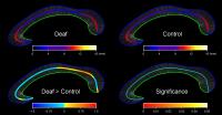
4423.
 |
84 |
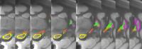
Congenital sensorineural hearing loss affects the development of
corpus callosum
Weiwei Men1, Tianbing Song2, Shuang
Xia3, Yaoyu Zhang1, Jing Che4,
and Jia-Hong Gao1
1Center for MRI Research, Academy for Advanced
Interdisciplinary Studies, Peking University, Beijing,
China, People's Republic of, 2Beijing
cancer hospital, Beijing, China, People's Republic of, 3Tianjing
First Central Hospital, Tianjing, China, People's Republic
of, 4Aerospace
Central Hospital, Beijing, China, People's Republic of
Congenital sensorineural hearing loss (CSHL) is a common
disease in newborns, which can affect the development of
corpus callosum (CC). In this study, a novel method of CC
thickness analysis was employed to compare the CC difference
between deaf and control groups. The results indicate that
after 24 months deaf group has thinner CC thickness in the
anterior splenium of CC compared to control group, which
means the development of deaf anterior splenium is slowed
down. Our study suggests that 12~24 month old is the best
time period for CSHL treatment and intervention.
|
|
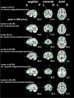
4417.
 |
78 |
White Matter Structural Alternations in Children with HIV
Infection and Exposure
Marcin Jankiewicz1, Paul A. Taylor1,2,3,
Martha Holmes1, Mark F. Cotton4,
Barbara Laughton4, Andre J.W. van der Kouwe5,
and Ernesta M. Meintjes1
1MRC/UCT Medical Imaging Research Unit,
Department of Human Biology, University of Cape Town, Cape
Town, South Africa, 2Scientific
and Statistical Computing Core, National Institutes of
Health, Bethesda, MD, United States, 3African
Institute for Mathematical Sciences, Muizenberg, South
Africa, 4Children’s
Infectious Diseases Clinical Research Unit, Department of
Pediatrics and Child Health, Stellenbosch University, Cape
Town, South Africa, 5Athinoula
A. Martinos Center for Biomedical Imaging, Massachusetts
General Hospital, Charlestown, MA, United States
In this work we examine WM alterations in HIV infected
children at age 7 years and compare those who initiated ART
before and after 12 weeks of age.
|
|
4435.
 |
96 |
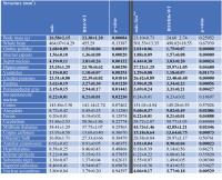
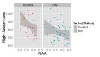
Effects of pediatric HIV/antiretroviral therapy on basal ganglia
metabolite-volume relationships
Frances C Robertson1, Martha J Holmes1,
Emmanuel C Nwosu1, Francesca Little2,
Mark F Cotton3, Els Dobbels3, Andre JW
van der Kouwe4,5, Barbara Laughton3,
and Ernesta M Meintjes1
1Department of Human Biology, University of Cape
Town, Cape Town, South Africa, 2Department
of Statistical Sciences, University of Cape Town, Cape Town,
South Africa, 3Department
of Paediatrics & Child Health, Stellenbosch University,
Stellenbosch, South Africa, 4A.A.
Martinos Centre for Biomedical Imaging, Massachusetts
General Hospital, Charlestown, MA, United States, 5Department
of Radiology, Harvard Medical School, Boston, MA, United
States
HIV is associated with structural deficits in the basal
ganglia (BG). Volumes from structural MRI may relate to
metabolic changes measurable with magnetic resonance
spectroscopy. We investigated the relationship between BG
NAA and Glutamate/Glutamine and caudate, putamen, nucleus
accumbens and subcortical gray matter (GM) volumes in 7-year
old HIV-infected children on antiretroviral therapy and
uninfected controls. Higher NAA was associated with smaller
accumbens and left putamen in all children. Higher
Glutamate/Glutamine was associated with greater subcortical
GM in controls, but not HIV-infected children. Relationships
between brain metabolites and volumes add to the description
of effects of HIV/ART on the BG.
|
|
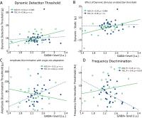
4429.
 |
90 |
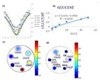
Reduced GABA levels and altered sensory function in children
with Autism Spectrum Disorder
Nicolaas AJ Puts1,2, Ericka L Wodka3,4,
Ashley D Harris1,2,5,6, Deana Crocetti7,
Mark Tommerdahl8, Richard AE Edden1,2,
and Stewart H Mostofsky3,7,9
1Radiology and Radiological Science, Johns
Hopkins University, Baltimore, MD, United States, 2F.M.
Kirby Center for Functional Brain Imaging, Kennedy Krieger
Institute, Baltimore, MD, United States,3Center
for Autism and Related Disorders, Kennedy Krieger Institute,
Baltimore, MD, United States, 4Psychiatry
and behavioral sciences, Johns Hopkins University,
Baltimore, MD, United States, 5Alberta
Children's Hospital Research Institute, University of
Calgary, Calgary, AB, Canada, 6Radiology,
University of Calgary, Calgary, AB, Canada, 7Laboratory
for Neurocognitive and Imaging Research, Kennedy Krieger
Institute, Baltimore, MD, United States, 8Biomedical
Engineering, University of North Carolina at Chapel Hill,
Chapel Hill, NC, United States, 9Neurology,
Johns Hopkins University, Baltimore, MD, United States
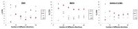
Children with Autism often show difficulties processing
sensory stimuli, but the underpinnings are poorly
understood. Multiple lines of evidence suggest that GABA,
the main inhibitory neurotransmitter in the brain, plays a
role in the pathophysiology of ASD. Here we show reduced
GABA levels in children with ASD, which is associated with
abnormal performance on vibrotactile tasks related to
inhibition. We show that alterations in GABA can contribute
to alterations in sensory processing in ASD.
|
|
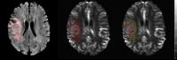
4434.
 |
95 |
Amide proton transfer imaging of neonatal brain development and
brain injury: a preliminary study -
Permission Withheld
Yang Zheng1, Xiaoming WANG1, Xuna Zhao2,
and Jinyuan Zhou3
1Department of Radiology, Shengjing Hospital of
China Medical University, Shenyang, China, People's Republic
of, 2Philips
Healthcare, Beijing, China, Beijing, China, People's
Republic of, 3Division
of MR Research, Department of Radiology, Johns Hopkins
University, Maryland, USA, Baltimore, MD, United States
Yang Zheng M.D. Department of Radiology, Shengjing Hospital
of China Medical University, No. 36, Sanhao Street, Heping
District, Shenyang 110004,PR China E-mail address:
jingshenbing0702@gmail.com Tel.: +86 13889830846
|
|
4419.
 |
80 |
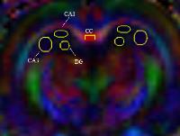
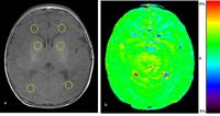
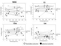
Absolute metabolite concentration of Creatine in the deep gray
matter measured using short echo 1H-MRS predict long-term
prognosis of neonatal hypoxic-ischemic encephalopathy as
excellent as NAA concentration
Noriko Aida1,2, Jun Shibasaki3, Moyoko
Tomiyasu1,2, Yuri Nishi1,4, Naho
Morisaki4, Takeo Fujiwara4, Katsuaki
Toyoshima3, and Takayuki Obata2
1Radiology, Kanagawa Children's Medical Center,
Yokohama, Japan, 2Research
Center for Charged Particle Therapy, National Institute of
Radiological Sciences, Chiba, Japan, 3Neonatology,
Kanagawa Children's Medical Center, Yokohama, Japan, 4Social
Medicine, National Research Institute for Child Health and
Development, Tokyo, Japan
Absolute metabolite concentrations of N-acethylaspartate (NAA),
Choline(Cho) and Creatine(Cr) in the deep gray matter of 44
near term neonates with hypoxic-ischemic encephalopathy (HIE),
measured using PRESS method short echo 1H-MRS within 2 weeks
after birth, showed excellent prognostic values (AUC; NAA:
0.98, Cho: 0.96, Cr: 0.99) with the adverse outcomes having
significantly lower measurements compared to those with
favorable outcomes, while Lactate was less efficient (AUC
0.74). Moreover NAA and Cr concentrations measured at 24-96
hours revealed perfect prognostic values (AUC 1.00). Early
measurement of absolute Cr and NAA concentrations can be
excellent biomarkers of infants suffered with neonatal HIE.
|
|
4418.
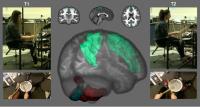 |
79 |
Drum Training induces MR visible changes in the Cerebellum and
Cortex
Muriel M.K. Bruchhage1, Ali Amad1,
Stephen B. Draper2, Jade Seidman1,
Flavio Dell'Acqua3, Luis Lacerda3,
Pedro Luque Laguna3, Ruth G. Lowry4,
Andrew Robertson5, Marcus S. Smith4,
and Steven C.R. Williams1
1Department of Neuroimaging, King's College
London, The Institute of Psychiatry, Psychology and
Neuroscience, London, United Kingdom, 2School
of Sport and Exercise, University of Gloucestershire,
Chichester, United Kingdom, 3NatBrainLab,
Department of Neuroimaging, King's College London, The
Institute of Psychiatry, Psychology and Neuroscience,
London, United Kingdom, 4Department
of Sport and Exercise, University of Chichester, Chichester,
United Kingdom, 5Centre
for Digital Music, School of Electronic Engineering and
Computer Science, Queen Mary University, London, United
Kingdom
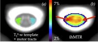
Cerebellar networks show long-term plasticity and motor
training has been shown to change cerebellar microstructure
and cortical thickness. We used a combination of
neuroimaging measures to visualise plastic changes in
drumming - a demanding multilimb training method: cerebellar
lobular volume and shape analysis, cortical thickness and
diffusion tensor imaging. Drum training reorganises and
reshapes the posterior cerebellum, expanding to connected
parietal and prefrontal cortical structures through the
inferior cerebellar white matter pathway. Thus, it may offer
a novel method for cerebellar and cortical plasticity,
relevant as an intervention method for psychiatric disorders
connected to cerebellar dysfunction, including autism
spectrum disorder.
|
|