|
Exhibition Hall 14:30 - 15:30 |
|
|
|
Computer # |
|
4508.
 |
73 |
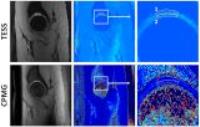
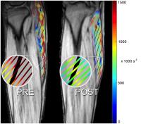
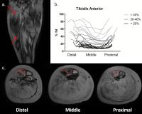
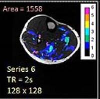
Functional Changes in Medial Gastrocnemius from Unilateral Limb
Suspension Induced Acute Atrophy: a 2D Strain Rate Study during
Isometric Contraction 
Vadim Malis1, Usha Sinha2, Robert
Csapo3, and Shantanu Sinha3
1Physics, University of California at San Diego,
San Diego, CA, United States, 2Physics,
San Diego State University, San Diego, CA, United States, 3Radiology,
University of California at San Diego, San Diego, CA, United
States
Unilateral limb suspension is a controlled method to
generate acute atrophy. The loss of muscle force with acute
atrophy may be due to changes in contractile elements and
extracellular matrix (ECM); a study of the strain rate (SR)
patterns could provide information on these changes.
Subjects were assessed at baseline (pre-ULLS) and post-ULLS
using dynamic velocity encoded phase contrast MR imaging.
The indices extracted from the SR tensor show at post-ULLS (i)
a decrease in the asymmetry of deformation in the fiber
cross-section and (ii) larger SR-muscle fiber angles. These
findings may reflect a loss of integrity in the ECM.
|
|
4509.
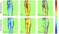 |
74 |
Physiological insights into medial gastrocnemius function during
eccentric contraction in normal and in acute atrophy –
Quantification of 2D strain rate indices from Velocity Encoded
Phase Contrast MR Imaging.
Usha Sinha1, Vadim Malis2, Robert
Csapo3, and Shantanu Sinha3
1Physics, San Diego State University, San Diego,
CA, United States, 2Physics,
University of California at San Diego, San Diego, CA, United
States, 3Radiology,
University of California at San Diego, San Diego, CA, United
States
In-vivo studies of muscle function under different motion
paradigms can elucidate the physiology of acute atrophy.
This study maps the 2D strain rate tensor in subjects
performing eccentric contractions before and after
Unilateral Limb Suspension induced acute atrophy. As
expected, strain rate values are smaller during eccentric
compared to isometric contractions, since in the eccentric
mode, muscle contraction occurs under lengthening conditions
resulting in a net smaller local elongation. Changes of SR
indices with atrophy are negligible possibly due to a
balance of force loss from atrophy and greater force
generation from a potentially stiffer matrix.
|
|
4510.
 |
75 |
Feasibility study of interleaved multi-nuclear acquisitions on a
3 T clinical NMR scanner without hardware modifications
Alfredo Liubomir Lopez Kolkovsky1,2, Benjamin
Marty1,2, Eric Giacomini1, and Pierre
G Carlier1,2
1NMR Laboratory, Institut of Myology, Paris,
France, 2NMR
Laboratory, CEA/DSV/I2BM/MIRCen, Fontenay-aux-Roses, France
NMR allows to investigate multiple aspects of physiological
parameters like regional perfusion, blood and tissue
oxygenation, intracellular pH or high-energy phosphate
metabolism. In the past, interleaved multiparametric
multinuclear dynamic NMR imaging and spectroscopy of
skeletal muscle was developed on prototype scanners. Here we
evaluated an interleaved pulse sequence combining the NMR
acquisition of a 1H
image and 31P
spectrum on a clinical system without any hardware
modifications from the customer. Having the possibility to
run interleaved multinuclear sequences on unmodified
clinical systems will greatly facilitate simultaneous
measurements of tissue perfusion, oxygen content and
mitochondrial ATP production in clinical research studies.
|
|
4511.
 |
76 |
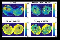
Comparison of T2-prepared 3D TSE with multi-echo spin-echo
sequences for T2 mapping of thigh muscles in healthy volunteers
Elisabeth Klupp1, Dominik Weidlich2,
Thomas Baum2, Barbara Cervantes2,
Marcus Deschauer3, Hendrik Kooijman4,
Ernst J. Rummeny2, Claus Zimmer1, Jan
S. Kirschke1, and Dimitrios C. Karampinos2
1Neuroradiology, Technische Universität München,
München, Germany, 2Radiology,
Technische Universität München, München, Germany, 3Neurology,
Technische Universität München, München, Germany, 4Philips
Healthcare, Hamburg, Germany
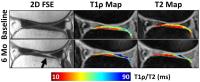
There is a growing interest for applying T2 mapping for
non-invasively tracking inflammatory changes in patients
with neuromuscular diseases. T2 has been traditionally
quantified using multi-echo spin-echo (MESE) sequences with
known problems related to the refocusing pulses in presence
of B1-inhomogeneity and slice profiles effects. The present
work proposes the combination of an adiabatic T2-preparation
with 3D TSE for B1-insenstive T2 mapping. The proposed
method is compared with 2D-MESE and 3D-MESE, in terms of
reproducibility on T2 quantification and sensitivity to B1
effects, in the thigh musculature of ten healthy subjects.
|
|
4512.
 |
77 |
T2* Mapping of Lower Leg Muscles Following Single Brief
Contractions at 3 T
Prodromos Parasoglou1, Tiejun Zhao2,
Oleksandr Khegai1, Xuejiao Che1, and
Jill M Slade3
1Department of Radiology, New York University
School of Medicine, New York, NY, United States, 2Siemens
Medical Solutions USA, Siemens Healthcare, New York, NY,
United States, 3Department
of Radiology, Michigan State University, East Lansing, MI,
United States
Microvascular function in the skeletal muscle can be
assessed through blood oxygenation level dependent (BOLD)
MRI signal changes after performing a brief exercise or
following a period of induced ischemia. Such BOLD related
relaxation changes are mainly attributed to intravascular
mechanisms, such as changes in the hemoglobin content and
oxygen saturation levels. In this work, we developed and
implemented a rapid echo-planar imaging (EPI) method to map T2*
changes, following a single maximum voluntary contraction on
a 3 T whole body clinical scanner.
|
|
4513.
 |
78 |
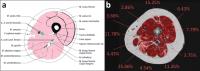
Quantitative Assessment of Muscle Fat in Sarcopenia Using
Magnetic Resonance Imaging (MRI) and Spectroscopy (MRS)
Alexandra Grimm1,2, Heiko Meyer2,
Mathias Nittka2, Esther Raithel2,
Andreas Friedberger1, Marc Teschler1,
Michael Uder3, Wolfgang Kemmler1,
Klaus Engelke1, and Harald H. Quick1,4
1Institute of Medical Physics, Erlangen, Germany, 2Product
Definition & Innovation, Siemens Healthcare GmbH, Erlangen,
Germany, 3Institute
of Radiology, University Hospital Erlangen, Erlangen,
Germany, 4Erwin
L. Hahn Institute for Magnetic Resonance Imaging, University
Duisburg-Essen, Essen, Germany
Sarcopenia describes muscle degeneration. In particular with
increasing age, muscle tissue is replaced by fatty
infiltrations. We developed an MRI sequence protocol (T1w
TSE, PDw SPACE, PDw TSE Dixon, q-Dixon, and HISTO) for
quantifying this degradation and applied it twice to 54
patients suffering from sarcopenia. Between both
measurements three months of whole body
electromyostimulation (EMS) training were performed. Initial
results show that image data can be used for muscle
segmentation and determination of muscle volumes, fat
fractions, and fat distribution within the muscles. Muscle
fat fractions correlate with muscle strength. In
spectroscopy accurate voxel repositioning is challenging.
|
|
4514.
 |
79 |
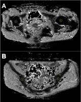
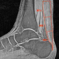
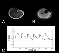
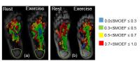
Foot Oximetry Angiosomes with MRI -
Permission Withheld
Jie Zheng1, David Muccigrosso1,
Xiaodong Zhang2, Hongyu An1, Andrew R
Coggan1, Charles F Hildebolt1, Chandu
Vemuri3, Patrick Geraghty3, Mary K
Hastings4, and Michael J Mueller4
1Radiology, Washington University in St. Louis,
St. Louis, MO, United States, 2Radiology,
Peking University First Hospital, Beijing, China, People's
Republic of, 3Surgery,
Washington University in St. Louis, St. Louis, MO, United
States, 4The
Program in Physical Therapy, Washington University in St.
Louis, St. Louis, MO, United States
The objective of this study was to develop a
non-contrast MRI based oximetry approach to assess the
skeletal muscle microcirculation in diabetic and healthy
feet. In both healthy and subjects with diabetes, the
feasibility of the foot oximetry was examined when the
subjects were at rest and during a toe-flexion isometric
exercise. The percent difference in the areas of the oxygen
extraction fraction within the 0.7 – 1.0 range between rest
and exercise was significantly different between healthy
subjects and subjects with diabetes. This is the first MRI
foot oximetry developed for assessing regional skeletal
muscle oxygenation.
|
|
4515.
 |
80 |
Correlation between sodium and T1? dispersion in human calf
muscle
Ping Wang1,2, Henry Zhu1,2, Hakmook
Kang3, and John C. Gore1,2
1Institute of Imaging Science, Vanderbilt
University Medical Center, Nashville, TN, United States, 2Department
of Radiology and Radiological Sciences, Vanderbilt
University Medical Center, Nashville, TN, United States, 3Department
of Biostatistics, Vanderbilt University Medical Center,
Nashville, TN, United States
Simultaneous acquisitions of sodium concentrations and T1ρ in
muscles from different aged individuals show that sodium
values increase with age and are accompanied by increases in
the dispersion of spin-lock relaxation rates (i.e. the
difference in R1ρ =
1/T1ρ at
low and high locking frequencies). A
previous study has suggested that such differences in R1ρ at
different fields reflects the contribution of chemical
exchange to relaxation, which is known to dominate
transverse relaxation at high fields, and potentially
reflects GAG concentration in cartilage. In
this study, we found ΔR1ρ in
muscle was smaller than in cartilage at 3T but was
measureable and showed a strong correlation with sodium
content in muscle. The
increase in sodium with age possibly corresponds to the loss
of muscle mass and increase in extracellular volume within a
voxel, but this appears to be accompanied by an increase in
exchangeable protons as well.
|
|
4516.
 |
81 |
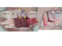
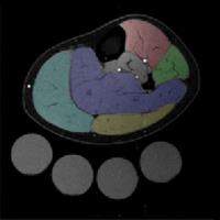
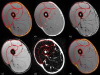
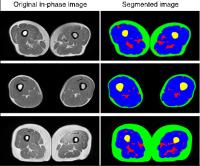
Automatic segmentation for volume quantification of quadriceps
muscle head in athletes during an extreme mountain
ultra-marathon
Benjamin Gilles1, Charles de Bourguignon2,
Pierre Croisille3, Grégoire Millet4,
Magalie Viallon3, and Olivier Beuf5
1LIRMM; CNRS (UMR 5506) Université de
Montpellier, Montpellier, France, 2Radiology
Dept, CHU de Saint Etienne, Saint Etienne, France, 3CREATIS,
Université de Lyon ; CNRS UMR5220 ; Inserm U1044 ; INSA-Lyon
; Université Claude Bernard Lyon 1, Saint Etienne, France, 4Institute
of Sport Sciences, University of Lausanne, Lausanne,
Switzerland, 5CREATIS,
Université de Lyon ; CNRS UMR5220 ; Inserm U1044 ; INSA-Lyon
; Université Claude Bernard Lyon 1, Villeurbanne, France
Acute loss of skeletal muscle mass is a common feature of
several pathologies such as stroke, cancer, chronic
obstructive pulmonary disease. Having a none invasive method
to accurately quantify muscle mass is of crucial interest to
follow procedure that could prevent muscle wasting and
restore physical capacity, mobility and optimize motor
recovery. The aim of the current study is to propose an
automatic segmentation technique to quantify muscle mass.
The automatic segmentation of 3D quadriceps volumes was
performed using a deformable registration technique applied
to 3D isotropic in-phase (IN), out-phase (OUT), and
calculated fat (F) and water (W) images obtained using a
double-echo gradient echo Dixon coronal acquisition in order
to test the best contrast channel for segmentation. The
method was tested in a longitudinal study in athletes
enrolled for the most extreme mountain ultra-marathon (The
Tor des Géants, Courmayeur, Italy: +24000 positive
elevation, 330km). 51 athletes were scans at departure, 27
finishers at the arrival and 2 days after recovery, leading
to 105 datasets that were segmented in total. The best
automatic segmentation accuracy was obtained when using the
calculated Water image (DSC=0,946).
|
|
4517.
 |
82 |
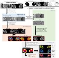
Simultaneous quantitative susceptibility, PDFF and transversal
relaxation time mapping in dystrophic skeletal muscle -
Video Not Available
Benjamin Leporq1,2, Arnaud Le Troter3,
Yann Le fur3, Emmanuelle Salort-Campana4,
Maxime Guye3, Olivier Beuf2, and David
Bendahan3,5
1Center of Research on inflammation; Inserm
U1149, Université Paris Diderot, Paris, France, 2CREATIS
CNRS UMR 5220; Inserm U1044, Université de Lyon,
Villeurbanne, France, 3CRMBM;
CNRS UMR 7339, Aix-Marseille University, Marseille, France, 4Genetique
Médicale et Génomique Fonctionelle; Inserm UMR S_910, Aix
Marseille University, Marseille, France, 5CEMEREM,
Hopital de la Timone, Pôle d’imagerie médicale, AP-HM,
Marseille, France
We have developed a dedicated algorithm allowing to
quantify, from a single MR acquisition fat and muscle
fractions together with magnetic susceptibility and
transverse relaxation time (T2*). This approach was linked
to a dedicated segmentation algorithm allowing to quantify
specific indices which could be of interest for the
assessment of disease severity and progression. Our results
showed the feasibility of quantitative susceptibility
mapping (QSM) in thigh muscles and demonstrated that its
implementation into the fat-water separation reconstruction
pipeline is possible. For dystrophies assessment, magnetic
susceptibility-related information might provide a useful
supplementary materials in comparison to relaxometry and fat
fraction measurements.
|
|
4518.
 |
83 |
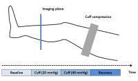
Evaluation of Cuff-Induced Skeletal Muscle Microvascular
Perfusion of Lower Extremity by ASL and IVIM MRI techniques
Qing Lu1, Shiteng Suo1, Hui Tang1,
Jianxun Qu2, Yong Zhang2, and Jianrong
Xu1
1Department of Radiology, Ren Ji Hospital, School
of Medicine, Shanghai Jiao Tong University, Shanghai, China,
People's Republic of, 2GE
Healthcare China, Shanghai, China, People's Republic of
Arterial spin labeling (ASL) and intravoxel incoherent
motion (IVIM) are both noninvasive MRI techniques that offer
quantitative perfusion measurements. The current study
showed that the ASL perfusion decreased while the IVIM
vascular volume fraction increased compared to baseline
under cuff compression paradigm in the lower extremity
muscle, indicating that the two MRI techniques based on two
completely distinct mechanisms provide complementary tissue
perfusion characteristics.
|
|
4519.
 |
84 |
Quantitative MR evaluation of fatty infiltration and edema-like
processes in skeletal muscles of Myotonic Dystrophy type 1
Linda Heskamp1, Marlies van Nimwegen2,
Barbara Janssen1, Baziel van Engelen2,
and Arend Heerschap1
1Department of Radiology and Nuclear Medicine,
Radboud university medical center, Nijmegen, Netherlands, 2Department
of Neurology, Radboud university medical center, Nijmegen,
Netherlands
We used quantitative MR to evaluate the extent of fatty
infiltration and edema-like processes in muscles of patients
with Myotonic Dystrophy type 1 (DM1). Fat fractions were
obtained using a DIXON method and the T2 of muscle water (T2water)
was calculated using a bi-component extended phase graph
model. The results show that fatty infiltration in DM1 is a
slow gradual process whereby the distal part of the muscle
is more heavily fat infiltrated than the proximal part. In
addition, muscles that are in an active process of fatty
infiltration have an elevated T2water, possibly
from reactive edema.
|
 |
4520.
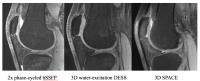 |
85 |
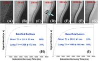
Rapid High Resolution 3D Musculoskeletal Imaging at 7T:
Contrast Optimization and Comparison of DESS, Phase-Cycled
bSSFP, and 3D SPACE
Meredith Taylor1, Haonan Wang1, Antony
JR Palmer2, Andrew J Carr2, Sion
Glyn-Jones2, Daniel Park2, and Neal K
Bangerter1
1Electrical Engineering, Brigham Young
University, Provo, UT, United States, 2Nuffield
Department of Orthopaedics, Rheumatology, and
Musculoskeletal Sciences, University of Oxford, Oxford,
United Kingdom
In this study, we (1) implemented a two-acquisition 3D
phase-cycled bSSFP protocol at 7 Tesla that achieves 0.31mm
isotropic resolution in under 9 minutes of scan time, (2)
implemented a 3D DESS protocol at 7 Tesla that achieves
0.36mm isotropic resolution in just under 7 minutes, (3)
performed a contrast optimization to identify flip angles
that maximize both cartilage/muscle and cartilage/synovial
fluid contrast, and (3) compared to a 3D SPACE acquisition
at 7T that achieves 0.55mm isotropic resolution in a scan
time of 11:37.
|
|
4521.
 |
86 |
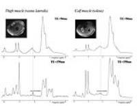
1H MRS can detect and quantify acetylcarnitine in
different human skeletal muscles at rest at 7T. -
Permission Withheld
Radka Tušková1,2,3, Ladislav Valkovic1,3,4,5,
Martin Gajdošík1,3, Thomas Heckmann6,
Norbert Bachl6, Harald Tschan6,
Siegfried Trattnig1,3, and Martin Krššák1,3,7
1High-Field MR Center, Department of Biomedical
Imaging and Image-Guided Therapy, Medical University of
Vienna, Vienna, Austria, 2Faculty
of Chemical and Food Technology, Department of NMR
Spectroscopy and Mass Spectrometry, Slovak University of
Technology in Bratislava, Bratislava, Slovakia, 3Christian
Doppler Laboratory for Clinical Molecular MR Imaging,
Vienna, Austria, 4John
Radcliffe Hospital, University of Oxford Centre for Clinical
Magnetic Resonance Research, University of Oxford, Oxford,
United Kingdom, 5Department
of Imaging Methods, Institute of Measurements Science,
Slovak Academy of Sciences, Bratislava, Slovakia, 6Center
of Sport Science and University Sport, University of Vienna,
Vienna, Austria, 7Division
of Endocrinology and Metabolism, Department of Internal
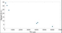
Medicine III, Medical University of Vienna, Vienna, Austria
Carnitine plays an important role in fat metabolism. A
long-echo time (TE of 350ms) proton magnetic resonance
spectroscopy protocol was implemented for detection of
skeletal muscle acetylcarnitine at rest on a clinical 7T
scanner in the calf (soleus) and thigh (vastus lateralis)
muscle. T2 relaxation times of the 2.13 ppm signal of
acetlylcarnitine at 7T were assessed as 137.8±47.7ms.
Concentrations of acetylcarnitine in vastus lateralis muscle
in four healthy volunteers were found to be 1.69±0.21mmol/kg
wet weight, whereas lower concentrations (i.e.,
0.54±0.19mmol/kg) were found in soleus muscle.
|
|
4522.
 |
87 |
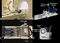
A Low-Cost MR Compatible Ergometer For Assessing Lower Leg
Muscle Metabolism
Xuejiao Che1, Ryan Brown 1,2,
Leeor Alon1,2, Ravinder R Regatte1,
and Prodromos Parasoglou1
1Department of Radiology, New York University
School of Medicine, New York, NY, United States, 2NYU
WIRELESS, Polytechnic Institute of New York University,
Brooklyn, NY, United States
In this work, we designed and constructed an inexpensive MR
compatible ergometer that can be used for studying lower leg
muscle metabolism. This ergometer allows subjects to perform
a plantar flexion exercise protocol while 31P-MR
data are acquired. The device is easy to use, and it can be
positioned inside the bore of the magnet in less than 10
min. The mechanical power exerted by the subject can be
estimated from force and angle displacement signals that are
continuously monitored, while the exersice intensity can be
varied by changing the number and/or the material of the
resistive elastic cords.
|
|
4523.
 |
88 |
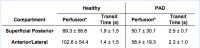
What is the Relationship between Vascular Disease Distribution
in PAD and Exercise-Induced Hyperemia Pattern in Calf Muscle?
Christopher J Hanrahan1, Jeff L Zhang1,
Gwenael Layec2, Corey Hart2, Michelle
Mueller3, Daniel Kim1, Kristi Carlston1,
Russell S Richardson2, and Vivian S Lee1
1Radiology, Utah Center for Advanced Imaging
Research (UCAIR), University of Utah School of Medicine,
Salt Lake City, UT, United States, 2Internal
Medicine, Division of Geriatrics, Utah Vascular Research Lab
(UVRL), University of Utah School of Medicine, Salt Lake
City, UT, United States, 3Surgery,
University of Utah School of Medicine, Salt Lake City, UT,
United States
Calf muscle perfusion by first-pass gadolinium MRI provides
objective measures to help understand the relationship
between vascular pathology and muscle dysfunction in
peripheral arterial disease (PAD) patients. We compared
perfusion in healthy and PAD subjects in exercise-recovery
and, in the same PAD patients, related muscle perfusion
pattern to hemodynamically significant vessel pathology
found at MR arteriography. We found no relation between
specific stenosis/occlusion and the expected muscle
perfusion downstream, but calf vascular pathology
significantly decreased perfusion in the superficial
posterior compartment muscles compared to
abdominopelvic/thigh vessel abnormality. Assessing muscle
perfusion shows promise in assessing PAD disease severity
and guiding treatment.
|
|
4524.
 |
89 |
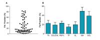
Magnetic resonance imaging estimates of muscle volume and
inter-muscular fat in the thigh in sarcopenia population:
correlation with physical performances
Yu Xin Yang1, Mei Sian Chong1, Laura
Tay1, Suzanne Yew1, Audrey Yeo1,
and Cher Heng Tan2
1Institute of Geriatrics and Active Ageing, Tan
Tock Seng Hospital, Singapore, Singapore, 2Department
of Diagnostic Radiology, Tan Tock Seng Hospital, Singapore,
Singapore
This study presented using MRI to quantify muscle and fat
volumes in thigh for sarcopenic and sarcopenic obese (SO)
populations. The correlation between different thigh
components and patients’ physical performances was also
assessed. Results show that MRI is a promising tool for
early detection of sarcopenia and SO. This may translate to
use in clinical trials and in clinical practice. MRI
measurement of inter-muscular fat volume is a valuable
component in thigh to monitor patients’ physical
performances.
|
|
4525.
 |
90 |
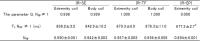
Comparison of relative RF power deposition for shoulder MRI at
3.0T and 7.0T using 3D dual echo steady state imaging -
Video Not Available
Marko Hoehne1, Andreas Graessl2, Antje
Els2, Thomas Herold3, and Thoralf
Niendorf4
1HELIOS Klinikum Berlin Buch, Radiology, Max
Delbrück Center for Molecular Medicine (MDC)
Berlin,Ultrahigh Field Facility (B.U.F.F.), Berlin, Germany, 2Max
Delbrück Center for Molecular Medicine (MDC), Ultrahigh
Field Facility (B.U.F.F.), Berlin, Germany, 3HELIOS
Klinikum Berlin Buch, Radiology, Berlin, Germany, 4Max
Delbrück Center for Molecular Medicine (MDC), Experimental
and Clinical Research Center (ECRC), Charite Campus Berlin
Buch, Humboldt University Berlin, Ultrahigh Field Facility
(B.U.F.F.), Berlin, Germany
Technology advances in ultra-high field systems improve
significantly diagnose of different musculoskeletal
structures. A challenge of this work examines relative RF
power deposition for shoulder MRI with dual echo steady
state imaging at 3.0T and 7.0T. Volunteers (n=10, mean age
36.5 ± 8.51 years) were investigated at 3.0 T and 7.0 T. The
flip angle was varied for each field strength. A comparison
of flip angle between 3.0T and 7.0T showed a SAR gain of
approximately 2.6 for the local RF coil setup used at 7.0 T
versus the body coil configuration employed at 3.0 T. It is
important to considering a right choice of sequences and
these parameters.
|
|
4526.
 |
91 |
Fat infiltration is non-uniform along the proximodistal muscle
axis in Duchenne Muscular Dystrophy
Melissa Hooijmans1, Nathalie Doorenweerd1,
Jedrek Burakiewicz1, Jan Verschuuren2,
Constantin Anastasopoulos1, Andrew Webb1,
Erik Niks2, and Hermien Kan1
1Radiology, Leiden University Medical Center,
Leiden, Netherlands, 2Neurology,
Leiden University Medical Center, Leiden, Netherlands
Progressive replacement of muscle tissue by fat is one of
the main characteristics of DMD. This muscle degeneration
process has been extensively studied in terms of differences
between individual muscles, but not as a function of
physical location within each individual muscle. This work
showed non-uniform fat infiltration along the proximodistal
muscle axis within individual muscles using the Dixon
water/fat technique. These observations provide new insight
into disease progression in DMD.
|
|
4527.
 |
92 |
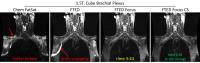
Towards Fast and Robust Bilateral Brachial Plexus Imaging
Kang Wang1, Ken-Pin Hwang2,3, Zac
Slavens4, Adriana Kanwischer5, Kevin
King5, Suchandrima Banerjee6, Pauline
Worters6, and Ersin Bayram2
1Global MR Applications & Workflow, GE
Healthcare, Madison, WI, United States, 2Global
MR Applications & Workflow, GE Healthcare, Houston, TX,
United States, 3Department
of Imaging Physics, University of Texas M.D. Anderson Cancer
Center, Houston, TX, United States, 4MR
Engineering, GE Healthcare, Waukesha, WI, United States, 5Global
MR Applications & Workflow, GE Healthcare, Waukesha, WI,
United States, 6Global
MR Applications & Workflow, GE Healthcare, Menlo Park, CA,
United States
MR imaging of bilateral brachial plexus has been challenging
due to various reasons, such as fat suppression failures
caused by B0 inhomogeneity,
arms wrapping in arms-down imaging for patient comfort, and
long scan time, etc. In this work, these aforementioned
challenges were addressed by combining and utilizing novel
MR imaging techniques, and a fast and robust protocol for
bilateral brachial plexus MR imaging is proposed.
|
|
4528.
 |
93 |
Fasciculation MR Imaging (faMRI) of the Lower Leg -
Permission Withheld
Nikolaus M. Szeverenyi1 and
Graeme M. Bydder1
1Radiology, University of California, San Diego,
San Diego, CA, United States
Fasciculations are brief spontaneous contractions affecting
a small number of muscle fibers. We investigated how
diffusion sensitized MR images were able to detect these
contractions in the lower leg of healthy volunteers. Large
intensity decreases were observed (at random times) in
random areas of muscle on images, acquired repeatedly using
single shot (diffusion sensitized) EPI acquisitions over the
course of several minutes. Signal intensity reductions were
attributed to intra-voxel incoherent-like motion due to
displacement of tissue. Quantification compared activated
areas to total muscle area and frequency of activation on a
per pixel basis. Results were expressed as a fasciculation
index parameter and in fasciculation frequency maps.
|
|
4529.
 |
94 |
Towards high temporal resolution Creatine Chemical Exchange
Saturation Transfer (Cr-CEST) during plantar flexion exercise:
Preliminary results at 7T
Esaú Poblador Rodriguez1, Marek Chmelík1,2,
Vladimír Mlynárik1,2, Siegfried Trattnig1,2,
and Wolfgang Bogner1
1High Field MR Centre, Department of Biomedical
Imaging and Image-guided Therapy, Medical University of
Vienna, Vienna, Austria, 2Christian
Doppler Laboratory for Clinical Molecular MR Imaging,
Vienna, Austria
Once the technical limitations are cleared, Cr-CEST could
replace 31P-MRS,
becoming a powerful tool for assessment of treatment
outcomes and diagnosis of muscular disorders, due to its
superior spatial resolution and sensitivity. Phantom
measurements show how Cr concentration and pH are linearly
correlated with CEST contrast maps. The preliminary in-vivo measurements,
with a time resolution of 13.1s per repetition, produce an
enhancement of gastrocnemius muscle of 12% during plantar
flexion exercise. However, a further increased time
resolution is anticipated for dynamic studies, close to
those routinely used in dynamic 31P-MRS.
|
|
4530.
 |
95 |
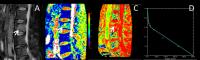
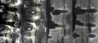
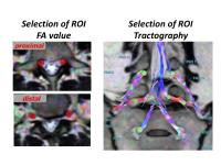
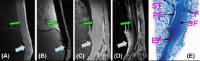
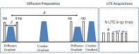
High-Resolution DTI of Distal Peripheral Nerves Using
Flow-Compensated Diffusion-Prepared 3D TSE
Barbara Cervantes1, Qinwei Zhang2, Kim
van de Ven3, Hendrik Kooijman4, Ernst
Rummeny1, Axel Haase5, Gustav J
Strijkers2, Jan S Kirschke6, Aart J
Nederveen2, and Dimitrios C Karampinos1
1Diagnostic and Interventional Radiology,
Technische Universität München, Munich, Germany, 2Radiology,
Academic Medical Center, Amsterdam, Netherlands, 3Philips
Healthcare, Best, Netherlands,4Philips
Healthcare, Hamburg, Germany, 5Zentralinstitut
für Medizintechnik, Garching, Germany, 6Neuroradiology,
Technische Universität München, Munich, Germany
Quantitative MRI is becoming a promising tool in the
assessment of peripheral nerve pathologies and anomalies.
Peripheral neuropathy is frequently accompanied by
neuropathic changes, which can be quantified with diffusion
tensor imaging (DTI). Given the small sizes and oblique
geometries of many peripheral nerves, peripheral-nerve DTI
requires an acquisition method that can provide
high-resolution, distortion-free images in acceptable
clinical scanning times. The present work demonstrates
isotropic- and sub-millimeter-resolution, artifact-free DTI
of the nerves in the lower extremity using flow-compensated
diffusion-prepared 3D turbo spin echo (TSE).
|
|
4531.
 |
96 |
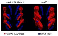
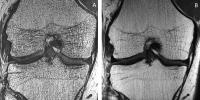
Volumetric Brachial Plexus Imaging at 3T with Dual-echo Dixon
TSE: comparison against 3D STIR and 3D SPAIR
Xinzeng Wang1, Crystal E. Harrison1,
Yogesh K. Mariappan2, Karthik Gopalakrishnan2,
Avneesh Chhabra1,3, Robert E. Lenkinski1,3,
and Ananth J. Madhuranthakam1,3
1Radiology, UT Southwestern Medical Center,
Dallas, TX, United States, 2Philips
Innovation Campus, Philips Healthcare, Bangalore, India, 3Advanced
Imaging Research Center, UT Southwestern Medical Center,
Dallas, TX, United States
Volumetric Brachial Plexus imaging at 3T often suffers from
incomplete fat suppression and reduced SNR with standard
STIR and SPAIR due to increased B1 and B0 inhomogeneities.
Dual-echo Dixon TSE has been shown to achieve uniform fat
suppression without increasing total scan time or SNR
penalty by acquiring two echoes in the same repetition. In
this work, we compared 3D dual-echo Dixon TSE against
current standard of care 3D STIR and 3D SPAIR for brachial
plexus imaging with respect to fat suppression, blood
suppression, nerve visualization and SNR at 3T. Overall, the
3D dual-echo Dixon TSE showed significantly improved
performance.
|
|