10:45
|
|
Assessing CNS Vasculature and inflammation using dual GBCA
and ferumoxytol-enhanced MRI 
Edward Neuwelt
Ferumoxytol as an MRI contrast can provide
additional information on CNS lesions. Pre-clinical
studies have used advanced neuroimaging techniques with
ferumoxytol to evaluate tumor changes after different
treatments in animal models as well as evaluation of
acute neuroinflammation. Clinically, ferumoxytol has
been used to differentiate tumor progression from
pseudoprogression and also to evaluate inflammatory and
vascular CNS lesions. Dual-contrast imaging may mark
the beginning of a multicontrast era when different
contrast agents are applied for specific purposes to
evaluate CNS lesions. Improved neuroimaging can
potentially be incorporated into standard of care for
assessing therapy-induced changes and tumor response to
therapy.
|
11:15
|
0082.
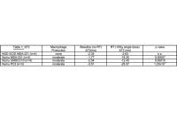 |
Radiation-induced inflammatory response in tumor-bearing
immune-compromised mice by SPIO-enhanced T2-MRI
Natalie Julie Serkova1, Kendra M Huber1,
Barbara Frederick2, Elizabeth R Kessler3,
Thomas W Flaig3, and Brian D Kabanagh2
1Anesthesiology, University of Colorado
Denver, Aurora, CO, United States, 2Radiation
Oncology, University of Colorado Denver, Aurora, CO,
United States, 3Medical
Oncology, University of Colorado Denver, Aurora, CO,
United States
Clinically, the radiation treatment (RT) is know to
trigger an inflammatory response which can be beneficial
for overall anti-cancer treatment efficacy. However, in
pre-clinical mouse models, the tumor response to the RT
is rather heterogenous. Our hypothesis is that
tumor-associated macrophages which drive the
pro-inflammatory response to the RT, are expressed
differently in various mouse strains based on their
genetic make-up. The goal of this study was to
non-invasively assess the tumor inflammatory response to
the RT based on iron oxide-induced changes in T2-MRI
after injection of SPIO nanoparticles in two different
mouse models with severely (NOD SCID) and moderately
(nu/nu athymic) compromised immune system.
|
11:27
|
0083.
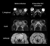 |
Neuroimaging of Nipah Virus in a Syrian Hamster Model of
Infection
Margaret R. Lentz1, Dima A. Hammoud2,
Yu Cong1, Oscar Rojas1, David
Thomasson1, Peter B. Jahrling1,3,
and Michael R. Holbrook1
1Integrated Research Facility, NIAID,
National Institutes of Health, Frederick, MD, United
States, 2Radiology
and Imaging Sciences, Clinical Center, National
Institutes of Health, Bethesda, MD, United States,3Emerging
Viral Pathogens Section, NIAID, National Insitutes of
Health, Frederick, MD, United States
The purpose of this study was to utilize MRI to assess
alterations in the brain that occur in a Golden Syrian
hamster infected with Nipah virus (NiV) via intranasal
inoculation. Within 9 days of exposure to NiV, signal
alterations were observed in the olfactory bulb in T2-weighted
and FLAIR images, suggestive of inflammation and edema
induced by NiV crossing the olfactory epithelium. The
identification of non-invasive imaging biomarkers of
acute NiV neurologic disease progression in this animal
model could aid in the examination of potential vaccines
and therapeutics.
|
11:39
|
|
Novel Imaging Tracers for Rapid and Noninvasive Assessment
of Bacterial Infections - Permission Withheld
Sanjay Jain1
1Johns Hopkins Medical Institute
We are developing novel imaging tracers for rapid and
noninvasive assessment of bacterial infections and to
study antimicrobial pharmacokinetics.
|
12:09
|
0084.
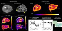 |
Diffusion and perfusion MR imaging indicate inflammation
followed by fibrosis in a hepatitis B infected humanized
mouse liver model
Prashant Chandrasekharan1, Dahai Zheng2,
Kavita Kaur D/O Ranjit Singh1, Qingfeng Chen2,
and Kai Hsiang Chuang1
1A*STAR, Singapore Bio Imaging Consortium,
Singapore, Singapore, 2A*STAR,
Institute of Molecular and Cell Biology, Singapore,
Singapore
Humanized mouse model of liver infection is essential to
understand the role of the immune system during disease
progression and therapeutic intervention. In this study
we have used MRI functional imaging bio-markers to
assess the pathology related to Hepatitis B infection in
a humanized mouse liver model.
|
12:21
|
0085.
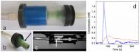 |
Identifying carotid plaque inflammation using high and low
molecular weight contrast agents
Jason Kraig Mendes1, Scott McNally1,
Seong-Eun Kim1, Bradley D. Bolster2,
Gerald S. Treiman3, and Dennis L. Parker1
1Radiology, University of Utah, SLC, UT,
United States, 2Siemens
Healthcare, SLC, UT, United States, 3Department
of Veterans Affairs, SLC, UT, United States
Carotid plaque inflammation can be measured with dynamic
contrast enhanced (DCE) MRI and is a marker for plaque
instability. Despite this, DCE has not become a
clinically viable tool in diagnosing carotid plaque
instability and the corresponding stroke risk. The
barrier to progress is a DCE protocol meeting
requirements for clinical use to monitor medical
treatment effect or failure. This project overcomes this
barrier by developing a reliable and inclusive dual
contrast DCE protocol to identify carotid plaque
inflammation.
|
12:33
|
|
Panel Discussion |
12:45
|
|
Adjournment & Meet the
Teachers |
|