| |
10:45
 |
0042.
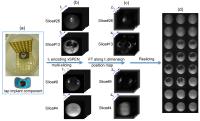 |
Evolution-time encoded single-scan cross spatiotemporal encoding
imaging near metal implants 
Zhiyong Zhang1,2, Amir Seginer1, and
Lucio Frydman1
1Chemical Physics, Weizmann Institute of Science,
Rehovot, Israel, 2Electronic
Science, Xiamen University, Xiamen, China, People's Republic
of
Magnetic resonance imaging (MRI) near metallic implants
remains an unmet need because of severe artifacts, which
mainly stem from large metal-induced field inhomogeneities.
The single-scan cross spatiotemporal encoding (xSPEN)
technique delivers in-plane distortion-free 2D images under
such large field inhomogeneity condition, while the
slice-plane displacement, “signal voids” and “pile-up”
effects are proposed to be solved by applying t1-evolution-time
encoding on the multi-slicing 2D xSPEN technique. Compared
to the popular “SEMAC” and “MAVIC” techniques, the
remarkable time efficiency of this t1-encoding
xSPEN thus enable many advanced MRI applications near metal
implants with another additional dimension, such as
diffusing MRI, function MRI.
|
| |
10:57
|
0043.
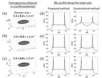 |
Fast Fourier transform-based susceptibility-to-B0 calculation
without aliasing artifacts
Lee Seungkyun1,2
1Center for Neuroscience Imaging Research (CNIR),
Institute for Basic Science (IBS), Suwon, Korea, Republic
of, 2Department
of Biomedical Engineering, Sungkyunkwan University (SKKU),
Suwon, Korea, Republic of
In the Fourier transform-based susceptibility-to-B0 calculation,
the dipolar field kernel (1/3-kz2/k2)
is discretely sampled in the k-space, which leads to
aliasing artifacts in the spatial domain. We show that
calculating and discretizing the dipolar field kernel in the
spatial domain, before the Fourier transform, can
effectively reduce the aliasing effect without resorting to
large zero-filled buffers. In particular, aliasing is
eliminated if the spatial-domain grid size is larger than
the combined dimensions of the susceptibility source and the
B0 target
regions. The new method can accelerate repeated calculations
of susceptibility-induced B0 fields.
|
| |
11:09
|
0044.
 |
Concomitant gradient effects on chemical shift encoded imaging
Timothy J Colgan1,2, Diego Hernando1,
Samir D Sharma1, Ann Shimakawa3, and
Scott B Reeder1,2,4,5,6
1Radiology, University of Wisconsin, Madison, WI,
United States, 2Medical
Physics, University of Wisconsin, Madison, WI, United
States, 3Global
Applied Science Lab, GE Healthcare, Menlo Park, CA, United
States, 4Biomedical
Engineering, University of Wisconsin, Madison, WI, United
States, 5Medicine,
University of Wisconsin, Madison, WI, United States, 6Emergency
Medicine, University of Wisconsin, Madison, WI, United
States
Quantitative chemical shift-encoded (CSE) MRI techniques
acquire complex-valued (magnitude and phase) images at
multiple echo times (TE), enabling simultaneous mapping of
fat-fraction, R2* (=1/T2*) and B0field.
Applications of CSE-MRI include tissue fat quantification,
iron quantification and quantitative susceptibility mapping
(QSM). Recently, phase shifts due to concomitant gradients
(CG) have been identified as a source of error for
quantitative CSE techniques, so their effects on
fat-fraction, R2* and B0 maps
are characterized in this study. CG correction of
experimental data demonstrates that the detrimental effects
of CG phase shifts can be removed before reconstruction to
produce more accurate estimates of the fat-fraction, R2*,
and field map measurements.
|
| |
11:21
|
0045.
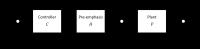 |
Real-Time Field Control Using Full 3rd-Order Matrix Pre-Emphasis
Yolanda Duerst1, Bertram J. Wilm1,
Benjamin E. Dietrich1, Simon Gross1,
Thomas Schmid1, David O. Brunner1, and
Klaas P. Pruessmann1
1ETH Zurich, Zurich, Switzerland
Update steps of real-time field control suffer from
imperfect shim responses which degrade control quality. By
including full 3rd-order matrix pre-emphasis as
an additional filter in the control loop, all self-term
responses are shaped to be equal and all cross-term
responses are directly suppressed. This leads to
disturbances being rejected faster and less noise
amplification. Thus enables better field control in
demanding situations such as caused by disturbance of high
spatial and temporal variability.
|
| |
11:33
|
0046.
 |
Reducing Brain MRI Artifacts Caused by Ferromagnetic Orthodontic
Appliances Using Permanent Magnets
Zhiyue J Wang1,2, Yong Jong Park1,2,
Youngseob Seo1,2, Michael C Morriss1,2,
and Nancy K Rollins1,2
1UT Southwestern Medical Center, Dallas, TX,
United States, 2Children's
Medical Center, Dallas, TX, United States
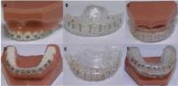
Stainless steel orthodontic appliances are commonly found in
adolescents undergoing clinical brain MRI examinations. They
cause severe magnetic susceptibility artifacts and failure
to obtain diagnostic information from many MR techniques.
The B0 shimming
capability present on clinical MR scanners cannot remove
these artifacts. We have constructed devices for the
correction of these artifacts at 1.5 T using small pieces of
permanent magnets mounted on intra-oral mouth guards or an
extra-oral mouth-band. The magnetic field from the permanent
magnets cancels the B0 inhomogeneity
induced by ferromagnetic orthodontic appliances, resulting
in drastic improvement of MR image quality.
|
| |
11:45
|
0047.
 |
Accelerated Imaging of Metallic Implants Using Model-Based
Nonlinear Reconstruction
Xinwei Shi1,2, Evan G Levine1,2, and
Brian A Hargreaves1,2
1Radiology, Stanford University, Stanford, CA,
United States, 2Electrical
Engineering, Stanford University, Stanford, CA, United
States
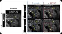
3D Multi-Spectral Imaging (MSI) methods, including SEMAC,
MAVRIC, and MAVRIC-SL, enable MRI near metallic implants by
correcting for the metal-induced off-resonance artifacts,
but their widespread application is limited by prolonged
scan time. In this work, we introduce a novel model-based
reconstruction method to accelerate 3D MSI. We demonstrate
in phantom and in vivo experiments that the proposed method
can accelerate MAVRIC-SL acquisitions by a factor of 4 when
used alone, and 13-17 when combined with parallel imaging
and half-Fourier acquisition. The images reconstructed by
the proposed method showed sharper details and lower level
of noise, compared with model-free L1-ESPIRiT.
|
| |
11:57
|
0048.
 |
Bayesian correction of bias field and Venetian blind for high
resolution ex vivo MRI with clinical scanners
Juan Eugenio Iglesias1, Pedro Manuel Paz-Alonso1,
Garikoitz Lerma-Usabiaga1, Ricardo Insausti2,
Karla Miller3, and César Caballero-Gaudes1
1Basque Center on Cognition, Brain and Language
(BCBL), Donostia - San Sebastián, Spain, 2Human
Neuroanatomy Laboratory, University of Castilla-La Mancha,
Albacete, Spain, 3Centre
for Functional MRI of the Brain, University of Oxford,
Oxford, United Kingdom
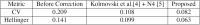
Multi-slab MRI enables the acquisition of ultra-high
resolution ex vivo MRI of the whole human brain with
clinical scanners, by overcoming their hardware limitations
(e.g., memory size). However, multi-slab MRI produces slab
boundary artifacts (SBA) that degrade the image quality and
bias subsequent image analyses. Here we propose a Bayesian
method that corrects for SBA and intensity inhomogeneities /
bias field (BF) simultaneously. The method, which combines a
probabilistic brain atlas and the Expectation Maximization
algorithm, takes advantage of the interplay between the two
artifacts to outperform state-of-the-art SBA and BF
correction algorithms (even when used in combination).
|
| |
12:09
|
0049.
 |
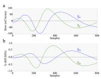
Breathing-induced B0 field fluctuations in the cervical spinal
cord at 7T
Signe Johanna Vannesjo1, Falk Eippert1,
Yazhuo Kong1, Stuart Clare1, Karla L
Miller1, and Irene Tracey1
1FMRIB centre, NDCN, University of Oxford,
Oxford, United Kingdom
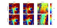
Spinal cord MRI at ultra-high field poses considerable
technical challenges, especially related to static and
dynamic B0 field
variations. We here investigated the magnitude and spatial
profile of breathing-induced B0 field
fluctuations in the cervical spinal cord at 7T, by comparing
field maps acquired during breath-holds in an expired vs.
inspired breathing state. Breathing-related field
fluctuations of up to 140Hz at the level of C7 were
observed. We further implemented a proof-of-principle shim
correction, demonstrating the feasibility of using the shim
system to compensate for the breathing-induced fields.
|
| |
12:21
|
0050.
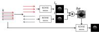 |
Robust Nyquist Ghost Correction by Incorporating Phase Errors
Correction in SENSE
Victor B. Xie1,2, Mengye Lyu1,2,
Yilong Liu1,2, Yangqiu Feng1,2, and Ed
X. Wu1,2
1Laboratory of Biomedical Imaging and Signal
Processing, The University of Hong Kong, Hong Kong SAR,
China, People's Republic of, 2Department
of Electrical and Electronic Engineering, The University of
Hong Kong, Hong Kong SAR, China, People's Republic of
In this abstract, we proposed a novel method that can fully
and robustly correct EPI Nyquist ghost by incorporating
high-order phase error correction into SENSE reconstruction.
More importantly, this method does not induce SNR loss,
greatly benefiting the final reconstructed images. Phantom
and in vivo imaging results clearly demonstrated the
efficacy of this method in ghost correct as well as its
superior SNR performance, particularly in accelerated data
set that can suffer from amplified noise problems. This
novel method has great potentials to be applied in all kinds
of EPI-based MRI studies, such as fMRI and DTI.
|
| |
12:33
|
0051.
 |
B0 Eddy Current Correction for Spiral MRI
Ryan K Robison1, Dinghui Wang1,
Zhiqiang Li1, and James G Pipe1
1Imaging Research, Barrow Neurological Institute,
Phoenix, AZ, United States
Eddy currents are a common source of artifacts in Spiral
MRI. Eddy currents that effect the k-space trajectory are
often the focus of eddy current correction. However, the
spatially uniform but time-varying B0 eddy currents can also
be a subtle but important source of artifacts in spiral
images. This work demonstrates the improvement in image
quality that can result from measuring and correcting the
phase produced by B0 eddy currents in spiral MRI.
|
|