| |
10:00
|
0587.
 |
A Joint Image Denoising Approach for Improved Precision and
Accuracy in Myocardial T1 Mapping 
Aurelien Bustin1,2,3, Pauline Ferry3,
Andrei Codreanu4, Anne Menini2, and
Freddy Odille3,5,6
1Department of Computer Science, Technische
Universität München, Munich, Germany, 2GE
Global Research, Munich, Germany, 3Imagerie
Adaptative Diagnostique et Interventionnelle, Universite de
Lorraine, Nancy, France, 4Centre
Hospitalier de Luxembourg, Luxembourg, Luxembourg, 5CIC-IT
1433, INSERM, Nancy, France, 6U947,
INSERM, Nancy, France
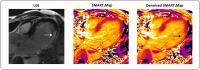
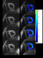
To improve precision and accuracy in myocardial T1 mapping
by combining saturation-recovery acquisitions with a joint
denoising method. The proposed method is shown to improve
mapping techniques by exploiting the spatiotemporal
correlations in the native T1-weighted images,
thus providing a promising tool for the measurement of
myocardial and blood T1 times.
|
| |
10:12
|
0588.
 |
Detecting diffuse cardiac fibrosis with T1? MRI
Joep van Oorschot1, Fatih Guclu2,
Peter Luijten1, Tim Leiner1, and Jaco
Zwanenburg1
1Radiology, University Medical Center Utrecht,
Utrecht, Netherlands, 2Cardiology,
University Medical Center Utrecht, Utrecht, Netherlands
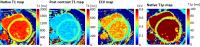
Native T1ρ-mapping is a promising non-contrast enhanced
method for fibrosis detection, that would overcome problems
associated with contrast agent use. In this work, we will
evaluate the performance of T1ρ-mapping versus ECV-m and
native T1 in DCM patients. Native T1, native T1ρ and
Contrast enhanced T1-maps were acquired in twelve DCM
patients, and 8 healthy volunteers. The T1ρ relaxation time
was significantly higher in the DCM patients (55.6 ± 3.0
ms), compared to the healthy control subjects (51.5 ± 1.2
ms), p<0.005. A significant correlation was found between
the T1ρ relaxation time and the Extracellular Volume
fraction in patients.
|
| |
10:24
 |
0589.
 |
Improved myocardial T1 mapping technique to eliminate
device-induced image artefacts for patients with implanted
cardiac devices
Jiaxin Shao1, Shams Rashid1, Kim-Lien
Nguyen2,3, and Peng Hu1,4
1UCLADepartment of Radiological Sciences, David
Geffen School of Medicine, University of California, Los
Angeles, CA, United States, 2Department
of Medicine, Division of Cardiology, David Geffen School of
Medicine, University of California, Los Angeles, CA, United
States, 3Division
of Cardiology, Veterans Affairs Greater Los Angeles
Healthcare System, Los Angeles, CA, United States, 4Biomedical
Physics Inter-Departmental Graduate Program, University of
California, Los Angeles, CA, United States
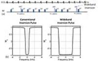
Current cardiac T1 mapping techniques, including the
modified Look-Locker inversion-recovery (MOLLI), cannot be
used effectively in patients with implanted cardiac devices
due to large off-resonance induced by the device. To
eliminate the device-induced image artefacts, we developed a
technique by modifying the MOLLI sequence to use spoiled
gradient echo readout and a wideband inversion pulse, with a
new acquisition scheme and T1 estimation algorithm. The
feasibility of our new technique was tested in phantom
studies and validated in eight healthy volunteers and ten
patients with implanted cardiac devices at 1.5 Tesla.
|
| |
10:36
|
0590.
 |
T2 mapping for non-invasive assessment of acute cardiac
allograft rejection in a mouse model of heterotopic heart
transplantation - Permission Withheld
Dagmar Hartung1,2, Rongjun Chen3,
Marcel Gutberlet1,2, Song Rong3,
Mi-Sun Jang3, Jan Hinrich Braesen4,
Martin Meier2,5, Hermann Haller3,
Frank Wacker1,2, Faikah Gueler3, and
Hueper Katja1,2
1Institute for Diagnostic and Interventional
Radiology, Hannover Medical School, Hannover, Germany, 2Rebirth,
Hannover, Germany, 3Clinic
for Nephrology, Hannover Medical School, Hannover, Germany,4Institute
for Pathology, Hannover Medical School, Hannover, Germany, 5Imaging
Center of the Central Animal Laboratory, Hannover Medical
School, Hannover, Germany
Acute cardiac allograft rejection is a frequent and
life-threatening complication during the first year after
heart transplantation (HTx) and therefore early detection is
most important. The standard of care for HTx recipients is
periodic rejection surveillance by endomyocardial biopsy. We
investigated whether T2 mapping allows non-invasive
detection of acute cardiac allograft rejection in mice. We
demonstrated that myocardial T2 is significantly increased
in allogenic HTx compared to isogenic HTx mice on day 6
after transplantation likely reflecting myocardial edema and
corresponds to the extent of T cell infiltration. Thus,
non-invasive T2 mapping might enable early and non-invasive
detection of acute cardiac allograft rejection.
|
| |
10:48
|
0591.
 |
Slice accelerated Double-Inversion Radial Fast-Spin-Echo for
myocardial black-blood MRI with T2 mapping
Mahesh Bharath Keerthivasan1, Sagar Mandava1,
Kevin Johnson2, Diego R Martin3, Ali
Bilgin1,3,4, and Maria I Altbach3
1Electrical and Computer Engineering, University
of Arizona, Tucson, AZ, United States, 2Siemens
Healthcare, Tucson, AZ, United States, 3Medical
Imaging, University of Arizona, Tucson, AZ, United States,4Biomedical
Engineering, University of Arizona, Tucson, AZ, United
States
A technique to increase slice coverage in dark blood fast
spin echo sequences by a multi-band excitation is presented.
The proposed technique can acquire multiple slices at the
exact null point of blood. The radial version of the single
slice sequence can generate black blood images, TE images
and T2 maps within a single breath-hold. In this work we
present a model based
reconstruction to generate TE images and T2 maps for upto 4
slices in a single breath-hold.
|
| |
11:00
|
0592.
 |
MRI Assessment of Coronary Endothelial Function using Native T1
Mapping with Nitric Oxide Synthase (NOS) Inhibition - Permission Withheld
Sophia Xinyuan Cui1 and
Frederick H. Epstein1,2
1Biomedical Engineering, University of Virginia,
Charlottesville, VA, United States, 2Radiology,
University of Virginia, Charlottesville, VA, United States
Endothelial nitric oxide synthase (eNOS)-mediated production
of NO is an important system regulating the
microvasculature, controlling both vessel diameter and
permeability. We hypothesized that T1 mapping of the heart
during NOS inhibition could detect increased water content
resulting from increased microvascular permeability,
providing a novel means to noninvasively probe eNOS
regulation of the coronary microvasculature. T1-mapping in
mice after intravenous NOS inhibition detected an increase
in myocardial T1 of 113±15 ms compared to baseline
(p<0.05). These methods are likely probing eNOS regulation
of coronary microvascular permeability, which may represent
a novel means of assessing the health of the coronary
endothelium.
|
| |
11:12
|
0593.
 |
Accuracy of cardiac magnetic resonance T1 mapping for detecting
diffuse myocardial fibrosis: comprehensive comparison with the
pathology in diabetic rabbits
Mu Zeng1, Nan Zhang1, Yi He1,
Jing An2, Andreas Greiser3, and
Zhanming Fan1
1Radiology, Beijing Anzhen Hospital,Capital
medical university, Beijing, China, People's Republic of, 2MR
Collaborations NE Asia, Siemens Healthcare, Beijing, China,
Beijing, China, People's Republic of, 3Siemens
AG Healthcare Sector MR, Erlangen, Germany
In recent years, use of the MRI T1 mapping technique to
detect diffuse myocardial fibrosis has received increasing
attention. Although previous studies have verified the
relationship between T1 mapping and pathological findings,
our study is the first to show continuity during the
observation of a single disease while avoiding interference
caused by other diseases. In addition, the pathology can be
fully verified in real time using animal experiments. The
main findings of this study were that (1) the ECV obtained
from the MRI T1 mapping sequence was highly correlated with
the CVF in terms of the degree of histologically diffuse
interstitial fibrosis; (2) the correlation between the
native T1 value and the CVF change was not strong; and (3)
the rabbit is a suitable model for cardiac magnetic
resonance research using clinical equipment.
|
| |
11:24
|
0594.
 |
Oxygen-enhanced T2* cardiac magnetic resonance imaging in
cardiomyopathy - Permission Withheld
Satoshi Kawanami1, Michinobu Nagao1,
Yuzo Yamasaki2, Takeshi Kamitani2,
Torahiko Yamanouchi2, Tomomi Ide3,
Ryohei Funatsu4, Hidetake Yabuuchi5,
Yuji Watanabe1, and Hiroshi Honda2
1Molecular Imaging & Diagnosis, Kyushu
University, Graduate School of Medical Sciences, Fukuoka,
Japan, 2Clinical
Radiology, Kyushu University, Graduate School of Medical
Sciences, Fukuoka, Japan,3Cardiovascular
Medicine, Kyushu University, Graduate School of Medical
Sciences, Fukuoka, Japan, 4Radiological
Technology, Kyushu University Hospital, Fukuoka, Japan, 5Health
Sciences, Kyushu University, Graduate School of Medical
Sciences, Fukuoka, Japan
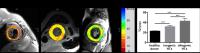
In this study, we analyzed T2* value in the mid-left
ventricular septum avid normoxia (T2*air) and hyperoxia
(T2*oxy) in cases with normal, hypertrophic cardiomyopathy
(HCM) and dilated cardiomyopathy (DCM). Oxygen-enhanced T2*
cardiac magnetic resonance (CMR) showed the different delta
T2* (T2*oxy – T2* air), reflecting myocardial blood-oxygen
dependent (BOLD) effect. Oxygen-enhanced T2* CMR has
potential to open up a new avenue for the study of the
pathophysiology of cardiomyopathy. The ΔT2* was prolonged in
DCM, stable in control and shortened in HCM, respectively.
Oxygen-enhanced T2* CMR can assess the oxygen metabolism in
the mid-left ventricular septum with various density of
capillaries and myocardial cells. We also note the
relationship between T2* value and late gadolinium
enhancement (LGE) or left ventricular ejection fraction
(LVEF).
|
| |
11:36
|
0595.
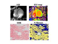 |
Myocardial extracellular volume fraction (ECV) quantified by T1
mapping can detect diffuse myocardial fibrosis in dilated
cardiomyopathy (DCM): Comparison with histological collagen
volume fraction by endomyocardial biopsy (EMB)
Yoshiaki Morita1, Naoaki Yamada1, Emi
Tateishi2, Teruo Noguchi2, Masahiro
Higashi1, and Hiroaki Naito1
1Department of Radiology, National Cerebral and
Cardiovascular Center, Suita, Osaka, Japan, 2Division
of Cardiology, National Cerebral and Cardiovascular Center,
Suita, Osaka, Japan
Diffuse interstitial fibrosis is frequently observed in
dilated cardiomyopathy (DCM). A non-invasive method that
could reliably quantify fibrosis would be preferable. In
this study, we demonstrated that the T1-map-derived ECV
reflects the myocardial collagen volume fraction in DCM.
Therefore, the ECV could be a useful and practical biomarker
for the detection of diffuse interstitial fibrosis that is
difficult to evaluate using only conventional LGE images.
|
| |
11:48
|
0596.
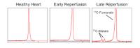 |
Assessment of Necrosis in the Ischemic Heart using
Hyperpolarized [1,4-13C2]Fumarate
Damian J Tyler1, Angus Lau1, Ferdia
Gallagher2, and Marie A Schroeder1
1DPAG, University of Oxford, Oxford, United
Kingdom, 2Radiology,
University of Cambridge, Cambridge, United Kingdom
The aim of this study was to evaluate the potential of
hyperpolarised [1, 4-13C2]fumarate, coupled with MRS, to
measure cardiac necrosis during ischemia and reperfusion.
Hyperpolarised [1, 4-13C2]fumarate was infused into rat
hearts at three time points, corresponding with the healthy
heart, early reperfusion after a 20 min ischemic period, and
late reperfusion. The amount of [1, 4-13C2]malate production
was measured using MRS and quantified to reflect degree of
cardiomyocyte necrosis. We observed a 3.8-fold increase in
[1,4-13C2]malate during the late reperfusion period but no
change in early reperfusion, suggesting that necrotic cell
death takes place during reperfusion only. This technique
shows potential to evaluate therapies targeting necrosis to
prevent cardiac remodeling into failure.
|
|