| |
08:00
 |
1108.
 |
Volume-Parcellated Quantitative Susceptibility Mapping 
Casey Anderson1, Andrew Nencka2, Tugan
Muftuler3, Kathleen Schmainda2, and
Kevin Koch2
1Biophysics, Medical College of Wisconsin,
Milwaukee, WI, United States, 2Radiology,
Medical College of Wisconsin, Milwaukee, WI, United States, 3Neurosurgery,
Medical College of Wisconsin, Milwaukee, WI, United States
Quantitative susceptibility maps are routinely compromised
by streaking artifacts. Here, we present a technique called
volume-parcellated quantitative susceptibility mapping (VP-QSM),
which performs independent susceptibility inversion on
multiple reduced field-of-view parcels over the entire
tissue field map. These parcels are combined to form a
composite susceptibility map. In this algorithm, streaking
artifacts are confined to individual parcels, improving the
quality of the susceptibility map without a dependence on
the underlying QSM inversion algorithm. In this study, VP-QSM
is demonstrated on a 7T human volunteer, as well as on 30
subjects participating in sports concussion and brain cancer
neuroimaging research protocols.
|
| |
08:12
|
1109.
 |
Mapping of magnetic fields due to current injection in the human
brain using MREIT: First measurements.
Aditya Kumar Kasinadhuni1, Munish Chauhan2,
Christopher Anderson1, Michael Schär3,
Aprinda Indahlastari2, Paul Carney1,
Rosalind Sadleir2, and Thomas Mareci1
1University of Florida, Gainesville, FL, United
States, 2Arizona
State University, Tempe, AZ, United States, 3Johns
Hopkins University, Baltimore, MD, United States
Magnetic resonance electrical impedance tomography (MREIT)
relies on phase changes resulting from
electric-current-induced magnetic fields in the direction of
static magnetic field of an MRI scanner. Therefore MREIT can
be employed to estimate conductivity/current density within
the object being imaged. Characterizing current density in
the brain is vital to improving our understanding of
neuromodulation techniques like transcranial direct current
stimulation (tDCS). In this study, to our knowledge, we
performed the first MREIT brain scans of healthy human
volunteers to localize the current-induced magnetic field
generated by tDCS-like currents. These measurements allow
estimation of current density in the human brain.
|
| |
08:24
|
1110.
 |
Relaxation based Conductivity Weighted Imaging (rCWI)
Jaewook Shin1, Min-Oh Kim1, Jun-Hyeong
Kim1, and Dong-Hyun Kim1
1Electrical and Electronic engineering, Yonsei
University, Seoul, Korea, Republic of
To reduce the noise amplification of the conductivity
imaging, the direct calculation of the Laplacian operator
was substituted by appropriate k-space weighted sampling
scheme by the combination of four TSE data with alternating
PE directions.
|
| |
08:36
|
1111.
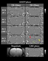 |
Phase Imaging with Multiple Phase-Cycled Pass-Band Balanced
Steady-State Free Precession at 9.4T
Jae-Woong Kim1, Seong-Gi Kim2,3, and
Sung-Hong Park1
1Korea Advanced Institute of Science and
Technology, Daejeon, Korea, Republic of, 2Center
for Neuroscience Imaging Research, Institute for Basic
Science, Suwon, Korea, Republic of, 3Departments
of Biomedical Engineering and Biological Sciences,
Sungkyunkwan University, Suwon, Korea, Republic of
Phase images of pass-band bSSFP were investigated at
multiple phase cycling (PC) angles at high field. Contrast
between white matter and gray matter in phase images of
pass-band bSSFP changed significantly with PC angle and was
twice as high as that of phase images of gradient recalled
echo at a specific PC angle. Phase images of pass-band bSSFP
clearly demonstrated white matter and small structures
presumed to be fiber bundles, which may not be easily
visualized in the conventional methods. Phase imaging with
pass-band bSSFP at multiple phase cycling angles may be a
good anatomical imaging method at ultrahigh field.
|
| |
08:48
|
1112.
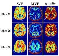 |
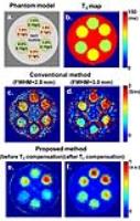
Whole brain in-vivo g-ratio mapping using neurite orientation
dispersion and density imaging (NODDI) and GRE myelin water
imaging (GRE-MWI)
Woojin Jung1, Yoonho Nam2, Hui Zhang3,
and Jongho Lee1
1Laboratory for Imaging Science and Technology,
Department of Electrical and Computer Engineering, Seoul
National University, Seoul, Korea, Republic of, 2Department
of Radiology, Seoul St. Mary's Hospital, College of
Medicine, The Catholic University of Korea, Seoul, Korea,
Republic of, 3Department
of Computer Science & Centre for Medical Image Computing,
University College London, London, United Kingdom
A new in-vivo g-ratio mapping method that combined neurite
orientation dispersion and density imaging (NODDI) and GRE
myelin water imaging (GRE-MWI) is proposed. The method is
substantially fast, taking 17 min for a 2 mm isotropic
resolution whole brain g-ratio mapping. The resulting map
reveals a reasonable range of g-ratio that has been reported
in histology studies.
|
| |
09:00
|
1113.
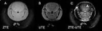 |
In Vivo Detection of Short T2* Lipid 1H in Mouse Brain with a
ZTE/UTE Subtraction Method (ZUS)
Yaotang Wu1,2, Michael Marcotrigiano3,
Hui Xue1,2,4, Robert V Mulkern1,2, and
Jeffrey Neil2,5
1Department of Radiology, Boston Children's
Hospital, Boston, MA, United States, 2Harvard
Medical School, Boston, MA, United States, 3Department
of Research, Boston Children's Hospital, Boston, MA, United
States, 4Sichuan
University, Chengdu, China, People's Republic of, 5Department
of Neurology, Boston Children's Hospital, Boston, MA, United
States
A new method, ZUS, utilizes ZTE to detect all signals with
T2* as short as a few hundred microseconds, including myelin
proton signals, and UTE to selectively detect signals with
longer T2* values, considered to be tissue water components.
The difference of these two types of images is used to
visualize signals from lipid 1H. In this study, the
feasibility of ZUS was demonstrated on a cholesterol phantom
(the major component of myelin) and on a live mouse. ZUS
images highlighted lipid, particularly myelin in the corpus
callosum, of mouse brain in vivo.
|
| |
09:12
|
1114.
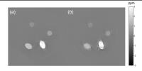 |
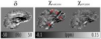
Quantitative susceptibility mapping of magnetic quadrupole
moments
Junghun Cho1, Dong Zhou2, Pascal
Spincemaille2, and Yi Wang1,2
1Biomedical Engineering, Cornell University, NEW
YORK, NY, United States, 2Radiology,
Weill Cornell Medical College, NEW YORK, NY, United States
In the study of quantitative susceptibility mapping, dipole
approximation is widely used where the magnetic field of
each voxel is approximated as dipole field. In general,
higher order field such as quadrupole field also exists,
especially for voxels with non-uniform subvoxel
magnetization/susceptibility distributions. We modeled the
magnetic field in MRI experiment up to quadrupole term and
used multiple orientation measurement to acquire both the
dipole (average susceptibility) and quadrupole
(susceptibility distribution) contributions. The feasibility
of the proposed method is demonstrated in an experimental
gadolinium water phantom study.
|
| |
09:24
|
1115.
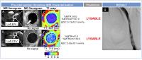 |
Multi-sequence non-contrast MRI characterization of deep vein
thrombosis in man
Alkystis Phinikaridou1, Prakash Saha2,
Marcelo Andia3, Alberto Smith2, and
René M Botnar1
1Biomedical Engineering, King's College London,
London, United Kingdom, 2Academic
Surgery, King's College London, London, United Kingdom, 3Radiology,
Pontificia Universidad Católica de Chile, Santiago, Chile
Deep vein thrombosis (DVT) affects 1 in 1000 people. Its
sequelae include post-thrombotic syndrome (PTS), which
affects up to 75% of patients within 5 years and is
characterised by persistent pain, swelling and ulceration.
Thrombolysis can reduce PTS by a third and is attempted in
patients with an ilio-femoral DVT and symptom onset of
<3weeks. Determining age and thrombus structure by history
alone is, however, subjective and there are no established
methods to quantify the abundance of matrix proteins, which
determines the response to lysis. This treatment is
therefore only effective in ~60% of patients, which may
unnecessarily exposes to haemorrhagic side effects. We have
developed a non-contrast enhanced magnetic resonance,
multi-sequence thrombus imaging (MSTI) technique that can
provide information about the structural composition of
experimental thrombus [1-2]. Here, we aim in translating the
MRI approach into man and determine whether it can help
guide venous intervention.
|
| |
09:36
|
1116.
 |
Positive visualization of interventional devices with
susceptibility mapping using the Turbo Spin Echo Sequence
caiyun shi1, guoxi xie1,2, xiaoyong
zhang1,3, min chen1, shi su1,
hairong zheng1, ying dong4, jim Ji4,
and xin liu1
1Shenzhen Institutes of Advanced Technology,
shenzhen, China, People's Republic of, 2Beijing
Center for Mathematics and Information Interdisciplinary
Sciences, beijing, China, People's Republic of, 3Centers
for Biomedical Engineering, College of Information Science
and Technology, University of Science and Technology of
China, hefei, China, People's Republic of, 4Department
of Electrical and Computer Engineering, Texas A&M
University, Texas, TX, United States
Susceptibility-based positive contrast MR imaging exhibits
excellent efficacy for visualizing the MR compatible
metallic devices, by taking advantage of their high magnetic
susceptibility. In this work, a novel method is developed to
accelerate the susceptibility-based positive contrast MR
imaging. The method is based on a modified turbo spin echo (TSE)
sequence and a kernel deconvolution algorithm with a
regularized l1 minimization to achieve positive contrast
imaging.
|
| |
09:48
|
1117.
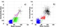 |
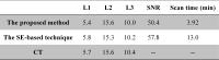
Correlation between MRI-derived water content and conductivity
in tumour and healthy tissue: how much cell water is active?
Ana-Maria Oros-Peusquens1, Yupeng Liao1,
and N. Jon Shah1
1INM-4, Research Centre Juelich, Juelich, Germany
About 80% of brain water is found inside the cells and a
large fraction of it is interfacial water with properties
substantially different from those of bulk water. Evidence
for a large osmotically unresponsive compartment, available
from literature, is substantiated by the finding that a very
large fraction of brain water does not contribute to its
electrical conductivity. This is determined by investigating
the correlation between conductivity and water content in
tumour patients in vivo. More than 80% of brain water is
found to be unresponsive, with variations reflecting tissue
and tumour type. This work describes a noninvasive method
for the characterisation of a deeply microscopic parameter
of the living tissue.
|
|