| |
08:00
|
1098.
 |
1H-MRS of the myocardium at 3T applying a 60-channel body array
coil – initial experiences 
Jürgen Machann1, Malte Niklas Bongers2,
Andreas Fritsche3, Hans-Ulrich Häring3,
Mike Notohamiprodjo4, Andreas Greiser5,
Konstantin Nikolaou4, and Fritz Schick2
1Section on Experimental Radiology, Department of
Diagnostic and Interventional Radiology, Institute for
Diabetes Research and Metabolic Diseases (IDM) of the
Helmholtz Center Munich, German Center for Diabetes Research
(DZD), Tübingen, Germany, 2Section
on Experimental Radiology, Department of Diagnostic and
Interventional Radiology, University Hospital Tübingen,
Tübingen, Germany, 3Department
of Endocrinology and Diabetology, Angiology, Nephrology and
Clinical Chemistry, Institute for Diabetes Research and
Metabolic Diseases (IDM) of the Helmholtz Center Munich,
German Center for Diabetes Research (DZD), Tübingen,
Germany, 4Department
of Diagnostic and Interventional Radiology, University
Hospital Tübingen, Tübingen, Germany, 5Siemens
Healthcare, Erlangen, Germany
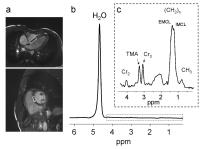
1H-MRS is increasingly applied in many organs for
non-invasive tissue characterization, e.g. for
quantification of ectopic lipids. Spectroscopic examinations
of the myocardium often suffer from limited spectral
dispersion, thus limiting the metabolic information content.
Applying a new 60-channel body-array receive coil, high
quality spectra with superior dispersion as compared to
previous setups are shown in this work. A single voxel PRESS
technique was applied in 10 subjects. After higher-order
shimming, linewidths of <20 Hz were obtained with high SNR
in a clinically acceptable measuring time. High
reproducibility and performance of the method may promote
1H-MRS applications in metabolic research and sports
medicine.
|
| |
08:12
 |
1099.
 |
Adiabatic excitation for 31P
spectroscopy in the human heart at 7T
Ladislav Valkovic1,2, William T Clarke1,
Benoit Schaller1, Lucian A B Purvis1,
Stefan Neubauer1, Ivan Frollo2,
Matthew D Robson1, and Christopher T Rodgers1
1Oxford Centre for Clinical Magnetic Resonance
Research, University of Oxford, Oxford, United Kingdom, 2Department
of Imaging Methods, Institute of Measurement Science, Slovak
Academy of Sciences, Bratislava, Slovakia
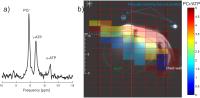
31P-MRS is of particular interest in
cardiovascular medicine, as the PCr/ATP ratio can serve as a
predictor of mortality. However, due to inherently low
signal-to-noise ratio (SNR), cardiac 31P-MRS
is not yet practical in the clinic. To increase SNR, the use
of 7T and dedicated receive arrays has been proposed.
However, the peak B1+ was
inadequate for the use of B1 insensitive
pulses, thus far. In this study, we demonstrate the
feasibility of homogeneous adiabatic excitation for cardiac 31P-MRS
using a novel quadrature 31P
transceiver at 7T. This constitutes an important step
towards absolute quantification of cardiac metabolites at
7T.
|
| |
08:24
|
1100.
|
Improvement of Quantification of 1H Cardiac MR Spectra Acquired
at 3T by the Use of Prior Knowledge
Ariane Fillmer1,2, Andreas Hock2,3,
and Anke Henning2,4
1Physikalisch Technische Bundesanstalt (PTB),
Berlin, Germany, 2Institute
for Biomedical Engineering, University and ETH Zurich,
Zurich, Switzerland, 3Department
of Psychiatry, Psychotherapy and Psychosomatics, Hospital of
Psychiatry, University of Zurich, Zurich, Switzerland, 4Max
Planck Institute for Biological Cybernetics, Tuebingen,
Germany
1H cardiac MRS is a promising tool for
investigation of human heart disease. In this context the
independent quantification of intramyocellular (IMCL) and
extramyocellular lipids (EMCL) is desired. Quantification
itself, however, remains challenging. This work
investigates, whether quantification of metabolite signals
within 1H
cardiac MR spectra could be improved by the use of prior
knowledge about the behavior of metabolite signals in the
quantification process.
|
| |
08:36
 |
1101.
 |
3D resolved human cardiac creatine kinase rate by 31P-MRS at 7T.
William Thomas Clarke1, Matthew D Robson1,
and Christopher T Rodgers1
1Oxford Centre for Clinical Magnetic Resonance
Research, University of Oxford, Oxford, United Kingdom
The creatine kinase (CK) forward rate constant kf is
a sensitive biomarker for heart failure. However, the low
SNR of 31P-MRS
at 1.5T and 3T has only allowed it to be measured at low
spatial resolution by 1D-CSI. Here, we show how cardiac 7T 31P-MRS
permits 3D resolved measurements for the first time. A 3D
variant of the FAST kfCK method was
combined with 31P
Bloch-Siegert B1+ mapping
to enable 3D-resolved measurements at 7T. The first
measurements of the creatine kinase rate in myocardium in
the interventricular septum are obtained from four subjects.
Our mean kf =
0.36±0.04 s-1 was
consistent with literature values.
|
| |
08:48
|
1102.
 |
Second-Order Motion-Compensated PRESS for Cardiac Spectroscopy
Maximilian Fuetterer1, Christian Torben Stoeck1,2,
and Sebastian Kozerke1,2
1Institute for Biomedical Engineering, University
and ETH Zurich, Zurich, Switzerland, 2Division
of Imaging Sciences and Biomedical Engineering, King's
College London, London, United Kingdom
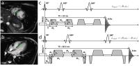
Second-order motion compensation for PRESS (PRESSmc)
is proposed to allow for robust single-voxel cardiac
spectroscopy throughout the entire cardiac cycle.
Motion-compensated spoiler gradients were designed and
implemented into a cardiac-triggered PRESS sequence. A
numerical 3D model of cardiac motion was used to optimize
and validate the gradient waveforms. In-vivo measurements in
healthy volunteers were obtained to assess SNR and
triglyceride-to-water ratio (TG/W). SNR gains and
variability of TG/W of PRESSmc were
evaluated against a conventional PRESS sequence with
optimized gradients. PRESSmc effectively
reduces cardiac-motion induced signal degradation during FID
spoiling providing higher SNR and less variability for TG/W
quantification.
|
| |
09:00
|
1103.
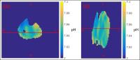 |
Mapping of pH in the human calf muscle at 7 T with 31P 3D
echo–planar spectroscopic imaging
Andreas Korzowski1 and
Peter Bachert1
1Medical Physics in Radiology, German Cancer
Research Center, Heidelberg, Germany
The tissue–pH value is an important parameter to assess
physiological function. The purpose of this work was to
explore the potential of three-dimensional 31P–{1H}
echo–planar spectroscopic imaging at B0 =
7 T for mapping of intracellular pH in the human calf muscle
with high spatial resolution. The acquired data demonstrate
that the proposed method allows the robust quantification of
intracellular pH value of voxels with less than 1 ml volume
and therefore may give insight into the pH heterogeneity of
different muscle groups.
|
| |
09:12
|
1104.
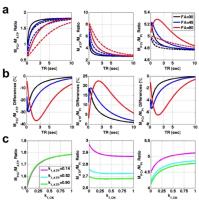 |
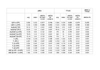
Rapid and Simultaneous Measurements for Reaction Kinetics and
Metabolite Pool Size Ratios using 31P Magnetization Saturation
Transfer Spectroscopy
Sang-Young Kim1,2, Wei Chen3, Dost
Ongur2, and Fei Du1,2
1McLean Imaging Center, McLean Hospital, Harvard
Medical School, Belmont, MA, United States, 2Psychotic
Disorders Division, McLean Hospital, Harvard Medical School,
Belmont, MA, United States, 3Center
for Magnetic Resonance Research, University of Minnesota,
Minneapolis, MN, United States
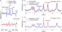
We demonstrates a novel strategy to simultaneously measure
metabolites pool sizes and kinetic constants of CK/ATPase
reactions using 31P-MST spectroscopy. Our method enables the
corrections for T1relaxation time and chemical
exchanges effects due to short TR. The most important
advantage of our proposed method is the reduction of TR for
complete measurements of both metabolites ratios and
reaction kinetics with high sensitivity. This can facilitate
future applications requiring high temporal and/or spatial
resolution.
|
| |
09:24
|
1105.
 |
Observation of 31P magnetization transfer at 3 Tesla using
asymmetric adiabatic inversion and two different fitting
strategies.
Bertrand Pouymayou1, Tania Buehler1,
Roland Kreis1, and Chris Boesch1
1Depts. Radiology and Clinical Research,
University of Bern, Bern, Switzerland
31P-MR spectroscopy inversion transfer (IT) is
increasingly investigated as a complementary method to study
ATP-synthesis and creatine kinase in vivo. Three aspects of
the IT experiment are studied here, in a test-retest design
(12 volunteers, resting vastus muscle): the ability to
produce an efficient half band inversion in vivo with a
short asymmetric adiabatic pulse, the repeatability of the
kinetic parameters estimation at 3T and the impact of two
different fitting strategies (individual spectrum vs.
two-dimensional fitting). As a result, k[Pi>γ-ATP] can be
reliably estimated within cohorts while k[PCr>γ-ATP] is
accurate enough to be distinguished between individuals.
|
| |
09:36
|
1106.
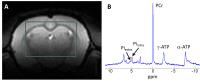 |
Localized 31P magnetization transfer in the rat brain to measure
ATP synthesis rate: inorganic phosphate comes in two pools
Brice Tiret1,2, Vincent Lebon1,2,
Emmanuel Brouillet1,2, and Julien Valette1,2
1CEA/DSV/I2BM/MIRCen, Fontenay-aux-Roses, France, 2CNRS
Université Paris-Saclay UMR 9199, Fontenay-aux-Roses, France
Localized 31P
MRS with progressive saturation transfer was performed in
the rat brain to estimate the exchange rate between
inorganic phosphate (Pi) and adenosine-tri-phosphate (ATP).
It was found that two Pi pools, tentatively intra and
extracellular pools, can be resolved at 11.7 T, and that
only the intracellular Pi signal varies with progressive
saturation, while the extracellular Pi signal remains
constant. Not resolving this extracellular Pi can cause a
significant bias in the estimation of the forward constant
rate of ATP synthesis.
|
| |
09:48
|
1107.
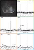 |
Dynamic 31P
MRSI with spiral readout for quantification of mitochondrial
capacity in muscles of the calf during plantar flexion exercise
at 7T - Permission Withheld
Ladislav Valkovic1,2,3,4, Marek Chmelík1,2,
Martin Meyerspeer1,5, Borjan Gagoski6,
Martin Krššák1,2,7, Christopher T Rodgers4,
Ivan Frollo3, Ovidiu C Andronesi8,
Siegfried Trattnig1,2,9, and Wolfgang Bogner1,2
1High-field MR Centre, Medical University of
Vienna, Vienna, Austria, 2Department
of Biomedical Imaging and Image-guided Therapy, Medical
University of Vienna, Vienna, Austria, 3Department
of Imaging Methods, Institute of Measurement Science, Slovak
Academy of Sciences, Bratislava, Slovakia, 4Oxford
Centre for Clinical Magnetic Resonance Research, University
of Oxford, Oxford, United Kingdom, 5Center
for Medical Physics and Biomedical Engineering, Medical
University of Vienna, Vienna, Austria, 6Fetal
Neonatal Neuroimaging and Developmental Science Center,
Boston Children's Hospital, Boston, MA, United States, 7Division
of Endocrinology and Metabolism, Department of Internal
Medicine III, Medical University of Vienna, Vienna, Austria, 8Athinoula
A. Martinos Center for Biomedical Imaging, Department of
Radiology, Massachusetts General Hospital, Harvard Medical
School, Boston, MA, United States, 9Christian
Doppler Laboratory for Clinical Molecular MR Imaging,
Vienna, Austria
Typically, only rough localization by the sensitive volume
of the surface coil is used for dynamic 31P-MRS.
However, such localization often mixes signals from several
muscle groups. Available single-muscle localization
techniques (e.g., semi-LASER or DRESS) provide only limited
coverage and current 31P-MRSI
techniques suffer from slow acquisition. To overcome the low
temporal resolution of the standard 31P-MRSI,
caused by slow Cartesian readout, we have developed, and
tested in healthy subjects at 7T, a 31P-MRSI
sequence using spiral readout trajectory. This sequence
enables spatially resolved quantification of mitochondrial
capacity in several investigated muscles (e.g., GM, GL and
SOL) simultaneously at 7T.
|
|