| |
16:00
|
0499.
 |
Multiparametric MRI Characterization of Magnetic Viral Complexes 
Alexander Joos1, Olga Mykhaylyk2,
Norbert Löwa3, Dietmar Eberbeck3,
Bernhard Gleich1, and Axel Haase1
1Zentralinstitut für Medizintechnik der
Technischen Universität München, Garching, Germany, 2Department
of Experimental Oncology, Klinikum rechts der Isar der TU
München, Munich, Germany, 3Physikalisch-Technische
Bundesanstalt, Berlin, Germany
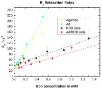
Magnetic nanoparticles can be used for magnetic drug
targeting while MRI can serve as non-invasive therapy
monitoring. We investigated the influence of the assembling
of magnetic nanoparticles with oncolytic viruses and their
uptake into cancer cells on the MRI relaxivities r1, r2 and r2* and
magnetically characterized all samples using magnetic
particle spectroscopy. Our results show that R2* measurements
seem most suitable for particle quantification while R2 is
sensitive to the uptake of the particles into the cells.
Magnetic particle spectroscopy proves to be an important
validation technique for MRI relaxometry.
|
| |
16:12
 |
0500.
 |
Fast Quantitative T2 Mapping using Simultaneous-Multi-Slice and
Model-Based Reconstruction
Tom Hilbert1,2,3, Jennifer Schulz4,
Lauren J. Bains4, José P. Marques4,
Reto Meuli2, Jean-Philippe Thiran2,3,
Gunnar Krueger2,3,5, David G. Norris4,
and Tobias Kober1,2,3
1Advanced Clinical Imaging Technology (HC CMEA
SUI DI BM PI), Siemens Healthcare AG, Lausanne, Switzerland, 2Department
of Radiology, University Hospital (CHUV), Lausanne,
Switzerland, 3LTS5,
École Polytechnique Fédérale de Lausanne, Lausanne,
Switzerland, 4Radboud
University Nijmegen, Donders Institute for Brain, Cognition
and Behaviour, Nijmegen, Netherlands, 5Siemens
Medical Solutions USA, Inc., Boston, MA, United States
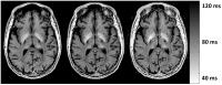
Long acquisition times of quantitative magnetic resonance
imaging (qMRI) are one obstacle that prevents qMRI to be
used in clinical routine. Acceleration methods, such as
simultaneous-multi-slice and model-based iterative
reconstruction proved in the past to allow high acceleration
factors in MRI. Here we suggest combining these two methods
to allow fast quantitative T2 mapping, yielding a
high-resolution (0.7x0.7x3mm³) whole brain (40 slices)
acquisition within a clinically acceptable acquisition time
of less than 3 minutes. T2 values of the proposed method are
similar to the values of the standard method as it is shown
on phantom experiments.
|
| |
16:24
|
0501.
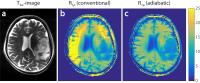 |
A robust T1?-mapping method for in-vivo glucose detection at 7T
whole-body scanners
Patrick Schuenke1, Moritz Zaiss1,
Christina Koehler2, Alexander Radbruch2,3,
Mark Edward Ladd1, and Peter Bachert1
1Medical Physics in Radiology, German Cancer
Research Center (DKFZ), Heidelberg, Germany, 2Department
of Neuroradiology, University of Heidelberg Medical Center,
Heidelberg, Germany, 3Department
of Radiology, German Cancer Research Center (DKFZ),
Heidelberg, Germany
Recently it was demonstrated that on–resonant
chemical–exchange–sensitive spin–lock (CESL) allows to
observe the uptake of administered D–glucose in
vivo and
thus could be used for glucose metabolism studies. However,
conventional spin–lock produces artifacts owing to B1–field
inhomogeneities, which are a common problem especially at
high-field whole-body MR scanners. Therefore we developed an
adiabatic water–T1ρ mapping
sequence which outperforms conventional spin-lock sequences
with respect to its insensitivity to B1–inhomogeneities;
its sensitivity to glucose in the millimolar range as well
as its applicability to in
vivo studies
is proven.
|
| |
16:36
|
0502.
 |
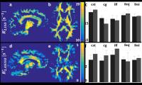
Imaging Subcortical White Matter by High Resolution 7 T MRI in
vivo: Towards Potential U-Fiber Density Mapping in Humans - Permission Withheld
Evgeniya Kirilina1,2, Juliane Dinse1,
Pierre-Louise Bazin1, Carsten Stueber3,4,
Stefan Geyer1, Robert Trample1,
Andreas Deistung5, Juergen R Reichenbach5,
and Nikolaus Weiskopf1,6
1Neurophysics, Max Plank Institute for Human
Cognitive and Brain Science, Leipzig, Germany, 2Neurocomputation
and Neuroimaging Unit, Department of Educational Science and
Psychology, Free University Berlin, Berlin, Germany, 3Department
of Radiology, Weill Cornell Medical College, New York, NY,
United States, 4Department
of Neurology, Yale School of Medicine, Yale University, New
Haven, CT, United States,5Medical Physics Group,
Jena University Hospital - Friedrich Schiller University
Jena, Jena, Germany, 6Wellcome
Trust Centre for Neuroimaging, University College London,
London, United Kingdom
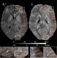
Subcortical white matter (SWM) incorporates U-fibers, the
intra-hemispheric connections between adjacent gyri. Despite
their importance for cortical connectivity little is known
about the U-fiber distribution in humans due to the lack of
appropriate imaging methods. Herein we investigate SWM using
high-resolution in-vivo MRI at 7T. A clear-cut
discrimination of SWM from the adjacent brain regions was
obtained based on higher qR2*, qR2 and
susceptibility in-vivo. These new findings may pave the way
for future in-vivo segmentation strategies for this crucial
brain region as well as potential U-fiber density mapping in
humans.
|
| |
16:48
|
0503.
 |
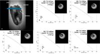
Mapping orientation dependent and independent components of
R2star in the human white matter - an in vivo study - Permission Withheld
Diana Khabipova1,2, Rita Gil2, Marcel
Zwiers2, and José Pedro Marques2
1CIBM-AIT, EPFL, Lausanne, Switzerland, 2Centre
for Cognitive Neuroimaging, Donders Institute, Nijmegen,
Netherlands
Anisotropic microstructure of the white matter causes the
apparent transverse relaxivity, $$$R_2^*$$$, to depend on
the orientation of white matter fibres in respect to the
applied magnetic field. Using the fibre orientation prior
knowledge from DTI orientation dependent $$$R_{2,ANISO}^*$$$
and independent $$$R_{2,ISO}^*$$$ components of $$$R_2^*$$$
were calculated. For all studied WM fibres a consistency for
the (an)isotropic components between both hemispheres was
present. The isotropic component showed higher variability
compared to the anisotropic component.
|
| |
17:00
|
0504.
 |
Gradient echo signal decay of bone material at high field
requires a gaussian augmentation of the mono-exponential model
for T2* determination
Weiqiang Dou1 and
Arend Heerschap1
1Radiology, Radboud University Medical Centre,
Nijmegen, Netherlands
Previously reported T2*quantification
for calcium phosphate cement (CPC), a widely used bone
material, remained unsatisfactory with a mono-exponential
(ME) fit. A recently proposed Gaussian augmentation of the
mono-exponential (GAME) model was reported to have robust
fit for gradient echo (GRE) signals. To accurately evaluate
GRE-signal decay of CPC, GAME and ME fits were applied in
this study for multi-echo time GRE signals acquired at
11.7T. Compared to ME, GAME showed optimal fitting with
significantly smaller sum of squared errors and larger
R-squared values. Therefore, GAME model is demonstrated to
be suitable for GRE signal modeling in CPC at ultra-high
field.
|
| |
17:12
|
0505.
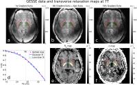 |
Simultaneous estimation of reversible and irreversible
transverse relaxation rates in the basal ganglia at 7T:
implications for brain iron deposition studies
Mukund Balasubramanian1,2, Jonathan R. Polimeni2,3,
and Robert V. Mulkern1,2
1Department of Radiology, Boston Children's
Hospital, Boston, MA, United States, 2Harvard
Medical School, Boston, MA, United States, 3Athinoula
A. Martinos Center for Biomedical Imaging, Department of
Radiology, Massachusetts General Hospital, Charlestown, MA,
United States
Reversible and irreversible transverse relaxation rates were
measured at 7T, using the GESSE pulse sequence, in basal
ganglia structures in 11 volunteers (ages: 23-81 years). We
found that, with a judicious choice of echo times,
irreversible rates (R2) in the globus pallidus
were conspicuous for all subjects. Furthermore, both
reversible and irreversible rates increased with age in a
manner consistent with prior postmortem studies of iron
concentration in these structures. Since these rates are
differentially affected by field perturbations at different
spatial scales, their consideration may provide information
about the microscopic and mesoscopic distribution and
concentration of iron in tissue.
|
| |
17:24
|
0506.
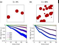 |
A General Solution for Transverse Signal Decay Under the Weak
Field Approximation: Theory and Validation with Spherical
Perturbers
Avery J.L. Berman1,2 and
Bruce Pike2
1Montreal Neurological Institute, McGill
University, Montreal, QC, Canada, 2Department
of Radiology and Hotchkiss Brain Institute, University of
Calgary, Calgary, AB, Canada
This study presents a closed-form analytical solution that
describes transverse signal relaxation using the weak field
approximation (WFA). The closed-form solution (CFS) fully
describes the net signal dynamics under any train of 180°
refocusing pulses, and we show that it is in close agreement
with a commonly employed mono-exponential expression of the
WFA. We compared the CFS to simulations from a medium
containing spherical perturbers, with a focus on modelling
red blood cells. The CFS and simulations were in close
agreement but the results systematically varied depending on
whether or not the spheres were allowed to overlap. This
theory can be applied in areas such as tissue iron imaging
or relaxometry of blood.
|
| |
17:36
|
0507.
|
Rotating frame MRI in human subjects with Multiple Sclerosis
Silvia Mangia1, Alena Svatkova2,3,
Peter Bednarik1,3, Igor Nestrasil2,
Lynn E. Eberly4, Adam Carpenter5, and
Shalom Michaeli1
1Radiology, CMRR, University of Minnesota,
Minneapolis, MN, United States, 2Department
of Pediatrics, University of Minnesota, Minneapolis, MN,
United States, 3Central
European Institute of Technology, Masaryk University, Brno,
Czech Republic, 4Division
of Biostatistics, University of Minnesota, Minneapolis, MN,
United States, 5Neurology,
University of Minnesota, Minneapolis, MN, United States
Rotating frame MRI methods including adiabatic T1ρ, T2ρ, and
RAFF4 were here employed for characterizing the white matter
(WM) of relapsing-remitting Multiple Sclerosis (MS)
patients. We calculated relaxograms from subcortical WM of
MS patients (excluding lesions) and age-matched controls,
and compared them with histograms of DTI outcomes. T1ρ, T2ρ
and TRAFF4 were significantly different in the WM of MS
patients vs controls, while DTI outcomes did not detect
group differences. These findings are supported by recent
validation studies using demyelination/dysmyelination animal
models, where RAFF4 exhibited exceptional ability to probe
myelin content/integrity which we attribute to enhanced
sensitivity to slow/ultra-slow motion.
|
| |
17:48
|
0508.
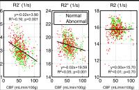 |
R2’ is the Best Transverse Relaxation Rate for Oxygenation
Mapping: Experience in Moyamoya Disease with Acetazolamide
Challenge
Wendy Ni1,2, Thomas Christen2, and
Greg Zaharchuk2
1Department of Electrical Engineering, Stanford
University, Stanford, CA, United States, 2Department
of Radiology, Stanford University, Stanford, CA, United
States
Transverse MR spin relaxation rates, R2*, R2 and
R2’ have all been considered sensitive to brain
tissue oxygenation. In this study, we focus on a cohort of
pre-operative Moyamoya disease patients and simultaneously
map all three rates in addition to cerebral blood flow, both
before and after the injection of the vasodilatory drug,
acetazolamide. We found our measurements to be consistent
with physiology and previous studies, and to support the use
of R2’ for oxygenation mapping instead of R2*
and R2.
|
|