| |
13:30
|
0408.
 |
3D black-blood thrombus imaging (BTI) for the diagnosis of deep
vein thrombosis: initial clinical experience 
Guoxi Xie1, Hanwei Chen2, Zhuonan He2,
Jianke Liang2, Xueping He2, Qi Yang3,4,
Xin Liu1, Debiao Li3, and Zhaoyang Fan3
1Shenzhen Institutes of Advanced Technology,
Chinese Academy of Sciences, Shenzhen, China, People's
Republic of, 2Department
of Radiology, Guangzhou Panyu Central Hospital, Guangzhou,
China, People's Republic of, 3Biomedical
Imaging Research Institute, Cedars-Sinai Medical Center, Los
Angeles, CA, United States, 4Xuanwu
Hospital, Beijing, China, People's Republic of
Deep vein thrombosis (DVT) is a common but elusive illness
that can lead to fatal pulmonary embolism and sudden death.
Effective treatment of DVT requires accurate evaluation of
thrombus distribution and stage. n this work, we further
accommodated the DANTE-SPACE technique to the deep vein
system and conducted preliminary clinical validation.
Experiment results demonstrated that DANTE-SPACE could
provide excellent venous blood signal suppression and
definitive thrombus detection and the technique may
outperform conventional SPACE, MPRAGE, and and become a
non-contrast alternative to CEMRV for the diagnosis of DVT.
|
| |
13:42
|
0409.
 |
A Combined Saturation and Imaging RF-Pulse for Fast and
Continuous Black-Blood Preparation in Dynamic Imaging
Simon Reiss1, Axel Joachim Krafft1,2,3,
Marius Menza1, Constantin von zur Mühlen4,
and Michael Bock1
1Dept. of Radiology - Medical Physics, University
Medical Center Freiburg, Freiburg, Germany, 2German
Cancer Consortium (DKTK), University Medical Center Freiburg,
Heidelberg, Germany, 3German
Cancer Research Center (DKFZ), Heidelberg, Germany, 4Department
of Cardiology and Angiology I, University Heart Center,
Freiburg, Germany
Black-blood preparation is a tool for improved contrast
generation in cardiovascular MRI to assess vessel wall
constitution, to delineate plaques and to characterize
myocardial tissue. Conventionally, black-blood MRI can be
done with dual inversion recovery pulses so that selective
signal nulling of the inflowing blood is achieved. The
inversion delays required to establish the black-blood
contrast can be favorably integrated into ECG-triggered
diastolic cardiac measurements, but they are by far too
time-consuming for dynamic measurements that cover the total
cardiac cycle. In this work we investigate the use of
conventional saturation pulses for black-blood imaging. We
propose a very time-efficient pulse implementation that
combines the saturation and the imaging RF pulse into a
single pulse structure and enables black-blood contrast in
dynamic measurements.
|
| |
13:54
|
0410.
 |
A Novel Method for Contact-Free Cardiac Synchronization Using
the Pilot Tone Navigator - Permission Withheld
Lea Schroeder1, Jens Wetzl1,2, Andreas
Maier1,2, Lars Lauer3, Jan Bollenbeck4,
Matthias Fenchel3, and Peter Speier3
1Pattern Recognition Lab, Department of Computer
Science, Friedrich-Alexander-Universität Erlangen-Nürnberg,
Erlangen, Germany, 2Erlangen
Graduate School in Advanced Optical Technologies,
Friedrich-Alexander-Universität Erlangen-Nürnberg, Erlangen,
Germany, 3Magnetic
Resonance, Product Definition and Innovation, Siemens
Healthcare GmbH, Erlangen, Germany, 4Magnetic
Resonance, Research and Development, Hardware, Siemens
Healthcare GmbH, Erlangen, Germany
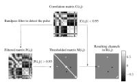
We evaluate the information content of externally generated
Pilot Tone signals, received with standard MR local coils,
with respect to cardiac motion. Free-breathing and
breathhold fluoroscopic measurements were performed with
applied electrocardiogram leads to provide ground truth.
Average mean correlation between RR intervals of our method
and the ground truth was 0.95. Our early results indicate
that locally generated PT signals contain information about
cardiac motion and suggest that the proposed method could be
developed into an electrocardiogram replacement by providing
a continuous signal for retrospective gating with minimal
hardware requirements.
|
| |
14:06
|
0411.
 |
Free-Breathing, Self-Navigated Isotropic 3-D CINE Imaging of the
Whole Heart Using Cartesian Sampling
Jens Wetzl1,2, Felix Lugauer1,
Michaela Schmidt3, Andreas Maier1,2,
Joachim Hornegger1,2, and Christoph Forman3
1Pattern Recognition Lab, Department of Computer
Science, Friedrich-Alexander-Universität Erlangen-Nürnberg,
Erlangen, Germany, 2Erlangen
Graduate School in Advanced Optical Technologies,
Friedrich-Alexander-Universität Erlangen-Nürnberg, Erlangen,
Germany, 3Magnetic
Resonance, Product Definition and Innovation, Siemens
Healthcare GmbH, Erlangen, Germany
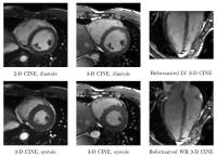
We present a method for free-breathing, isotropic 3-D CINE
imaging of the whole heart, demonstrated with experiments in
7 healthy volunteers. Respiratory information for
retrospective gating is derived directly from the imaging
data. Ventricular function parameters were compared to
reference 2-D CINE acquisitions. Excellent image quality and
match to ground truth ventricular function parameters could
be achieved in an acquisition time similar to multi-slice
2-D CINE with equivalent coverage. Cartesian sampling
combined with dual-GPU acceleration enabled a fast
reconstruction in under 5 minutes for left-ventricular and
under 7 minutes for whole heart coverage.
|
| |
14:18
|
0412.
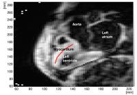 |
Using intrinsic Cardiac Shear Waves to measure Myocardial
Stiffness: Initial results on a Patient Cohort with Heart
failure with preserved Ejection Fraction
Jessica Webb1, Ondrej Holub1, Rachel
Clough1, Gerald Carr-White2, Reza
Razavi1, and Ralph Sinkus1
1King's College London, London, United Kingdom, 2Guys
and St Thomas' NHS Trust, London, United Kingdom
Heart Failure with preserved Ejection Fraction (HFpEF) is
common and associated with high morbidity and mortality.
There are challenges in diagnosing HFpEF and a non invasive
technique to detect myocardial stiffness would have an
enormous clinical impact.
We have developed a novel non invasive technique to
quantify myocardial stiffness in vivo using transient
Magnetic Resonance Elastography (tMRE). The technique relies
on accurately identifying the aortic valve closure time. The
speed of the propagating shear wave, created by the valve
closure, is measured using a short navigated free breathing
MRI sequence. Increased myocardial stiffness results in
increased speed of shear wave propagation.
|
| |
14:30
|
0413.
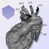 |
Three-Dimensional Modelling of the Fetal Vasculature from
Prenatal MRI using Motion-Corrected Slice-to-Volume Registration
David F A Lloyd1,2, Bernhard Kainz3,
Joshua F P van Amerom1, Kuberan Pushparajah1,2,
John M Simpson2, Vita Zidere2, Owen
Miller2, Gurleen Sharland2, Tong Zhang1,
Maelene Lohezic1, Joanne Allsop1,
Matthew Fox1, Christina Malamateniou1,
Mary Rutherford1, Jo Hajnal1, and Reza
Razavi1,2
1Division of Imaging Sciences and Biomedical
Engineering, King's College London, London, United Kingdom, 2Evelina
Children's Hospital, London, United Kingdom, 3Department
of Computing (BioMedIA), Imperial College London, London,
United Kingdom
The diagnosis of potentially life-threatening vascular
abnormalities in the fetus can be difficult with ultrasound
alone. MRI is one of the few safe alternative imaging
modalities in pregnancy; however to date it has been limited
by unpredictable fetal and maternal motion during
acquisition. We present six antenatal cases, four with
important structural congenital heart disease, in which we
employed a novel algorithm for motion-corrected slice-volume
registration, producing a navigable 3D volume of the fetal
thoracic vasculature. The anatomical findings in each case
were then correlated to fetal echocardiographic findings,
and finally displayed as interactive surface rendered
models.
|
| |
14:42
 |
0414.
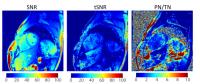 |
The Physiological Noise Contribution to Temporal Signal-to-Noise
Increases with Decreasing Resolution and Acceleration in
Quantitative CMR
Terrence Jao1 and
Krishna Nayak2
1Biomedical Engineering, University of Southern
California, Los Angeles, CA, United States, 2Electrical
Engineering, Los Angeles, CA, United States
Advances in MR hardware, pulse sequences, and calibration
have made quantitative CMR a reality. Quantitative maps
(e.g. T1, T2, ECV) are formed from multiple images, which
make them susceptible to errors caused by signal
fluctuations from cardiac or respiratory motion, termed
physiological noise. Reproducibility of quantitative CMR
maps is critical for future clinical adoption and depends on
the ratio of signal amplitude to physiological noise, termed
temporal SNR. In this study, we measure temporal SNR in
bSSFP quantitative CMR to characterize physiological noise
for a range of image resolutions, acceleration factors, and
post inversion delays.
|
| |
14:54
|
0415.
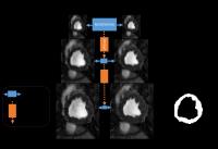 |
Multi-Resolution Registration and Segmentation for cardiac BOLD
MRI
Ilkay Oksuz1,2, Rohan Dharmakumar3,4,
and Sotirios A. Tsaftaris2,5
1Diagnostic Radiology, Yale University, New
Haven, CT, United States, 2IMT
Institute for Advanced Studies Lucca, Lucca, Italy, 3Biomedical
Imaging Research Institute, Cedars Sinai Medical Center, Los
Angeles, CA, United States, 4University
of California, Los Angeles, CA, United States, 5The
University of Edinburgh, Edinburgh, United Kingdom
Cardiac Phase-resolved Blood Oxygen-Level-Dependent
(CP-BOLD) MRI is a new contrast and stress-free approach for
detecting myocardial ischemia, that identifies the ischemic
myocardium by examining changes in myocardial signal
intensity patterns as a function of cardiac phase. But,
these changes coupled with cardiac motion, challenge
automated standard CINE MR myocardial segmentation and
registration techniques resulting in a significant drop of
segmentation and registration accuracy. We propose a
dictionary learning based multi-resolution registration
scheme for supervised learning and sparse representation of
the myocardium. Our results show an improvement of 15%
myocardial segmentation w.r.t. the state of the art.
|
| |
15:06
|
0416.
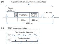 |
Optimized Cardiac CEST MRI for Assessment of Metabolic Activity
in the Heart
Zhengwei Zhou1,2, Yuhua Chen3, Yibin
Xie1, Christopher Nguyen1, Mu Zeng4,
James Dawkins5, Zhanming Fan4, Eduardo
Marbán5, and Debiao Li1,2,5
1Biomedical Imaging Research Institute,
Cedars-Sinai Medical Center, Los Angeles, CA, United States, 2Department
of Bioengineering, University of California, Los Angeles,
Los Angeles, CA, United States,3Department of
Computer and Information Science, University of
Pennsylvania, Philadelphia, PA, United States, 4Department
of Radiology, Anzhen Hospital, Capital Medical University,
Beijing, China, People's Republic of, 5Heart
Institute, Cedars-Sinai Medical Center, Los Angeles, CA,
United States
In this work, we developed an optimized cardiac CEST method
to detect myocardial metabolic change with significantly
reduced scan time. Our initial results in porcine model with
chronic myocardial infarction show that scar region has
lower metabolic activity compared to healthy myocardium,
using LGE as reference. This study also shows the
feasibility of cardiac CEST imaging in a patient, for the
first time.
|
| |
15:18
|
0417.
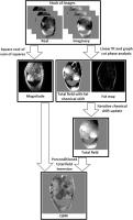 |
In vivo Quantitative Susceptibility Mapping (QSM) in cardiac MRI
Yan Wen1, Thanh D. Nguyen2, Zhe Liu1,
Pascal Spincemaille2, Dong Zhou2,
Alexey Dimov1, Youngwook Kee2, Jiwon
Kim3, Jonathan W. Weinsaft3, and Yi
Wang1,2
1Biomedical Engineering, Cornell University, New
York, NY, United States, 2Physics
in Radiology, Weill Cornell Medicine, New York, NY, United
States, 3Medicine,
Weill Cornell Medicine, New York, NY, United States
Quantitative Susceptibility Mapping (QSM) has yet to be
applied on cardiac patients due to the challenges from
motion artifacts and background fields. In this first
attempt to apply QSM in cardiac MRI, we overcome these data
acquisition and processing challenges by using robust graph
cut phase analysis and a novel preconditioned inversion of
total field. Our preliminary results demonstrate high
quality susceptibility maps, and the measured heart chamber
blood oxygenation level is consistent with reported values
from literature.
|
|