| |
10:00
 |
0597.
 |
Transient Oxygen Extraction Fraction as a Measure of
Cerebrovascular Reserve - Permission Withheld
Charles G Cantrell1, Parmede Vakil1,2,
Donald R Cantrell3, Yong Jeong1,
Sameer A Ansari3, and Timothy J Carroll1,3
1Biomedical Engineering, Northwestern, Chicago,
IL, United States, 2College
of Medicine, University of Illinois, Chicago, IL, United
States, 3Radiology,
Northwestern, Chicago, IL, United States
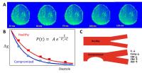
We have found that MR-PARSE has detectable sensitivity to
frequency shifts induced by transient alterations in de-oxyhemoglobin
through the cardiac cycle. Our initial studies have shown,
through the use of ICA, a statistically significant
hemispheric difference between healthy and compromised
regions. Our approach to quantifying cerebrovascular
reactivity represents a new and simple, non contrast
approach to stratifying patients toward therapies to prevent
stroke.
|
| |
10:12
 |
0598.
 |
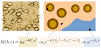
Ex-vivo Quantitative Imaging and Qualitative Plaque Type
Classification of Intracranial Atherosclerotic Plaque using High
Resolution MRI 
Yuanliang Jiang1, Chengcheng Zhu2,
Andrew J Degnan3, Wenjia Peng1,
Luguang Chen1, Xinrui Wang1, Qi Liu1,
Yang Wang4, Zhenzhen Xiang4, Zhongzhao
Teng5, David Saloner2, and Jianping Lu1
1Radiology, Changhai Hospital, Shanghai, China,
People's Republic of, 2Radiology,
University of California, San Francisco, San Francisco, CA,
United States, 3Radilogy,
University of Pittsburgh, Pittsburgh, PA, United States, 4Pathology,
Changhai Hospital, Shanghai, China, People's Republic of, 5Radiology,
University of Cambridge, Cambridge, United Kingdom
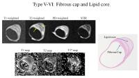
The first ex vivo measurement of T1, T2, and T2* relaxation
times of intracranial plaque components at 3T is reported.
The ability of multi-contrast MRI to characterize plaque
type was evaluated with histological validation. Plaque
components could be differentiated based on relaxation
times. Specifically, lipid core had significantly lower T2
values than fibrous cap. MRI and histology correlation was
consistent across specimens and locations, and MRI showed a
high sensitivity and specificity for identifying plaque
features previously associated with high-risk. Therefore,
MRI has the potential to characterize intracranial plaque
composition and improve patient risk stratification.
|
| |
10:24
|
0599.
 |
Acceleration-selective Arterial Spin Labeling (AccASL) MR
Angiography for Visualization of Distal Cerebral Arteries in
Moyamoya Disease
Osamu Togao1, Akio Hiwatashi1, Makoto
Obara2, Koji Yamashita1, Kazufumi
Kikuchi1, and Hiroshi Honda1
1Department of Clinical Radiology, Graduate
School of Medical Sciences, Kyushu University, Fukuoka,
Japan, 2Philips
Electronics Japan, Tokyo, Japan
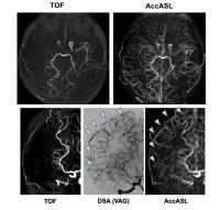
In this study, we demonstrated the utility of intracranial
MR angiography (MRA) using acceleration-selective arterial
spin labeling (AccASL) technique in Moyamoya disease. The
AccASL-MRA markedly improved the visualization of arteries
distal to the steno-occlusive site reflecting collateral
flow via LMA in Moyamoya disease in comparison with
time-of-flight (TOF)-MRA.
|
| |
10:36
|
0600.
 |
Serial Quantification of Brain Oxygenation using
Streamlined-qBOLD in Acute Stroke Patients
Alan J Stone1, George WJ Harston2,
Davide Carone2, Mmua Ngwako2, Radim
Licenik2, James Kennedy 2,
and Nicholas P Blockley1
1FMRIB, Nuffield Department of Clinical
Neurosciences, University of Oxford, Oxford, United Kingdom, 2Acute
Stroke Programme, Radcliffe Department of Medicine,
University of Oxford, Oxford, United Kingdom
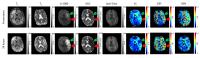
Streamlined-qBOLD is applied to an exploratory cohort of
acute stroke patients in a serial imaging study to map brain
oxygen metabolism. Quantitative brain oxygenation parameters
are demonstrated to vary between regions with different
tissue outcomes and this imaging approach is shown to have
the potential to refine the identification of the ischemic
penumbra.
|
| |
10:48
|
0601.
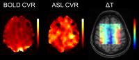 |
Relationship between Cerebrovascular Reserve and Brain
Temperature following Acetazolamide Challenge in Patients with
Chronic Steno-Occlusive Disease - Permission Withheld
Seena Dehkharghani1, Candace C. Fleischer2,
Deqiang Qiu1, Sang-Eon Park2, Junjie
Wu1, and Fadi Nahab3
1Radiology and Imaging Sciences, Emory
University, Atlanta, GA, United States, 2Biomedical
Engineering, Emory University and Georgia Institute of
Technology, Atlanta, GA, United States, 3Neurology,
Emory University, Atlanta, GA, United States
Methods for characterizing misery
perfusion to
predict stroke are largely limited to positron emission
tomography, which suffers from high radiation exposure.
Magnetic resonance imaging (MRI) and spectroscopy (MRS)
offer non-invasive alternatives to explore cerebral
hemodynamics and brain temperature regulation, a poorly
understood physiologic variable at the intersection of
perfusion and metabolism. We detail the first reported use
of MRI/MRS to relate cerebrovascular reserve with
temperature in patients following acetazolamide challenge,
observing significant correlation between temperature
changes and cerebrovascular reserve. These findings will be
used to inform future MRI studies of perfusion and brain
temperature among patients with chronic steno-occlusive
disease.
|
| |
11:00
|
0602.
 |
Stroke Volume Evolution Following Endovascular Therapy on DWI
and FLAIR
Christian Federau1, Soren Christensen1,
Michael Mlynash1, Jenny Tsai1, Sun Kim1,
Greg Zaharchuk1, Matus Straka1,
Nishant Mishra1, Maarten Lansberg1,
and Greg Albers1
1Stanford University, Stanford, CA, United States
We studied the evolution of the infarct volume between an
early post-revascularization scan (within 24 h of symptom
onset) and day 5 in patients of the CRISP and DEFUSE 2
cohort studies. On the early post-revascularization scan,
FLAIR lesions were smaller compared to DWI, but were larger
at day 5. The early post-revascularization stroke volume on
DWI, compared to FLAIR, was closer, and correlated better
with the day 5 DWI and FLAIR lesion volumes. Together, our
findings suggest that DWI is a better early surrogate marker
of stroke volume.
|
| |
11:12
|
0603.
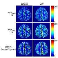 |
Quantitative Susceptibility Mapping (QSM) based Cerebral
Metabolic Rate of Oxygen (CMRO2) Mapping: Eliminating Blood Flow
Challenge with Minimal Local Variance (MLV)
Jingwei Zhang1,2, Dong Zhou2, Sarah
Eskreis-Winkler2, Thanh Nguyen2,
Pascal Spincemaille2, Ajay Gupta2, and
Yi Wang1,2
1Biomedical Engineering, Cornell University, New
York, NY, United States, 2Radiology,
Weill Cornell Medical College, New York, NY, United States
We propose a cerebral metabolic rate of oxygen consumption
(CMRO2) mapping method without blood flow challenge using
quantitative susceptibility mapping, cerebral blood flow and
a regularization of minimal local variance (MLV) within the
same type of tissue. Getting rid of blood flow challenge
would vastly increase the clinical utility of MRI CMRO2. The
MNV CMRO2 maps were very similar to CMRO2 maps using
caffeine as challenge, with no significant bias in value.
|
| |
11:24
|
0604.
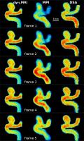 |
Flow dynamics in a 3D printed brain aneurysm model assessed by
magnetic particle imaging, magnetic resonance imaging and
dynamic subtraction angiography
Jan Sedlacik1, Andreas M. Frölich1,
Johanna Spallek2, Nils D. Forkert3,
Tobias D. Faizy1, Franziska Werner4,5,
Tobias Knopp4,5, Dieter Krause2, Jens
Fiehler1, and Jan-Hendrik Buhk1
1Neuroradiology, UKE, Hamburg, Germany, 2Product
Development and Mechanical Engineering Design, TUHH,
Hamburg, Germany, 3University
of Calgary, Calgary, AB, Canada, 4Biomedical
Imaging, UKE, Hamburg, Germany, 5Biomedical
Imaging, TUHH, Hamburg, Germany
Magnetic particle imaging (MPI) was compared with dynamic
magnetic resonance imaging (MRI) and dynamic subtraction
angiography (DSA) in a realistic 3D printed aneurysm model.
All three methods clearly depicted a distinct pulsatile flow
pattern and a delayed contrast agent outflow from the
aneurysm. Despite the disadvantages of a much lower temporal
resolution of the dynamic MRI and the 2D projection of the
DSA, all three methods are valid tools for characterizing
the hemodynamics of aneurysms. Especially the radiation
free, 3D, high temporal resolution MPI method seems to be a
very promising tool for imaging and characterization of
hemodynamics.
|
| |
11:36
|
0605.
 |
Microstructure Parameters in Acute Stroke: A Bayesian Approach
to diffusion-weighted MRI
Elias Kellner1, Karl Egger2, Valerij G
Kiselev2, Horst Urbach2, and Marco
Reisert1
1Department of Radiology, Medical Physics,
University Medical Center Freiburg, Freiburg, Germany, 2Department
of Neuroradiology, University Medical Center Freiburg,
Freiburg, Germany
In a recent study, we proposed a method for fast estimation
of microstructural tissue parameters such as
intra/extraaxonal volume fraction and diffusivities based on
a Bayesian approach and machine learning. In this study, we
report the application to cases of acute ischemic stroke. We
show that the parameters are able to outline the infarct
core qualitatively better than standard DTI. The results are
in line with the currently accepted picture of axonal
beading.
|
| |
11:48
|
0606.
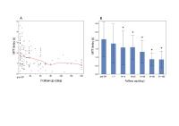 |
Chronological evaluation of Cerebral Hemodynamics by Dynamic
Susceptibility Contrast Magnetic Resonance Imaging after
Indirect Bypass Surgery for Moyamoya Disease
Yosuke Ishii1,2, Tadashi Nariai1, Yoji
Tanaka1, HIroshi Aihara2, Yoshio
Suyama2, Shinichi Wakabayashi2, and
Taketoshi Maehara1
1Neurosurgery, Tokyo Medial and Dental
University, Tokyo, Japan, 2Neurosurgery,
Suiseikai Kajikawa Hospital, Hiroshima, Japan
We used dynamic susceptibility contrast (DSC)-MRI to
evaluate the chronological changes in hemodynamics after
indirect bypass surgery for moyamoya disease. Twenty five
patients who underwent indirect bypass surgery and repeated
DSC–MRI measurement within the first 6 postoperative months
were included. We analyzed mean transit time (MTT) delay
using the cerebellum as control. Mean MTT delay in the
anterior circulation area gradually decreased soon after
surgery and stabilized after 3 postoperative
months. Postoperative MTT delay values were significantly
decreased compared with preoperative values from 1 to 2
weeks onwards. These results suggested DSC–MRI detected
angiogenesis during the early postoperative stages.
|
|