| |
10:00
 |
0317.
 |
4D flow MRI-Derived Hemodynamic Atlases of the Left Ventricle
with Hypertrophic Cardiomyopathy Demonstrate Abnormally Elevated
Blood Flow Velocities 
Pim van Ooij1, Alex J Barker2, Henk A
Marquering3, Gustav J Strijkers3,
James C Carr2, Michael Markl2,4, and
Aart J Nederveen5
1Radiology, Academic Medical Center, Amsterdam,
Netherlands, 2Radiology,
Northwestern University, Chicago, IL, United States, 3Biomedical
Engineering & Physics, Academic Medical Center, Amsterdam,
Netherlands, 4Biomedical
Engineering, Northwestern University, Chicago, IL, United
States, 5Academic
Medical Center, Amsterdam, Netherlands
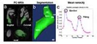
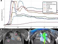
Altered hemodynamics in the left ventricle (LV) may
contribute to heart failure in hypertrophic cardiomyopathy (HCM).
The aim of this was study was to employ 4D flow MRI to
identify regions with altered velocity in HCM patients based
on the concept of 'LV flow heat maps' comparing velocity
fields in HCM patients with an atlas derived from healthy
controls. In the ejection phase, abnormally elevated
velocity was found in the LV outflow tract, whereas the
filling phase showed elevated velocity in the LV apex.
|
| |
10:12
|
0318.
 |
Characterization of aortic blood flow after aortic valve
replacement by 4D flow MRI
Alex S Hong1, Emilie Bollache1, Pim
van Ooij1, James C Carr1, Alex J
Barker1, Jeremy D Collins1, and
Michael Markl2
1Department of Radiology, Northwestern
University, Chicago, IL, United States, 2Department
of Radiology, Department of Biomedical Engineering,
Northwestern University, Chicago, IL, United States
Aortic valve replacement (AVR) is an effective surgical
approach to treating aortic valvular disease, but it is
unclear if and what type of prosthesis can fully reproduce
physiologically normal flow characteristic of a native
aortic valve. We utilized 4D flow MRI to systematically
compare blood flow in the thoracic aorta in post-AVR (bioprosthetic
vs. mechanical) patients and healthy controls. Both
bioprosthetic and mechanical valves were found to produce
higher peak systolic flow velocities and peak wall shear
stress in the ascending aorta than native valves,
demonstrating the presence of significant changes in aortic
blood flow in AVR patients.
|
| |
10:24
|
0319.
 |
Pressure Gradient Measurement in the Coronary Artery Using Phase
Contrast (PC)-MRI: Initial Patient Results Towards Noninvasive
Quantification of Fractional Flow Reserve
Zixin Deng1,2, Sangeun Lee3, Zhaoyang
Fan1, Christopher Nguyen1, Iksung Cho3,
Qi Yang1, Xiaoming Bi4, Byoung-Wook
Choi5, Jung-Sun Kim3, Daniel Berman1,
Hyuk-Jae Chang3, and Debiao Li1
1Biomedical Imaging Research Institute,
Cedars-Sinai Medical Center, Los Angeles, CA, United States, 2Bioengineering,
University of California, Los Angeles, Los Angeles, CA,
United States, 3Cardiology,
Severance Hospital, Yonsei Univeristy College of Medicine,
Seoul, Korea, Republic of, 4R&D,
Siemens Healthcare, Los Angeles, CA, United States, 5Radiology,
Severance Hospital, Yonsei Univeristy College of Medicine,
Seoul, Korea, Republic of
Fractional flow reserve is an invasive diagnostic tool to
evaluate the functional significance of a coronary stenosis
by quantifying the pressure gradient (ΔP) across the
stenosis. We proposed a non-invasive technique to derive ΔP
using Phase-contrast (PC)-MRI in conjunction with the
Navier-Stokes equations (ΔPMR). Excellent
correlation was observed between derived ΔPMR and
measure ΔP from a pressure transducer in a small caliber
phantom model. A significant increase in ΔPMR was
seen in the patient group vs. healthy controls. Preliminary
results suggested that noninvasive quantification of ΔPMR in
coronary arteries is feasible.
|
| |
10:36
|
0320.
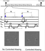 |
Cine Phase Contrast Simultaneous Multi-Slice imaging of blood
flow and CSF motion.
David A Feinberg1,2, Alexander Beckett1,
An T Vu1,2, and Liyong Chen2
1Helen Wills Neuroscience Institute, University
of California, Berkeley, CA, United States, 2Advanced
MRI Technologies, Sebastopol, CA, United States
The purpose was to develop and evaluate a novel approach to
MR phase imaging of blood flow and CSF flow by combining
cine phase contrast (cine-PC) with simultaneous multi-slice
(SMS) technique to measure velocity in several slice planes
simultaneously. Comparisons were made between SMS 2-4 and
conventional single-slice 2D cine-PC GE imaging. The
velocity curves measured in internal carotid (ICA) and
vertebral arteries and jugular veins and aqueductal CSF were
similar between SMS and conventional single-slice cine-PC.
In ICA correlations (R=0.92-0.98) in 6 subjects. This new
ability for simultaneous cross-sectional hemodynamic
quantification may be useful for medical diagnoses.
|
| |
10:48
|
0321.
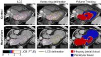 |
Vortex-ring mixing as a measure of diastolic function of the
human heart: phantom validation and initial observations in
healthy volunteers and patients with heart failure
Johannes Töger1,2, Mikael Kanski1, Per
M Arvidsson1, Marcus Carlsson1, Sándor
J Kovács3, Rasmus Borgquist4, Johan
Revstedt5, Gustaf Söderlind2, Håkan
Arheden1, and Einar Heiberg1,2,6
1Department of Clinical Physiology, Lund
University Hospital, Lund University, Lund, Sweden, 2Department
of Numerical Analysis, Centre for Mathematical Sciences,
Lund University, Lund, Sweden, 3Department
of Internal Medicine, Washington University School of
Medicine, St Louis, MO, United States, 4Department
of Arrhythmias, Lund University Hospital, Lund University,
Lund, Sweden, 5Department
of Energy Sciences, Lund University, Faculty of Engineering,
Lund, Sweden, 6Department
of Biomedical Engineering, Lund University, Faculty of
Engineering, Lund, Sweden
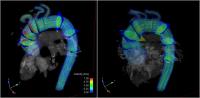
Diastolic dysfunction of the left ventricle (LV) of the
heart is a severe condition associated with poor prognosis.
However, objective and reproducible assessment of diastolic
function remains a challenge. We propose a new method using
4D flow MR by quantification of blood mixing within the LV
diastolic vortex-ring. Phantom validation showed fair
agreement between 4D flow MR and planar laser-induced
fluorescence (PLIF). Quantitative vortex-ring mixing differs
between healthy controls and patients with heart failure,
which demonstrates its potential as a marker of diastolic
dysfunction.
|
| |
11:00
|
0322.
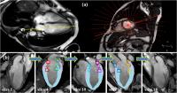 |
Dynamic assessment of atrioventricular junction (AVJ) based on
radial long-axis cine cardiac MR imaging
Shuang Leng1, Shuo Zhang2, Xiaodan
Zhao1, Baoru Leong1, Yiying Han1,
Yasutomo Katsumata3, Stuart Cook1,4,
Ru San Tan1,4, and Liang Zhong1,4
1National Heart Centre Singapore, Singapore,
Singapore, 2Philips
Healthcare Singapore, Singapore, Singapore, 3Philips
Healthcare Japan, Tokyo, Japan, 4Duke-NUS
Graduate Medical School Singapore, Singapore, Singapore
We have developed a semi-automatic tracking system of
atrioventricular junction (AVJ) deformation with two-,
three-, and four-chamber cardiovascular magnetic resonance
(CMR) long-axis images 1.
In this study, we applied the feature-tracking technique in
18 radial rotational long-axis cine CMR planes and evaluated
the motion of 36 evenly located AVJ points. Results have
shown that 1) the obtained average AVJ velocities (Sm, Em
and Am) and maximal displacements are independent of the
number of AVJ points selected, and 2) the routinely acquired
CMR imaging generated in clinical practice are sufficient
enough for dynamic assessment of AVJ deformation.
|
| |
11:12
|
0323.
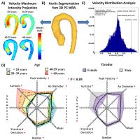 |
3D Blood Flow Velocity Distribution in the Normal Aorta: Effect
of Age and Gender Across 101 Subjects
Julio Garcia1, Roel L.F. van der Palen2,
Alex J. Barker1, Jeremy D. Collins1,
James C. Carr1, Joshua Robinson3,
Cynthia Rigsby3, and Michael Markl1,4
1Radiology, Northwestern University, Chicago, IL,
United States, 2Pediatric
Cardiology, Leiden University Medical Center, Leiden,
Netherlands, 3Department
of Medical Imaging, Ann & Robert H. Lurie Children’s
Hospital of Chicago, Chicago, IL, United States, 4Biomedical
Engineering, Northwestern University, Evanston, IL, United
States
The systematic characterization of effects in aortic disease
patients and healthy controls is important to improve
disease diagnosis. 4D flow MRI can be applied for the
analysis of altered hemodynamics in cardiovascular disease.
However, data analysis can be time consuming and often data
are not fully utilized by analysis based on 2D planes. This
study aimed to systematically apply flow distribution
analysis in the entire volume of the aorta to establish
normative reference values across a wide age range from
pediatric to adult subjects.
|
| |
11:24
 |
0324.
 |
High Quality Preclinical 4D-Flow Phase Contrast Imaging
Moritz Braig1, Jochen Leupold1, Ko
Cheng-Wen2, Marius Menza1, Juergen
Hennig1, Jan Korvink3, and Dominik von
Elverfeldt1
1University Medical Center Freiburg, Freiburg,
Germany, 2Dept.
Computer Science and Engineering, National Sun Yat-sen
University, Kaohsiung, Taiwan, 3Institute
of Microstructure Technology, Karlsruhe Institute of
Technology, Karlsruhe, Germany
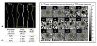
So far preclinical 4D-Flow MRI has not been able to deliver
an analysis of complex flow due to low resolution. The
presented framework and improvements allow high quality data
acquisitions with a reduced measurement time and the
possibility to visualize regional flow abnormalities. An
automatic magnitude segmentation in every timeframe combines
anatomic information with the underlying blood flow showing
even small vessels. It will draw new conclusions in mouse
models of cardiovascular diseases as a valuable tool for
preclinical researchers.
|
| |
11:36
|
0325.
 |
Ultra-High-Dimensional Flow Imaging (N-D Flow)
Joseph Y. Cheng1, Tao Zhang1, Marcus
T. Alley1, Michael Lustig2, John M.
Pauly3, and Shreyas S. Vasanawala1
1Radiology, Stanford University, Stanford, CA,
United States, 2Electrical
Engineering & Computer Sciences, University of California,
Berkeley, CA, United States, 3Electrical
Engineering, Stanford University, Stanford, CA, United
States
Volumetric cardiac-resolved flow imaging (4D flow) can
enable the assessment of flow, function, and anatomy from a
single sequence. Here, 4D flow is extended to higher
dimensional space as N-D flow. By resolving different
dynamics such as respiration or contrast enhancement, more
diagnostic information can be extracted for a
single-sequence protocol. Furthermore, this potentially
improves image quality and quantification accuracy. N-D flow
is enabled by a compressed-sensing and parallel imaging
based acquisition and reconstruction. The feasibility of
this approach is demonstrated for pediatric imaging.
|
| |
11:48
|
0326.
 |
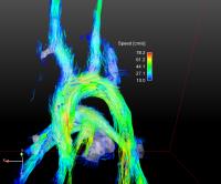
In vitro validation of Cartesian 4D flow mapping using
patient-specific 3D printed total cavo-pulmonary connection
models
Zachary Borden1, Peng Lai2, Ann
Shimakawa2, Alejandro Roldan-Alzate1,3,
and Christopher J Francois1
1Department of Radiology, University of
Wisconsin-Madison, Madison, WI, United States, 2GE
Healthcare, Menlo Park, CA, United States, 3Department
of Mechanical Engineering, University of Wisconsin-Madison,
Madison, WI, United States
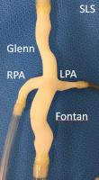
Congenital heart disease is a common disease process which
benefits from MRI 4D flow analysis. In a total
cavo-pulmonary connection model, Cartesion 4D Flow mapping
using k-t acceleration and variable density signal averaging
correlates well with US flow probe data and 2D PC
measurements. The improved post processing efficiency of
Cartesian acquisition may allow more widespread adoption of
4D flow technology for analyzing congenital heart disease.
|
|