| |
13:30
 |
0959.
 |
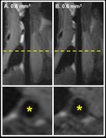
High resolution 3D diffusion imaging of carotid vessel wall
using stimulated echo based diffusion prepared turbo spin echo
sequence 
Qinwei Zhang1, Barbara Cervantes2,
Dimitrios C. Karampinos2, Bram F. Coolen1,
Aart J. Nederveen1, and Gustav J. Strijkers3
1Department of Radiology, Academic Medical
Center, University of Amsterdam, Amsterdam, Netherlands, 2Department
of Diagnostic and Interventional Radiology, Technische
Universität München, Munich, Germany, 3Biomedical
Engineering and Physics, Academic Medical Center, University
of Amsterdam, Amsterdam, Netherlands
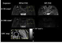
Diffusion imaging is becoming a promising alternative to
contrast enhanced imaging in detecting lipid core and
hemorrhage in atherosclerotic plaques. Diffusion prepared
turbo spin echo sequence (DP-TSE) has been proven to be
feasible to acquire 3D diffusion images of carotid vessel
wall, but it has critical requirement on the eddy currents.
This study demonstrates that using stimulated echo based DP-TSE
sequence, together with m1 nulling diffusion gradients, and
MLEV refocusing RF pulses, high resolution 3D carotid vessel
wall diffusion imaging can be achieved in the presence of
eddy current, motion and B1-inhomogeneity.
|
| |
13:42
|
0960.
 |
DCE-MRI reveals more extensive vasa vasorum in patients with
cardiovascular events
Huijun Chen1, Juan Wang2, Jie Sun3,
Daniel S Hippe3, Xihai Zhao1, and
Hongbing Liu2
1Biomedical Engineering, School of Medicine,
Tsinghua University, Beijing, China, People's Republic of, 2Cardiology,
People’s Liberation Army General Hospital, Beijing, China,
People's Republic of, 3Radiology,
University of Washington, Seattle, WA, United States
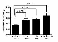
Pharmacokinetic modeling of DCE-MRI can quantify the
adventitial vasa vasorum of carotid atherosclerotic lesions
using the transfer constant (Ktrans).
However, the relationship between the DCE-MRI quantified
carotid adventitial vasa vasorum and cardiovascular events
remains unclear. In this study, we found that the
adventitial Ktrans of
carotid artery measured by DCE-MRI was associated with
cardiovascular events (cerebral ischemic events and coronary
artery events), suggesting that the carotid adventitial vasa
vasorum is not merely a local risk factor but also a
promising systemic marker for cardiovascular risk. DCE-MRI
may be valuable for identifying high risk patients in
clinical practice.
|
| |
13:54
|
0961.
 |
Texture-Based Classification of Advanced Carotid Atherosclerotic
Lesions on Multi-contrast Black-blood MRI at 3.0 Tesla: A Pilot
Study
Huilin Zhao1, Shiteng Suo1, Peipei Hao1,
Xiaosheng Liu1, Xihai Zhao2, Yongming
Dai3, Chun Yuan4, and Jianrong Xu1
1Radiology, Renji Hospital, Shanghai Jiao Tong
University School of Medicine, Shanghai, China, People's
Republic of, 2Center
for Biomedical Imaging Research, Tsinghua University School
of Medicine, Beijing, China, People's Republic of, 3Philips
Healthcare, Shanghai, China, People's Republic of, 4Radiology,
University of Washington, Seattle, WA, United States
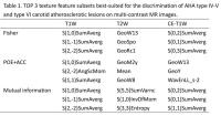
Texture analysis with the combined set of texture features
may be useful in discriminating vulnerable plaque. This
study sought to determine the feasibility of texture
analysis for the classification of American Heart
Association (AHA) type IV-V and type VI carotid
atherosclerotic lesions at multi-contrast black-blood MR
images. Our results suggest that texture-based
classification of type IV-V and type VI lesions is feasible
on precontrast T1-weighted images. This preliminary
evaluation indicates that carotid plaque texture analysis is
a potentially useful adjunct tool for quantitative
evaluation of atherosclerotic plaque vulnerability.
|
| |
14:06
|
0962.
 |
3D Carotid Wall Imaging: Stack-of-stars Trajectory for
Multi-contrast Atherosclerosis Characterization (STAR-MATCH)
Xiaoming Bi1, Zhaoyang Fan2, Yutaka
Natsuaki1, Debiao Li2, and Gerhard
Laub1
1Siemens Healthcare, Los Angeles, CA, United
States, 2Cedars-Sinai
Medical Center, Los Angeles, CA, United States
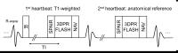
The recently developed MATCH technique integrates multiple
3D image sets into a single measurement and it is a
promising method for carotid plaque characterization. One of
the remaining challenges is the gross motion of carotid
arteries that originates from pulsation, breathing and
swallowing. In this work, a motion robust stack-of-stars
sampling trajectory was implemented into the MATCH sequence
(STAR-MATCH). Preliminary studies from volunteers and
patient demonstrate it is feasible to characterize carotid
plaque using the STAR-MATCH sequence with improve motion
robustness.
|
| |
14:18
|
0963.
 |
Coronary Atherosclerosis T1-weighed Characterization with
Integrated Anatomical Reference (CATCH): Comparison with
High-risk Plaque Features on OCT
Yibin Xie1, Young-Jin Kim2, Jianing
Pang1, Qi Yang1, Jung-Sun Kim3,
Christopher T. Nguyen1, Zixin Deng1,
Byoung Wook Choi2, Zhaoyang Fan1,
Daniel S. Berman1, Hyuk-Jae Chang3,
and Debiao Li1
1Cedars-Sinai Medical Center, Los Angeles, CA,
United States, 2Department
of Radiology, Severance Hospital, Yonsei University College
of Medicine, Seoul, Korea, Republic of, 3Division
of Cardiology, Severance Cardiovascular Hospital, Yonsei
University College of Medicine, Seoul, Korea, Republic of
The aim of this work is to investigate the nature of
pre-contrast and post-contrast T1w plaque hyper-intensity by
comparing with coronary plaque morphology assessed by
intracoronary optical coherence tomography (OCT). We scanned
13 healthy subjects and 30 stable angina patients using our
recently developed whole-heart T1w coronary plaque
characterization framework (CATCH). Compared with the
classification based on OCT, we found that pre-contrast
plaque to myocardial ratio (PMR) was significantly higher in
the presence of large lipids, macrophages, and cholesterol
crystals, whereas post-contrast PMR was significantly higher
in the presence of macrophages and microvessels.
|
| |
14:30
|
0964.
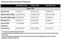 |
Increased Coronary Vessel Wall Thickness in Hyper IgE Syndrome
Patients; Depiction by Magnetic Resonance Vessel Wall Imaging
and Pathological Correction - Permission Withheld
Khaled Z. Abd-Elmoniem1, Nadine Z. Ramos1,
Saami Yazdani2, Ahmed M. Ghanem1,3,
Steven M. Holland4, Alexandra F. Freeman4,
and Ahmed M Gharib1
1Biomedical and Metabolic Imaging Branch, NIDDK,
Bethesda, MD, United States, 2University
of Southern Alabama, Mobile, AL, United States, 3Electrical
Engineering, Suez Canal University, Ismailia, Egypt,4NIAID,
Bethesda, MD, United States
In this study, coronary wall MRI is used to assess the
coronary wall thickness of patients with autosomal dominant
hyper-IgE (AD-HIES) or Job's syndrome; a primary
immunodeficiency caused by mutations in STAT3. Supported by
post-mortem histology, MRI coronary wall of AD-HIES patients
was thicker than in healthy subjects but comparable to CAD
patients. These findings suggest that coronary arteries in
Job’s syndrome are affected with atherosclerosis, contrary
to prior beliefs and study findings. Direct histologic
evaluation confirms the presence of atherosclerosis with
lack of needed supportive adventitial thickening and elastic
components. These findings suggest mechanisms for weakened
vessel wall that may lead to coronary dilation and aneurysm
in AD-HIES.
|
| |
14:42
|
0965.
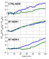 |
Evaluation of endothelial barrier function in atherosclerosis
induced rabbits using S-nitroso human serum albumin (S-NO-HSA) -
blood pool agent compound and dynamic contrast-enhanced
(DCE)-MRI
Peter Opriessnig1, Gunter Almer1,
Harald Froehlich1, Claudia Cabella2,
Rudolf Stollberger3, Seth Hallstroem4,
Gerd Hoerl4, and Harald Mangge1
1Clinical Institute for Medical and Chemical
Laboratory Diagnosis, Medical University of Graz, Graz,
Austria, 2CRB
Bracco Imaging SpA, Colleretto Giacosa, Torino, Italy, 3Institute
of Medical Engineering, Graz University of Technology, Graz,
Austria, 4Institute
of Physiological Chemistry, Medical University of Graz,
Graz, Austria
Endothelial dysfunction plays a key role in the progression
and pathogenesis of atherosclerosis (AS). DCE-MRI in
combination with a special nitric oxide donor S-nitroso
human serum albumin (S-NO-HAS) blood pool agent (B22956/1)
compound could be an additional measure that provides
information on the influence of plaque burden on the
vascular permeability and vasomotion. In this work, we
demonstrate the feasibility to investigate endothelial
barrier function and NO induced endothelium-independent
vasomotor response of the abdominal aorta in control and AS
induced rabbits simultaneously. Relative vessel wall signal
enhancement and change in lumen area were measured using a
double-inversion-recovery turbo-spin-echo sequence.
|
| |
14:54
|
0966.
 |
Impact of exercise intervention on vascular function in PAD
Erin K Englund1, Michael C Langham2,
Thomas F Floyd3, Felix W Wehrli2, and
Emile R Mohler4
1Department of Bioengineering, University of
Pennsylvania, Philadelphia, PA, United States, 2Department
of Radiology, University of Pennsylvania, Philadelphia, PA,
United States, 3Department
of Anesthesiology, Stony Brook University, Stony Brook, NY,
United States, 4Department
of Medicine, University of Pennsylvania, Philadelphia, PA,
United States
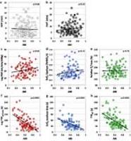
Peripheral vascular function can be interrogated by
measuring recovery dynamics following induced ischemia. In
this study, 136 patients with peripheral artery disease were
randomized into supervised exercise rehabilitation (SER) or
standard medical care (SMC). Each patient’s leg was scanned
before and after the intervention period. MRI data were
acquired throughout an ischemia-reperfusion paradigm with
PIVOT, a method to simultaneously and dynamically measure
perfusion, venous oxygen saturation, and skeletal muscle T2*.
Patients randomized to SER had a significant increase in
peak perfusion from baseline to follow-up when averaged
across the entire cross-section of the leg and in the
peroneus muscle.
|
| |
15:06
|
0967.
 |
MRI biomarkers associated with guide wire puncture forces
required to cross ex-vivo human peripheral arterial chronic
total occlusions
Trisha Roy1,2, Garry Liu1, Noor Shaikh1,
Kevan Anderson1, Nicolas Yak1, Xiuling
Qi1, Andrew Dueck1,2, and Graham
Wright1,3
1Schulich Heart Program and the Sunnybrook
Research Institute, Sunnybrook Health Sciences Centre,
Toronto, ON, Canada, 2Division
of Vascular Surgery, University of Toronto, Toronto, Canada, 3Department
of Medical Biophysics, University of Toronto, Toronto,
Canada
Percutaneous vascular interventions (PVI) for treating
peripheral arterial disease (PAD) have poor outcomes with
high re-intervention and failure rates. Not all lesions are
amenable to PVI, but predicting failure is difficult. While
CT can identify heavily calcified lesions, current imaging
offers limited differentiation between hard and soft PAD
plaques, which impacts procedural success. This study
demonstrates the feasibility of using MRI biomarkers to
characterize plaque components in ex-vivo human peripheral
arteries with histologic and microCT validation. We
demonstrate that significantly higher puncture forces are
required to cross non-calcified “hard” chronic total
occlusions (CTOs) compared to “soft” CTOs, as classified by
these MRI biomarkers.
|
| |
15:18
|
0968.
 |
Optimization of 3 dimensional (3D), high resolution T2 weighted
SPACE for carotid vessel wall imaging on a 7T whole-body
clinical scanner
Claudia Calcagno1,2, Bram Coolen3, Bei
Zhang1,2, Gilles Boeykens 3,
Philip Robson1,2, Venkatesh Mani1,2,
Aart J Nederveen3, Willem Mulder1,2,
and Zahi Fayad1,2
1Department of Radiology, Icahn School of
Medicine at Mount Sinai, New York, NY, United States, 2Translational
and Molecular Imaging Institute, Icahn School of Medicine at
Mount Sinai, New York, NY, United States, 3Department
of Radiology, Academisch Medisch Centrum, Amsterdam,
Netherlands
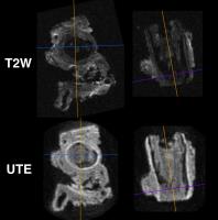
Accurate morphological measurements and classification of
carotid plaques require imaging with high spatial
resolution, and may benefit from the increased signal
intrinsically available on ultra-high field (7T) magnets.
Several studies have already investigated carotid vessel
wall imaging at 7T and compared it with state-of-the-art 3T
protocols. These initial investigations have focused on 2
dimensional (2D), multi-slice imaging. Better than this
approach, 3 dimensional (3D) vessel wall imaging allows
characterizing extensive vascular territories while
minimizing partial volume artifacts in plaque-prone regions,
such as the carotid bulb and bifurcation. Here, we
demonstrated the feasibility of performing 3D carotid vessel
wall imaging on a whole body 7T clinical magnet using a
custom made carotid coil.
|
|