| |
08:00
|
1068.
 |
Time-Resolved Non-Contrast-Enhanced MR Angiography with Static
Tissue Suppression using Velocity-Selective Pulse Trains - Permission Withheld
Qin Qin1,2, Guanshu Liu1,2, Ye Qiao1,
and Dexiang Liu1,2,3
1Radiology, Johns Hopkins University, Baltimore,
MD, United States, 2F.M.
Kirby Research Center for Functional Brain Imaging, Kennedy
Krieger Institute, Baltimore, MD, United States, 3Radiology,
Panyu District Central Hospital, Guangzhou, China, People's
Republic of
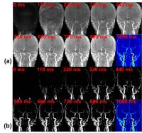
Time-resolved non-contrast-enhanced MR angiography (NCE-MRA),
by providing hemodynamic flow patterns, is promising for
many vascular disorders. Conventional techniques remove
tissue background using various arterial spin labeling (ASL)
approaches with paired subtraction of control and label
scans. Here a new multi-phase MRA method is introduced that
achieves background suppresstion by applying a tissue mask,
which is derived from thresholding a velocity-selective MRA
(VSMRA) obtained at the end of the cycle. The feasibility of
this new single-scan dynamic approach was demonstrated on
extracranial and intracranial vasculatures of healthy
volunteers at 3T.
|
| |
08:12
|
1069.
 |
Nonenhanced hybridized arterial spin-labeled magnetic resonance
angiography of the extracranial carotid arteries at 3 Tesla
using a fast low-angle shot readout 
Ioannis Koktzoglou1,2, Matthew T Walker1,2,
Joel R Meyer1,2, Ian G Murphy1,3, and
Robert R Edelman1,3
1Radiology, NorthShore University HealthSystem,
Evanston, IL, United States, 2University
of Chicago Pritzker School of Medicine, Chicago, IL, United
States, 3Northwestern
University Feinberg School of Medicine, Chicago, IL, United
States
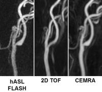
Nonenhanced hybridized arterial spin labeling (hASL)
magnetic resonance angiography (MRA) using a fast low-angle
shot (FLASH) readout was used to image the extracranial
carotid arteries at 3 Tesla. Comparisons were made with 2D
time-of-flight (TOF) MRA and contrast-enhanced MRA. Image
quality obtained hASL FLASH MRA was found to be superior to
that 2D TOF, with the method also providing improved
inter-rater agreement, quantification of arterial
cross-sectional area, and vessel sharpness.
|
| |
08:24
|
1070.
 |
Isotropic 3D Black Blood MRI of Abdominal Aortic Aneurysm:
Comparison with CT Anigography
Chengcheng Zhu1, Bing Tian2, Florent
Seguro1, Joe Leach1, Qi Liu2,
Jianping Lu2, Luguang Chen2, Michael
Hope1, and David Saloner1
1Radiology, University of California, San
Francisco, San Francisco, CA, United States, 2Radiology,
Changhai Hospital, Shanghai, China, People's Republic of
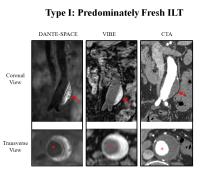
Computed Tomography angiography (CTA) is the gold standard
for abdominal aortic aneurysm (AAA) imaging, but requires
radiation and iodinated contrast. We previously developed an
isotropic 3D black blood technique (DANTE-SPACE) for AAA
imaging. In this study we validated 3D MRI against CTA for
AAA diameter and volume measurement, and found excellent
accuracy and reproducibility. Features of intra-luminal
thrombus (ILT) composition that are possibly related to
faster AAA growth can be identified on 3D MRI but not on CTA.
3D black blood MRI can be used as a non-invasive tool for
AAA serial monitoring and ILT evaluation and has the
potential to improve patient risk stratification.
|
| |
08:36
|
1071.
 |
High Resolution MRI for Characterization of Inflammation within
Abdominal Aortic Aneurysm
Chengcheng Zhu1, Thomas Hope1, Henrik
Haraldsson1, Farshid Faraji1, David
Saloner1, and Michael Hope1
1Radiology, University of California, San
Francisco, San Francisco, CA, United States
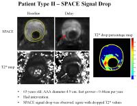
Abdominal aortic aneurysms (AAAs) with focal inflammation
(identified by USPIO uptake) have been reported to predict
faster growth. Previous 2D T2* mapping method is limited by
spatial resolution. This study evaluated 3D high-resolution
techniques (up to 1.3mm isotropic) for inflammation imaging
of AAAs. Experiments were preformed using both USPIO
phantoms and in vivo patient studies. We found the signal
characteristics of 3D DANTE-SPACE images had good agreement
with T2* value drop, and it provided higher resolution and
possible information on USPIO concentration. Therefore, 3D
high resolution methods may help risk stratify patients with
AAA disease by characterizing and quantifying inflammation.
|
| |
08:48
|
1072.
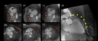 |
Robust large-volume fat suppression in whole-heart
free-breathing self-navigated coronary MR angiography at 3T
using lipid insensitive binomial off-resonant excitation (LIBRE)
pulses
Jessica AM Bastiaansen1, Davide Piccini1,2,
Ruud B van Heeswijk1,3, and Matthias Stuber1,3
1Department of Radiology, University hospital
(CHUV) and University of Lausanne (UNIL), Lausanne,
Switzerland, 2Advanced
Clinical Imaging Technology, Siemens Healthcare, Lausanne,
Switzerland, 3Center
for Biomedical Imaging, Lausanne, Switzerland
Large volume fat suppression is increasingly challenging at
high magnetic field strengths due to B0 and
B1 inhomogeneities.
In this study, we developed a novel lipid-insensitive
binomial off-resonant (LIBRE) radiofrequency excitation
pulse to achieve near-complete fat suppression in large 3D
volumes and applied it to whole-heart coronary imaging at
3T. In 6 healthy volunteers, we performed free-breathing
self-navigated whole-heart 3D radial coronary MRA, and
quantitatively compared the results to more commonly used
methods for lipid nulling. We show that LIBRE significantly
improves the signal nulling of lipid resonances resulting in
both improved blood pool delineation for self-navigation and
increased vessel conspicuity in the final images.
|
| |
09:00
 |
1073.
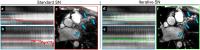 |
An Iterative Approach to Respiratory Self-Navigation Allows for
Improved Image Quality and 100% Scan Efficiency in
Contrast-Enhanced Inversion-Recovery Whole-Heart Coronary MRA at
3T; a First Patient Study
Giulia Ginami1, Davide Piccini1,2,
Pierre Monney3, Pier Giorgio Masci3,
and Matthias Stuber1,4
1University Hospital (CHUV) and University of
Lausanne (UNIL), Lausanne, Switzerland, 2Advanced
Clinical Imaging Technology, Siemens Healthcare, Lausanne,
Switzerland, 3Division
of Cardiology and Cardiac MR Center, University Hospital of
Lausanne (CHUV), Lausanne, Switzerland, 4Center
for Biomedical Imaging (CIBM), Lausanne, Switzerland
The performance of Self-Navigation (SN) for respiratory
motion compensation in 3T whole-heart coronary MRA may be
compromised by contrast variations secondary to
slow-infusion of a contrast agent. In this study, we
quantitatively and successfully tested the hypothesis that
an Iterative approach to SN (IT-SN) leads to improved
performance during slow infusion.
|
| |
09:12
|
1074.
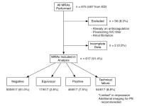 |
Six month clinical outcomes following pulmonary contrast
enhanced magnetic resonance angiography for the primary workup
of pulmonary embolism
Mark L. Schiebler1, Michael D. Repplinger2,
Christopher Lindholm3, John Harringa2,
Christopher J. François1, Karl K. Vigen1,
Azita G. Hamedani2, Thomas M. Grist1,4,5,
Scott B. Reeder1,2,4,6, and Scott K. Nagle1,5,7
1Radiology, UW-Madison, Madison, WI, United
States, 2Emergency
Medicine, UW-Madison, Madison, WI, United States, 3UW
Madison School of Medicine, UW-Madison, Madison, WI, United
States, 4Biomedical
Engineering, UW-Madison, Madison, WI, United States, 5Medical
Physics, UW-Madison, Madison, WI, United States, 6Medicine,
UW-Madison, Madison, WI, United States, 7Pediatrics,
UW-Madison, Madison, WI, United States
The aim of this study was to determine the effectiveness of
pulmonary magnetic resonance angiography (PE-MRA) for the
primary diagnosis of pulmonary embolism (PE). We
retrospectively reviewed the electronic medical records of
675 consecutive patients who underwent PE-MRA. Adverse
events (venous thromboembolism (VTE), bleeding or
death) that were potentially related either to over or
under treatment of PE during the subsequent 6 months were
extracted from the electronic medical record. The negative
predictive value for this test was found to be 97%. Based
upon these outcomes, PE-MRA performs similarly to CTA as a
primary test to exclude clinically significant pulmonary
embolism in patients presenting acutely with dyspnea.
|
| |
09:24
|
1075.
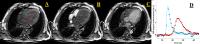 |
Model-based characterization of the transpulmonary circulation
by DCE-MRI
Salvatore Saporito1, Ingeborg H.F. Herold 1,2,
Patrick Houthuizen3, Jacques A. den Boer1,
Harrie C.M. van den Bosch4, Hendrikus H.M.
Korsten 1,2,
Hans C. van Assen1, and Massimo Mischi1
1Department of Electrical Engineering, Eindhoven
University of technology, Eindhoven, Netherlands, 2Department
of Anesthesiology and Intensive Care, Catharina Hospital
Eindhoven, Eindhoven, Netherlands,3Department of
Cardiology, Catharina Hospital Eindhoven, Eindhoven,
Netherlands, 4Department
of Radiology, Catharina Hospital Eindhoven, Eindhoven,
Netherlands
Objective measures to assess pulmonary circulation status
would improve heart failure patient care. We propose a
method for the characterization of the transpulmonary
circulation by DCE-MRI. Parametric deconvolution was
performed between contrast agent first passage
time-enhancement curves derived from the right and left
ventricular blood pool. The transpulmonary circulation was
characterized as a linear system with impulse response
modelled as local density random walk model. We tested the
method on 32 heart failure patients and 19 healthy
volunteers; patients presented longer transpulmonary transit
times and more skewed transpulmonary impulse responses.
|
| |
09:36
|
1076.
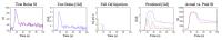 |
Predictive Bolus Tailoring of Gd-Based Contrast Agents for
Optimized Contrast-Enhanced MRA
Jeffrey H Maki1 and
Gregory J Wilson1
1Radiology, University of Washington, Seattle,
WA, United States
Gadolinium contrast for CE-MRA is typically injected at a
fixed, relatively fast (1.5 – 2.0 mL/s) rate. This results
in a peaked bolus profile such that vascular signal
intensity (SI) decays during latter k-space acquisition,
leading to blurring and ringing artifacts. A “tailored”
test bolus-based predictive algorithm was developed to
determine a patient-individualized multi-phase injection to
achieve any arbitrary arterial SI “plateau” duration. This
technique was tested on 10 patients and compared to 10
patients receiving a fixed 1.6 mL/s contrast injection. The
tailored bolus plateau duration was 24 vs. 9 s (p < 0.01)
with only a 20% SI loss.
|
| |
09:48
 |
1077.
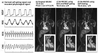 |
Cardiac and Respiratory Self-Gated 4D Multi-Phase Steady-State
Imaging with Ferumoxytol Contrast (MUSIC)
Fei Han1, Ziwu Zhou1, Takegawa Yoshida1,
Kim-Lien Nguyen1,2, Paul J Finn1, and
Peng Hu1
1Radiology, University of California, Los
Angeles, Los Angeles, CA, United States, 2Division
of Cardiology, Veterans Affairs Greater Los Angeles
Healthcare System, Los Angeles, CA, United States
We proposed a cardiac and respiratory self-gated, 4D
multi-phase steady-state imaging with contrast (MUSIC)
technique for detailed evaluation of cardiovascular
anatomies. A rotating cartesian k-space sampling pattern was
designed that integrates frequently sampled k-space
centerline as self-gating signal and allows retrospective
data-binning based on derived motion signal. Phantom and
in-vivo results on 7 clinical indicated pediatric CHD
patients show that the proposed self-gated MUSIC could
potentially eliminates the need of external physiological
signal for motion gating, has increased scan efficiency
while maintaining or exceeding the image quality of the
original MUSIC.
|
|