| |
16:00
|
0790.
 |
False positive bundles in tractography - Permission Withheld
Maxime Descoteaux1, Jasmeen Sidhu1,
Eleftherios Garyfallidis1, Jean-Christophe Houde1,
Peter Neher2, Bram Stieltjes3, and
Klaus H. Maier-Hein2
1Computer Science, Université de Sherbrooke,
Sherbrooke, QC, Canada, 2German
Cancer Research Center, Heindeberg, Germany, 3Basel
University, Basel University Hospital, Switzerland
This work provides novel insights in false positive bundles
produced by tractography using a highly realistic diffusion
MRI phantom with known underlying white matter ground truth
anatomy. This MRI phantom was used in the ISMRM 2015
Tractography Challenge. We show that regardless of the
tractography pipeline used, many invalid bundles with dense
and meaningful structures are found in the tractograms.
|
| |
16:20
 |
0791.
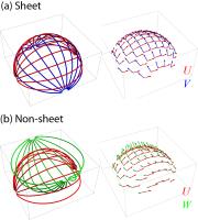 |
Mapping the brain’s “Sheet Probability Index” (SPI) with
diffusion MRI: Sheet happens?! 
Chantal Tax1,2, Tom Dela Haije3,
Andrea Fuster3, Carl-Fredrik Westin2,
Max A. Viergever1, Luc Florack3, and
Alexander Leemans1
1Image Sciences Institute, University Medical
Center Utrecht, Utrecht, Netherlands, 2Department
of Radiology, Brigham and Women's Hospital, Harvard Medical
School, Boston, MA, United States, 3Mathematics
and Computer Science, Eindhoven University of Technology,
Eindhoven, Netherlands
The prevalence of sheet structure in the brain has been a
debated issue since its proposal. This structure can be
analyzed by means of the Lie bracket, which can be derived
from diffusion MRI (dMRI) data. Due to the occurrence of
noise, however, it is difficult to quantify to what degree
the local structure effectively resembles a sheet. In this
work, we propose a new and robust local measure based on the
Lie bracket that can be interpreted as the sheet probability
index (SPI).
|
| |
16:40
|
0792.
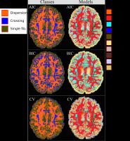 |
To be Dispersed or Not to be Dispersed: A Study Using HCP Data
Aurobrata Ghosh1, Daniel C Alexander1,
and Hui Zhang1
1Centre for Medical Image Computing, University
College London, London, United Kingdom
We conduct model comparison experiments on the widely
available HCP dataset to assess the importance of fibre-dispersion
when modelling the brain’s tissue-microstructure from
diffusion MRI (dMRI). Although many fibre dispersion
configurations have been identified in the brain, most dMRI
methods only model parallel or crossing fibres. To highlight
the importance of dispersion, we design k-fold
cross-validation experiments, on two HCP subjects, and
compare ten compartment-based models using three metrics. We
find that up to 50% of the brain-voxels, including white
matter regions, support dispersion models over crossing
models. Hence we conclude that it is important to model
dispersion in dMRI.
|
| |
17:00
|
0793.
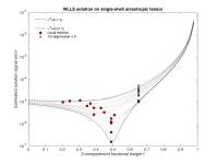 |
Challenges in solving the two-compartment free-water diffusion
MRI model
Ørjan Bergmann1,2, Carl-Fredrik Westin1,
and Ofer Pasternak1
1Dept of Radiology, Brigham and Women's Hospital,
Harvard Medical School, Boston, MA, United States, 2Norwegian
Competency Center for MS, Haukeland University Hospital,
Bergen, Norway
In this work we explore the solution space of the
two-compartment free-water problem under different noise
levels. Based on the shape of the solution space we show
that solving this model in an intuitive and straightforward
manner may result in solutions which are sensitive to noise,
and that are biased towards neglecting the free-water
component. Although multi-shell techniques improve the
situation we show that more advanced methods are required to
further stabilize the solution.
|
| |
17:20
|
0794.
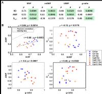 |
Quantification of demyelination and remyelination with diffusion
MRI: specific in vivo White Matter Tract Integrity metrics agree
with electron microscopy-derived features
Ileana O Jelescu1, Magdalena Zurek1,
Kerryanne V Winters1, Jelle Veraart1,
Anjali Rajaratnam1, Nathanael S Kim1,
James S Babb1, Timothy M Shepherd1,
Dmitry S Novikov1, Sungheon G Kim1,
and Els Fieremans1
1Center for Biomedical Imaging, Radiology, New
York University School of Medicine, New York, NY, United
States
White Matter Tract Integrity (WMTI) metrics derived from
diffusion data provide a compartment-specific
characterization of white matter. Here, we evaluated the
specificity of the axonal water fraction (AWF) and
extra-axonal radial diffusivity (De,-)
by assessing their correlations to metrics derived from
electron microscopy (EM), in the splenium of control,
cuprizone-treated and recovering mice. As the model
predicted, the WMTI-derived AWF correlated very strongly
with the EM-derived AWF, but not with the g-ratio,
while De,- correlated
with the g-ratio,
but not with the EM-derived AWF. WMTI parameters are
therefore promising biomarkers for specific biophysical
aspects of white matter pathology in
vivo.
|
| |
17:40
 |
0795.
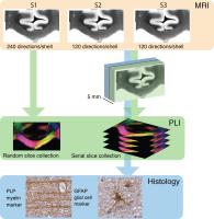 |
Exploring fibre orientation dispersion in the corpus callosum:
Comparison of Diffusion MRI, Polarized Light Imaging and
Histology
Jeroen Mollink1,2, Michiel Kleinnijenhuis1,
Stamatios N Sotiropoulos1, Michiel Cottaar1,
Anne-Marie van Cappellen van Walsum2, Menuka
Pallebage Gamarallage3, Olaf Ansorge3,
Saad Jbabdi1, and Karla L Miller1
1FMRIB centre, University of Oxford, Oxford,
United Kingdom, 2Donders
Institute for Brain, Cognition and Behaviour, Department of
Anatomy, Radboud University Medical Centre, Nijmegen,
Netherlands,3Department of Neuropathology,
University of Oxford, Oxford, United Kingdom
In this study we explored fibre orientation dispersion in
the corpus callosum using diffusion-weighted MRI, Polarized
Light Imaging and Histology. Microscopic fibre orientations
were derived from Polarized Light Imaging and histological
myelin and glial cell staining, with the aim of
understanding the microstructural features that correlate
with the diffusion signal.
|
|