| |
10:00
|
0307.
 |
Cerebral Cortex Parcellation by Fusion of Local and Global
Functional Connectivity Feature 
Alexander Schaefer1, Ru Kong1, Evan M.
Gordon2, Timothy Laumann 3,
Simon B. Eickhoff4,5, Xi-Nian Zuo6,
Avram J. Holmes7, and B.T. Thomas Yeo1
1Department of Electrical and Computer
Engineering, ASTAR-NUS Clinical Imaging Research Centre,
Singapore Institute for Neurotechnology and Memory Networks
Program, National University of Singapore, Singapore,
Singapore, 2VISN
17 Center of Excellence for Research on Returning War
Veterans, Waco, TX, United States, 3Department
of Neurology, Washington University in St. Louis, St. Louis,
MO, United States,4Institut for Clinical
Neuroscience, Heinrich Heine University, Düsseldorf,
Germany, 5Institute
for Neuroscience and Medicine, Research Center Jülich,
Jülich, Germany, 6Lab
for Functional Connectome and Development, Division of
Cognitive and Developmental, Chinese Academy of Sciences,
Beijing, China, People's Republic of, 7Department
of Psychology, Yale University, New Haven, CT, United States
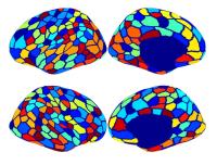
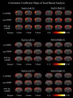
Current approaches to cerebral cortex parcellation with
resting-state functional connectivity MRI (fcMRI) can be
divided into local (e.g., fcMRI gradients) and global (e.g.,
clustering) approaches. Previous work suggests that local
and global approaches capture complementary aspects of brain
organization. Here we propose a novel hidden Markov Random
Field model that fuses local connectivity gradients with
global functional connectivity similarities. The resulting
parcellation compares favorably with a state-of-the-art
parcellation in terms of (1) parcel homogeneity in two
different datasets and (2) agreement with cytoarchitectonic
and visuotopic boundaries.
|
| |
10:12
|
0308.
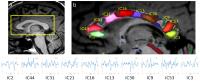 |
Track-weighted dynamic functional connectivity (TWdFC): a new
method to study dynamic connectivity
Fernando Calamante1,2, Robert Elton Smith1,
Xiaoyun Liang1, Andrew Zalesky3, and
Alan Connelly1,2
1The Florey Institute of Neuroscience and Mental
Health, Melbourne, Australia, 2Florey
Department of Neuroscience and Mental Health, The University
of Melbourne, Melbourne, Australia, 3Melbourne
Neuropsychiatry Centre and Melbourne School of Engineering,
The University of Melbourne, Melbourne, Australia
There is great interest in the study of brain connectivity
(structural and functional), and on the development of
methods that facilitate these investigations. In functional
connectivity (FC), there is also growing interest in
characterising the dynamic changes (dynamic-FC, dFC).
Track-weighted FC (TWFC) was proposed as a means to combine
the structural and (static) functional information into a
single image, by integrating a functional network with a
diffusion MRI tractogram. Here we propose TW-dynamic-FC (TWdFC),
by extending TWFC in two ways: first, it does not rely on an
a-priori FC network; second, it allows studying dFC.
|
| |
10:24
 |
0309.
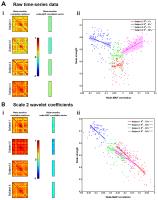 |
Beat-to-beat blood pressure fluctuations are present in
time-frequency dynamics of resting-state fMRI
Joseph R Whittaker1, Molly G Bright1,2,
Ian D Driver1, and Kevin Murphy1
1CUBRIC, School of Psychology, Cardiff
University, Cardiff, United Kingdom, 2Sir
Peter Mansfield Imaging Centre, University of Nottingham,
Nottingham, United Kingdom
A pilot study of fMRI time-frequency dynamics, characterized
using a maximal overlap discrete wavelet transform,
demonstrates matched frequency correlations with
beat-to-beat mean arterial blood pressure fluctuations.
Voxel-wise correlations between fMRI and blood pressure
wavelet coefficients, on a frequency scale centred at 0.1Hz,
reveal distributed and structured spatial variance across
the brain. We demonstrate that functional connectivity
methods that include time-frequency representations of fMRI
data are likely very sensitive to these blood pressure
fluctuations.
|
| |
10:36
|
0310.
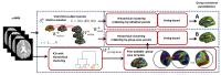 |
A cortical and sub-cortical parcellation clustering by intrinsic
functional connectivity
Ying-Chia Lin1, Tommaso Gili2,3,
Sotirios A. Tsaftaris 1,4,
Andrea Gabrielli5, Mariangela Iorio3,
Gianfranco Spalletta3, and Guido Caldarelli1
1IMT Institute for Advanced Studies Lucca, Lucca,
Italy, 2Enrico
Fermi Centre, Rome, Italy, 3IRCCS
Fondazione Santa Lucia, Rome, Italy, 4Institute
of Digital Communications, School of Engineering, The
University of Edinburgh, Edinburgh, United Kingdom, 5ISC-CNR,
UOS Sapienza, Dipartimento di Fisica, Universita Sapienza,
Rome, Italy
Network analysis of resting-state fMRI (rsfMRI) has been
widely utilized to investigate the functional architecture
of the whole brain. Here we propose a robust parcellation
method that first divides cortical and sub-cortical regions
into sub-regions by clustering the rsfMRI data for each
subject independently, and then merges those individual
parcellations to obtain a global whole brain parcellation.
To do so our method relies on majority voting (to merge
parcellations of multiple subjects) and enforces spatial
constraints within a hierarchical agglomerative clustering
framework to define parcels that are spatially homogeneous.
|
| |
10:48
 |
0311.
 |
Low Frequency Optogenetic Stimulation of Dentate Gyrus Enhances
Brain Functional Connectivity Revealed by Resting-State fMRI
Russell W Chan1,2, Alex TL Leong1,2,
Patrick P Gao1,2, Y S Chan3, W H Yung4,
Kevin K Tsia2, and Ed X Wu1,2
1Laboratory of Biomedical Imaging and Signal
Processing, The University of Hong Kong, Hong Kong, China,
People's Republic of, 2Electrical
and Electronic Engineering, The University of Hong Kong,
Hong Kong, China, People's Republic of, 3School
of Biomedical Sciences, The University of Hong Kong, Hong
Kong, China, People's Republic of, 4School
of Biomedical Sciences, The Chinese University of Hong Kong,
Hong Kong, China, People's Republic of
Low frequency coherent rsfMRI signals (<0.1Hz) do not match
the bandwidth of established neuronal oscillations,
highlighting a gap in our knowledge regarding the neuronal
basis of rsfMRI underlying long-range brain networks. In
this study, optogenetics and rsfMRI were combined to
investigate the neuronal basis of rsfMRI connectivity by
probing alternations of brain functional connectivity
before, during and after low frequency stimulation in dorsal
dentate gyrus. Our results demonstrated that low frequency
optogenetic stimulation enhanced brain functional
connectivity. This indicated that low frequency neuronal
oscillations contribute and underlie the synchronized
long-range rsfMRI brain functional networks.
|
| |
11:00
|
0312.
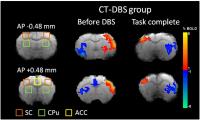 |
Functional MRI reveals striatal–thalamic connectivity in
cognitive neural behavior altered by central thalamic deep brain
stimulation
Hsin-Yi Lai1, Hui-Ching Lin2,3,
Yu-Chun Lo4, Lun-De Liao5,6, Wei-Che
Wei7, and You-Yin Chen7
1Interdisciplinary Institute of Neuroscience and
Technology (ZIINT), Zhejiang University, Hangzhou City,
China, People's Republic of, 2Department
and Institute of Physiology, National Yang-Ming University,
Taipei, Taiwan, 3Brain
Research Center, National Yang Ming University, Taipei,
Taiwan, 4Center
for Optoelectronic Biomedicine, National Taiwan University
College of Medicine, Taipei, Taiwan, 5Institute
of Biomedical Engineering and Nanomedicine, National Health
Research Institutes, Miaoli County, Taiwan, 6Singapore
Institute for Neurotechnology, National University of
Singapore, Singapore, Singapore, 7Department
of Biomedical Engineering, National Yang-Ming University,
Taipei, Taiwan
This study demonstrates neuronal striatal–thalamic
connectivity modulated by direct stimulating the central
thalamus in rats. Our results indicate that the CT-DBS
modulate the neuronal activity in bilateral anterior
cingulate cortex, caudate-putamen and somatosensory cortex
and increases in functional connectivity between the
striatum and parafascicular thalamic nucleus, hippocampus
and primary motor cortex to shorten the cognitive related
behavior task. CT-DBS fMRI has potential to explore
functional connectivity in the brain and monitor functional
plasticity changes in a specific neuroanatomical pathway in
vivo.
|
| |
11:12
|
0313.
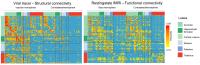 |
The structural basis for supporting functional connectivity in
mice
Joanes Grandjean1, Valerio Zerbi2,
Nicole Wenderoth2, and Markus Rudin1
1University and ETH Zurich, Zurich, Switzerland, 2ETH
Zurich, Zurich, Switzerland
Connectomics holds promise to foster our understanding of
the healthy and disordered brain. MRI has been the method of
choice for such analysis, combining diffusion weighted with
functional imaging to resolve structural and functional
connectivity, respectively. However, both methods are
indirect measures prone to bias and artifacts. In mice,
structural connectivity has been reconstructed with high
spatial resolution by mapping the distribution of viral
tracers following local injections at multiple sites
offering a unique opportunity to compare functional
connectivity with detailed mono-synaptic projections. Such
comparisons should help bridging functional and structural
connectivity in rodents with implications for human studies.
|
| |
11:24
|
0314.
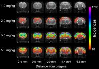 |
Characterization of acute phencyclidine-induced dose-dependent
schizophrenic symptoms in rat: relationship between functional
connectivity, hemodynamic response, behavior, and
neurotransmitter levels
Jaakko Paasonen1, Raimo A Salo1, Jouni
Ihalainen2, Juuso Leikas2, Katja
Savolainen2, Markus M Forsberg2, and
Olli Gröhn1
1Department of Neurobiology, University of
Eastern Finland, Kuopio, Finland, 2School
of Pharmacy, University of Eastern Finland, Kuopio, Finland
Schizophrenia is a disorder that lack effective medication.
In order to improve treatments, better disease models are
required. Here, phencyclidine (PCP)-induced schizophrenic
symptoms were investigated in rats with fMRI. Results were
compared with microdialysis measurements and behavioral
tests. At PCP doses ≥ 3 mg/kg, characteristics for psychotic
symptoms were detected in functional connectivity (FC),
having good correspondence with locomotor and dopamine
activity. With PCP doses ≤ 2 mg/kg, markers for psychotic
symptoms were absent. The FC of mesolimbic pathway was still
affected, and social and cognitive deficits were confirmed
in behavioral tests. Thus, PCP ≤ 2 mg/kg induces
specifically the social and cognitive schizophrenic
deficits.
|
| |
11:36
|
0315.
 |
ACC GABA levels predict activity and connectivity in the
fronto-striatal network during interference inhibition in
borderline personality disorder
Guoying Wang1, Julia van Eijk1, Traute
Demirakca1, Markus Sack1, Sylvia
Cackowski2, Annegret Krause-Utz2,
Christian Schmahl2, and Gabriele Ende 1
1Neuroimaging, Central Institute of Mental
Health, Mannheim, Germany, 2Psychosomatic
Medicine and Psychotherapy, Central Institute of Mental
Health, Mannheim, Germany
By combining the MRS and fMRI technique, we tested whether
ACC GABA levels would predict the activity and connectivity
in fronto-striatal networks during interference inhibition
(Simon task) in BPD patients. BPD patients showed a
significant positive correlation between ACC GABA levels and
BOLD responses in fronto-striatal regions during
interference inhibition. Additionally, ACC GABA levels in
BPD patients were positively related to ACC-caudate
functional connectivity during the incongruent condition.
Our findings highlight that the GABAergic system in the ACC
plays an important role in the modulation of impulsivity via
regulating the local neural activity and remote connectivity
between key regions.
|
| |
11:48
|
0316.
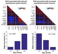 |
Fluctuations in Functional Connectivity Predict Shifts in
Arousal State
Chenhao Wang1, Ju Lynn Ong1, Amiya
Patanaik1, Juan Zhou1,2, and Michael
W. L. Chee1
1Neuroscience and Behavioral Disorders Program,
Duke-NUS Graduate Medical School Singapore, Singapore,
Singapore, 2Clinical
Imaging Research Center, Agency for Science, Technology and
Research, Singapore, Singapore
To elucidate relationship between fluctuation in functional
connectivity and behavior we estimated dynamic connectivity
states (DCS) from task-free fMRI obtained from
sleep-deprived healthy young adults. Using spontaneous eye
closures as a proxy for vigilance, we identified two DCS
that were associated with high and low arousal respectively.
DCS exhibiting similar connectivity patterns were also
observed when individuals were performing an auditory
vigilance task. Dwell time in high or low arousal DCS
predicted task performance. Additionally, fluctuations in
DCS and task response time were correlated. Fluctuations in
functional connectivity appear to be related to spontaneous
changes in arousal that affect vigilance.
|
|