| |
10:45
|
0072.
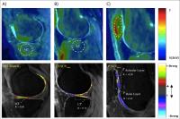 |
Imaging Cartilage-Bone Interactions in Osteoarthritis using
Simultaneous 18F-NaF PET-MR imaging– the “Bone-Cartilage
Connectome” 
Dragana Savic1,2, Valentina Pedoia1,
Youngho Seo1, Matthew Bucknor1,
Benjamin Franc1, and Sharmila Majumdar1
1University of California San Francisco, San
Francisco, CA, United States, 2University
of Oxford, Oxford, United Kingdom
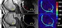
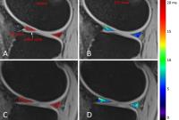
This first in human study evaluated cartilage biochemistry
and bone function in sixteen knee osteoarthritis patients
using simultaneous Time-Of-Flight (TOF) PET/MR imaging.
Bone turnover and blood flow was studied using 18F
Sodium Fluoride (NaF) and quantitative voxel by voxel MR
derived T1ρ relaxation
times characterizing the biochemical cartilage degeneration.
Increased degeneration of cartilage, was associated with
increased turnover in the adjoining bone as well as in the
non-adjoining compartments. These observations highlight the
complex biomechanical and biochemical interactions in the
whole knee joint, alluding to a “bone-cartilage connectome”,
that potentially changes during the natural history of the
disease.
|
| |
10:57
 |
0073.
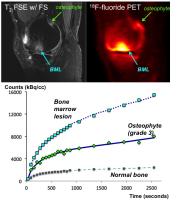 |
Dynamic analysis of [18F]-sodium fluoride uptake in knee
osteoarthritis with PET-MRI
Audrey P Fan1, Feliks Kogan1, Aleema
Patel1, Edwin HG Oei2, Andrew Quon1,
and Garry E Gold1
1Radiology, Stanford University, Stanford, CA,
United States, 2Erasmus
MC: University Medical Center Rotterdam, Rotterdam,
Netherlands
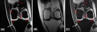
This study investigates dynamic uptake of [18F]-fluoride in
bone marrow lesions (BMLs) and osteophytes observed on MRI
of patients with knee osteoarthritis. Through kinetic
modeling, we characterized rate constants of bone metabolism
in bone pathology relative to healthy bone. BMLs and
higher-grade osteophytes showed higher total bone metabolism
Ki (P <
0.01) and higher bone mineralization rate k3 (P <
0.01) relative to grade 1 osteophytes and normal bone. While
a similar trend was observed for blood flow, the differences
from normal tissue were subtler suggests that rate of
mineralization k3 and
not blood flow is a key driver of [18F]-fluoride
accumulation in OA lesions. These new physiological
parameters may help differentiate between different grades
of OA lesions or identify which lesions are active parts of
the disease process.
|
| |
11:09
|
0074.
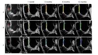 |
Longitudinal Evaluation of Cartilage Component of
Matrix-Associated Autologous Chondrocyte Transplants using
Biochemical MR Imaging
Xian Xu1, Ningyu An1, Panli Zuo2,
and Esther Raithel3
1Department of Radiology, Chinese PLA General
Hospital, Beijing, China, People's Republic of, 2Siemens
Healthcare, MR Collaborations NE Asia, Beijing, China,
People's Republic of, 3Siemens
Healthcare GmbH, Berlin, Germany
This study combined T2 mapping and delayed
gadolinium-enhanced MRI of cartilage (dGEMRIC) technique to
evaluate the repair cartilage tissue after Matrix-associated
autologous chondrocyte implantation (MACI). We found that
the T2 and ΔR1 values of the repair tissue were
significantly higher than the native tissue at 1, 3 and 6
months after MACI, but showed a downward trend and showed no
difference with native tissue at 12 months, which suggested
that the integrity of the collagen and GAG of repair tissue
was similar to native cartilage.
|
| |
11:21
|
0075.
 |
Loaded MRI – A Surrogate Measurement of in vivo Knee Joint
Contact Mechanics
Matthew F. Koff1, Hongsheng Wang2,
Suzanne Maher2, Scott Rodeo3, and
Hollis G Potter1
1Department of Radiology and Imaging - MRI,
Hospital for Special Surgery, New York, NY, United States, 2Department
of Biomechanics, Hospital for Special Surgery, New York, NY,
United States, 3Sports
Medicine and Shoulder Service, Hospital for Special Surgery,
New York, NY, United States
The relationship between calculated articular cartilage
deformation when using an MR compatible loading device and
actual contact mechanics has not been assessed. This study
evaluated the accuracy of in vivo cartilage deformation as a
surrogate for in vivo contact mechanics. Meniscal allograft
transplantation patients underwent loaded MR pre-operatively
and direct stress measurement intra-operatively. Good
correlation, 0.72 (range: 0.56 to 0.85), between cartilage
deformation and contact stress measurements was found. In
vivo cartilage deformation may be a surrogate for in vivo
contact mechanics.
|
| |
11:33
|
0076.
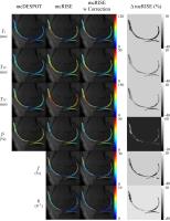 |
Incorporation of Finite Pulse Correction for Improved
MT-Corrected Multicomponent T2 analysis of Cartilage
Fang Liu1, Alexey Samsonov1, Wally
Block2, and Richard Kijowski1
1Department of Radiology, University of
Wisconsin-Madison, Madison, WI, United States, 2Department
of Medical Physics, University of Wisconsin-Madison,
Madison, WI, United States
Nuclear magnetic resonance studies have identified multiple
water components within cartilage tissue. Previous studies
using steady-state sequences based rapid method such as
mcDESPOT and mcRISE have shown feasibility of multicomponent
T2 analysis of cartilage. However, steady-state signal can
be influenced by the finite pulse effect which might lead to
biased parameter estimation. In this study, we incorporated
the finite pulse correction in the mcRISE model and
demonstrated the potential MT and finite pulse effect
in-sensitive T2 parameters for multicomponent cartilage
relaxometry analysis.
|
| |
11:45
|
0077.
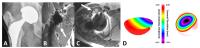 |
Correlation of MRI Appearance of Total Hip Arthroplasty With
Wear Metric and Histologic Evaluation
Matthew F. Koff1, Parina H. Shah1,
Mauro Miranda1, Christina Esposito2,
Elexis Baral2, Kara Fields3, Thomas
Bauer4, HSS Adult Reconstruction & Joint
Replacement Division5, Douglass Padgett5,
Timothy Wright2, and Hollis G. Potter1
1Department of Radiology and Imaging - MRI,
Hospital for Special Surgery, New York, NY, United States, 2Department
of Biomechanics, Hospital for Special Surgery, New York, NY,
United States, 3Healthcare
Research Institute, Hospital for Special Surgery, New York,
NY, United States, 4Cleveland
Clinic Foundation, Cleveland, OH, United States, 5Adult
Reconstruction and Joint Replacement Division, Hospital for
Special Surgery, New York, NY, United States
A majority of primary total hip arthroplasty (THA) function
well but implant failure may occur. We propose MRI to
evaluate adverse local tissue reactions (ALTRs) in patients
with THA. In this study, we correlate indirect measures of
ALTRs with direct measurements of implant wear. Greater
volumetric wear and visual damage was found in subjects with
ALTR on MR images. MR also correlated with histologic
metrics of implant wear. The results indicate that MRI
allows for accurate diagnosis of different synovial patterns
in THA, which correlate to wear analysis at retrieval.
|
| |
11:57
|
0078.
 |
In Vivo Evaluation of Low-grade Cartilage Defects in the Knee
using Sodium MRI at 7T
Stefan Zbyn1,2, Vladimir Mlynarik1,
Vladimir Juras1, Markus Schreiner1,3,
Didier Laurent4, Joerg Goldhahn4,
Nicole Getzmann4, Stefan Marlovits5,
and Siegfried Trattnig1
1Department of Biomedical Imaging and
Image-Guided Therapy, Medical University Vienna, Vienna,
Austria, 2CD
Laboratory for Clinical Molecular MR Imaging, Vienna,
Austria, 3Department
of Orthopaedics, Medical University Vienna, Vienna, Austria, 4Novartis
Institutes for Biomedical Research, Basel, Switzerland, 5Department
of Trauma Surgery, Medical University Vienna, Vienna,
Austria
To our best knowledge, this is the first report on employing
sodium (23Na) MRI for the in vivo evaluation of low-grade
cartilage defects in the knee joint. In this 7T study,
regions with chondral defect, weight-bearing, and
non-weight-bearing femoral cartilage were evaluated in
23Na-images of patients after knee injury. Test-retest
comparison showed high robustness and repeatability of
sodium data. 23Na-MRI allowed differentiation between
normal-appearing cartilage and low-grade chondral defects.
23Na-MRI can be used for noninvasive follow-up of changes in
GAG content associated with cartilage degeneration. This
method might be particularly useful for the evaluation of
cartilage regenerating therapies.
|
| |
12:09
|
0079.
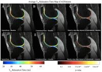 |
Local Analysis of T1?, T2, and R2–R1? Compositional MR Imaging
in Patients with ACL Injury Using Voxel-Based Relaxometry
Colin Russell1, Valentina Pedoia1,
Keiko Amano1, and Sharmila Majumdar1
1Radiology and Biomedical Imaging, University of
California, San Francsico, San Francisco, CA, United States
This multicenter study employs VBR as a novel technique to
analyze patients with ACL tears at the time of injury and 6
months after ACL reconstruction. T1ρ and
T2 analysis,
correlation, and dispersion difference (R2–R1ρ)
are three methods employed to highlight significant
cartilage changes. The most posterior region of the
posterior lateral tibia and the patella indicated partial
cartilage recovery 6 months after reconstruction,
demonstrated by decreasing T1ρ and
T2, decreased T1ρ T2 correlation
baseline to 6 months, and dispersion differences (R2–R1ρ).
The trochlea displayed symptoms of cartilage degeneration,
such as elevated T1ρ and
T2 and
dispersion differences.
|
| |
12:21
|
0080.
 |
In vivo assessment of T2* in menisci under loading conditions at
3 Tesla: preliminary results
Vladimir Juras1,2, Lenka Hornakova3,
Petr Kubovy3, Daniel Hadraba3,4, Pavel
Stursa5, David Gerych3, Pavol
Szomolanyi1, Karel Jelen3, and
Siegfried Trattnig1,6
1Department of Biomedical Imaging and
Image-Guided Therapy, High Field MR Centre, Medical
University of Vienna, Vienna, Austria, 2Department
of Imaging Methods, Institute for Measurement Science,
Bratislava, Slovakia, 3Department
of Anatomy and Biomechanics, Faculty Of Physical Education
and Sport, Prague, Czech Republic, 4Department
of Radiology, Hospital na Homolce, Prague, Czech Republic,5Academy
of Sciences of the Czech Republic, Prague, Czech Republic, 6Christian
Doppler Laboratory for Clinical Molecular MR Imaging,
Vienna, Austria
Meniscus behavior under loading in vivo has been studied
using parametric MR imaging. T2* has been acquired with vTE
using very short first TE = 0.8 ms to secure the precise
estimation. The knees of the subjects were loaded in situ
with custom made compression device and T2* mapping was
performed in 5 time points (without loading, and 4
consequent scans under the loading 7 min apart). The
increase in T2* was observed in all compartments,
significance was found in medial meniscus only. vTE T2*
mapping might be a prospective marker for detecting the
dynamic response of the meniscal tissue.
|
| |
12:33
|
0081.
 |
3D UTE Cones-IDEAL Imaging of the Knee and Ankle joints: Fast
Volumetric Imaging with Robust Fat/water Separation
Qun He1,2, Michael Carl3, Graeme
Bydder1, and Jiang Du1
1University of California, San Diego, San Diego,
CA, United States, 2Ningbo
Jansen NMR Technology Co., Ltd., Cixi, Zhejiang, China,
People's Republic of, 3Global
MR Applications & Workflow, General Electric, San Diego, CA,
United States
UTE sequences combined with IDEAL processing produces high
contrast images of short T2 tissues
or tissue components such as menisci, ligaments, and
tendons. In this work, we report the use of 3D UTE Cones
imaging and IDEAL processing (3D Cones-IDEAL) for volumetric
imaging of short T2 tissues
in the knee and ankle joints at 3T. High resolution
volumetric imaging of the knee and ankle joints, together
with robust fat/water separation, field map estimation, R2*/T2*
mapping and fat fraction mapping are demonstrated.
|
|