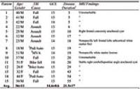
| |
10:30
|
0856.
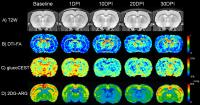 |
Comparable glucoCEST and 2DG autoradiography measures of glucose
metabolism in mild traumatic brain injury 
Tsang-Wei Tu1, Wael Ibrahim1, Neekita
Jikaria1, William Reid1, George Z.
Papadakis1, Dima Hammoud 1,
and Joseph A. Frank1
1Radiology and Imaging Sciences, National
Institutes of Health, Bethesda, MD, United States
The present glucose measurements from the brain are still
insufficient to provide the essential spatial-temporal
information. This study presents longitudinal glucose
chemical exchange saturation transfer (glucoCEST) MRI to
noninvasively detect the glucose metabolism in a rat model
of mild traumatic brain injury (mTBI) and compares to the
gold-standard 2-deoxyglucose (2DG) autoradiography. The
current glucoCEST results parallel with 2DG-autoradiography
results showing glucose uptake largely decreased after mTBI,
that persisted over time. GlucoCEST is capable of delivering
better image quality, higher image resolution and
sensitivity to identify the potential window for effective
treatments to increase the survival of injured brain.
|
| |
10:42
|
0857.
 |
Cortical neurometabolic alterations induces anxiety-like
behavior in rodent model of mild traumatic brain injury: A
1H-MRS and behavior study
Kavita Singh1, Seenu Haridas2, Kailash
Manda2, Richa Trivedi1, and Subash
Khushu1
1NMR, INMAS, DRDO, Delhi, Delhi, India, 2Neurobehavioral
lab, INMAS, DRDO, Delhi, Delhi, India
Mild traumatic brain injury (mTBI), (70-90% of all TBI)
shows consequences of anxiety-like behavioral alterations in
approximately 23% of cases. The present study assesses acute
anxiety-like behavior and its neurometabolic basis in a
rodent model of mTBI using 1H-MRS and neurobehavioral
analysis. At day5 reduced Tau/tCr levels in cortex was
observed in mTBI group as compared to control.
Neurobehavioral analysis showed increased anxiety-like
behavior with normal cognition at day5. This study provides
a putative neurometabolic basis of anxiety-like behavior in
mTBI model which closely mimics human concussion injury.
|
| |
10:54
 |
0858.
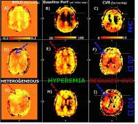 |
INDICATION OF IMPAIRED BASAL CEREBRAL BLOOD FLOW AND REACTIVE
CAPACITY IN CONCUSSED ATHLETES USING DUAL-ECHO PCASL
Clarisse Ildiko Mark1, Alex Bhogal2,
Douglas J Cook3, and Ingrid Johnsrude4
1Centre for Neuroscience Studies, Queen's
University, Kingston, ON, Canada, 2Radiology,
University Medical Center Utrecht, Utrecht, Netherlands, 3Department
of Surgery, Division of Neurosurgery, Queen’s University,
Kingston, ON, Canada, 4Brain
and Mind Institute, Department of Psychology, University of
Western Ontario, London, ON, Canada
Concussion can result in disability related to covert
symptoms and deficits that persist long after the initial
injury. A possible explanation for these observed phenomena
is sustained impairment of cerebrovascular autoregulation.
Here, we complement BOLD acquisition with simultaneous
cerebral blood flow (CBF) measurements during targeted
hypercapnic breathing challenges in varsity athletes during
the acute, early and late stages following injury. Changes
in basal CBF and cerebrovascular reactivity (CVR) were
observed over the first 2 weeks following injury compared to
matched un-concussed athletes. These biomarkers represent
promising tools to gauge the extent of brain injury and
monitor recovery.
|
| |
11:06
|
0859.
 |
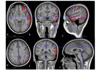
MRS and DTI Examination of Immature Rats Following Mild
Traumatic Brain Injury
Lesley May Foley1, Emin Fidan2, Henry
L Alexander2, Lee Ann New2, Patrick M
Kochanek2,3, T Kevin Hitchens1,4, and
Hulya Bayir2,3
1Pittsburgh NMR Center for Biomedical Research,
Carnegie Mellon University, Pittsburgh, PA, United States, 2Safar
Center for Resuscitation Research, University of Pittsburgh,
Pittsburgh, PA, United States,3Department of
Critical Care Medicine, University of Pittsburgh,
Pittsburgh, PA, United States, 4Animal
Imaging Center, University of Pittsburgh, Pittsburgh, PA,
United States
Recently we developed a closed-skull repeated mild (rm) TBI
model in postnatal day (PND) 18 rats. We hypothesized that
MRS and DTI can detect early microstructural changes of
brain and metabolite changes in the hippocampus. Alterations
in NAA and Ins after mTBI and rmTBI likely reflect
neuroaxonal damage and glial proliferation, respectively.
Reduced FA and increased AD in the white matter may reflect
a loss of integrity a possible indication of damage to
myelin/axonal membranes or demyelination. 1H-MRS
and DTI can identify subtle metabolic and structural
alterations in the hippocampus which appears normal on
histological analysis and conventional MR images.
|
| |
11:18
|
0860.
 |
Mapping axonal injury distribution in mild traumatic brain
injury with quantitative proton MR spectroscopy
Ivan Kirov1,2, Matthew S. Davitz1,2,
Assaf Tal3, James S. Babb1,2, Robert I
Grossman1,2, Yvonne W Lui1,4, and Oded
Gonen1,2
1Center for Advanced Imaging Innovation and
Research (CAI2R), New York University School of Medicine,
New York, NY, United States, 2Bernard
and Irene Schwartz Center for Biomedical Imaging, New York
University School of Medicine, New York, NY, United States, 3Chemical
Physics, Weizmann Institute of Science, Rehovot, Israel, 4Bernard
and Irene Schwartz Center for Biomedical Imaging, New York,
NY, United States
Since axonal injury is a primary outcome of traumatic brain
injury, our goal was to characterize its regional
distribution from a metabolic perspective. We set out to
identify regions prone to disproportionate injury, hence,
candidate targets in potential clinical applications of
proton MR spectroscopy (1H-MRS). We found that
metabolic axonal injury is diffusely distributed among
commonly injured tracts, but multivoxel 1H-MRS
may lack sensitivity for its detection on a regional basis.
These results motivate the use of 1H-MRS
approaches with higher sensitivity, such as global
averaging, or large "single voxels" in areas of white
matter, irrespective of placement location.
|
| |
11:30
|
0861.
 |
Proton MR spectroscopy identifies neuronal damage consistent
with gray/white matter interface involvement in mild traumatic
brain injury
Ivan Kirov1,2, Matthew S. Davitz1,2,
Assaf Tal3, James S. Babb1,2, Robert I
Grossman1,2, Yvonne W Lui1,4, and Oded
Gonen1,2
1Center for Advanced Imaging Innovation and
Research (CAI2R), New York University School of Medicine,
New York, NY, United States, 2Bernard
and Irene Schwartz Center for Biomedical Imaging, New York
University School of Medicine, New York, NY, United States, 3Chemical
Physics, Weizmann Institute of Science, Rehovot, Israel, 4Bernard
and Irene Schwartz Center for Biomedical Imaging, New York,
NY, United States
Basic science studies have posited that the mechanical force
associated with a traumatic brain injury disproportionately
affects the interface between the brain’s gray and white
matter (GM, WM); however, this has not yet been demonstrated
in vivo. In this study we used multivoxel proton MR
spectroscopy to compare metabolite levels of patients and
controls in voxels with different GM and WM partial volume,
on a continuum from “pure” GM to “pure” WM. The results
indicate that the largest amount of damage lies within
voxels representative of interface tissue.
|
| |
11:42
|
0862.
 |
Reduced Cortical and Thalamic Cerebral Blood Flow in Adolescents
with Chronic Post-Concussive Symptoms
Samuel Barnes1, Brenda Bartnik-Olson1,
Barbara Holshouser1, and Stephen Ashwal2
1Radiology, Loma Linda University, Loma Linda,
CA, United States, 2Pediatric
Neurology, Loma Linda University, Loma Linda, CA, United
States
Several studies have shown regions of hypoperfusion in
symptomatic patients in the chronic phase of mild TBI. In
this study we used whole-brain spatial mapping and a
voxel-wise statistical approach to investigate the extent
and anatomical distribution of cerebral hypoperfusion in
chronic symptomatic pediatric concussion subjects. Our
findings identified multiple areas of reduced CBF,
incorporating both the cerebral cortex and subcortical
regions. Compared to our previous results using region of
interest analysis, we detected a greater number of areas of
hypoperfusion suggesting that the use of whole-brain spatial
mapping and voxel-wise analysis improved detection of CBF
abnormalities. We speculate that hypoperfusion in these
regions may be implicated in cognitive deficits in these
subjects.
|
| |
11:54
 |
0863.
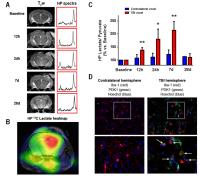 |
Hyperpolarized 13C Metabolic imaging of neuroinflammation in
Traumatic Brain Injury
Caroline Guglielmetti1,2, Austin Chou1,
Annemie Van der Linden2, Susanna Rosi1,
and Myriam M Chaumeil1
1University of California San Francisco, San
Francisco, CA, United States, 2University
of Antwerp, Antwerp, Belgium
This study demonstrates that 13C
MRS of hyperpolarized pyruvate can be used to detect
increased lactate production from pro-inflammatory
macrophages in a preclinical model of Traumatic Brain
Injury, hence providing a novel tool for in
vivo detection
of neuroinflammation.
|
| |
12:06
|
0864.
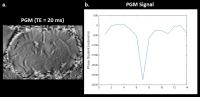 |
Gauging the Effectiveness of Traumatic Brain Injury Treatment
using MR Phase Gradient Mapping
Gregory Simchick1,2, Martha Betancur3,4,
Lohitash Karumbaiah3,4, and Qun Zhao1,2
1Physics, University of Georgia, Athens, GA,
United States, 2Bio-Imaging
Research Center, Athens, GA, United States, 3Animal
and Dairy Science, University of Georgia, Athens, GA, United
States, 4Regenerative
Bioscience Center, Athens, GA, United States
Due to both short-term and long-term effects, traumatic
brain injuries (TBIs) have been a growing topic of interest
over the last several years; therefore, research related to
the development of new methods to treat and monitor these
types of injuries has also gained interest. Presented here
is a non-invasive method using magnetic resonance (MR) phase
gradient mapping (PGM) to characterize TBI treatment in
relation to regional cerebral blood flow (rCBF) in
angiogenesis and tissue loss. In a rat moderate-to-severe
TBI model, increases between 16-29% in rCBF were seen in the
treatment group twenty weeks post TBI, while decreases
between 9-27% in rCBF were seen in the non-treatment group.
|
| |
12:18
|
0865.
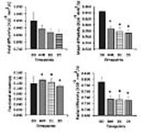 |
Evaluation of time-course of diffusivity changes and
inflammatory response in hippocampus post moderate traumatic
brain injury
Kavita Singh1, Richa Trivedi1, Maria M
D'souza2, and Subash Khushu1
1NMR, INMAS, DRDO, Delhi, Delhi, India, 2Molecular
imaging, INMAS, DRDO, Delhi, Delhi, India
Hippocampal atrophy is seen in traumatic brain injury even
when it is remote to the site of injury. Present study
assess acute microstructural and inflammatory changes
affecting hippocampal damage using diffusion tensor imaging
and Iba-1, GFAP immunostaining at D0, 4H, D1 and D5 in
rodent model of moderate TBI. Significantly reduced mean
diffusivity and radial diffusivity alongwith increased
fractional anisotropy at 4H, D1 and D5. Iba-1+ cells
significantly increased at D1 and D5 with GFAP+ cells
peaking at D5. Study provides temporal evaluation of
diffusion changes which may be due to underlying
inflammatory changes.
|
|