| |
13:30
 |
0418.
 |
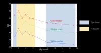
A differential arterial blood volume response during Lower Body
Negative Pressure measured using Pulsed Arterial Spin Labelling
with multiple short inversion times 
Joseph R Whittaker1, Molly G Bright1,2,
Ian D Driver1, Adele Babic1,3, Martin
Stuart1, and Kevin Murphy1
1CUBRIC, School of Psychology, Cardiff
University, Cardiff, United Kingdom, 2Sir
Peter Mansfield Imaging Centre, University of Nottingham,
Nottingham, United Kingdom, 3Department
of Anesthesia and Intensive Care Medicine, Cardiff
University School of Medicine, Cardiff, United Kingdom
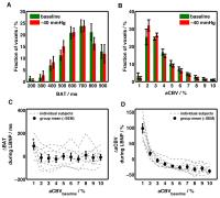
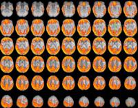
A custom made MRI compatible lower body negative pressure (LBNP)
chamber induced central hypovolemia in a group of healthy
volunteers. Pulsed ASL data with multiple short inversion
times was acquired during a baseline period and -40mmHg LBNP
in order to estimate arterial cerebral blood volume changes
related to cerebral autoregulation. We found a differential
response, in which arterial blood volume changes during LBNP
were dependent on vessel size. These data provide a useful
first step for fully understand the complex vascular changes
that occur in the brain to maintain perfusion during
systemic physiological perturbations.
|
| |
13:42
|
0419.
 |
Short-term cerebral blood flow reduction induced “apparent”
brain tissue density reduction
Qiu Ge1, Wei Peng1, Yong Zhang2,
Yu-Feng Zhang1, Thomas Liu3, Xuchu
Weng1, and Ze Wang1
1Hangzhou Normal University, Hangzhou, China,
People's Republic of, 2GE
Healthcare, MR Research China, Beijing, Shanghai, China,
People's Republic of, 3University
of California San Diego, San Diego, CA, United States
MRI-identified short-term brain tissue changes have been in
debate because of the lack of solid evidence of neurogenesis.
Cerebral blood flow (CBF) has been traced as one
contributing factor. We used caffeine to modulate CBF and to
subsequently examine brain tissue change using MRI. Both CBF
reduction and grey matter decrease were observed after
caffeine ingestion, which were further related to each other
in some brain regions. The data provide direct evidence for
the CBF contribution to the short-term apparent tissue
changes.
|
| |
13:54
|
0420.
 |
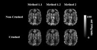
Rethinking macro-vascular artifacts from single post-label delay
ASL: can we extract a "free-lunch" arterial transit time metric?
Henk Mutsaerts1, Lena Vaclavu2,
Jan-Willem van Dalen2, Andrew Robertson1,
Paul Groot2, Mario Masellis1, Edo
Richard2, Aart J Nederveen2, and
Bradley MacIntosh1
1Sunnybrook Research Institute, Toronto, ON,
Canada, 2Academic
Medical Center, Amsterdam, Netherlands
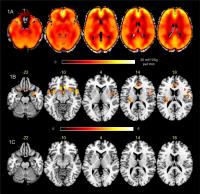
In this work, we propose a novel method to infer an ATT
estimate from the spatial signal distribution of single-time
point ASL CBF maps, using a spatial Coefficient of Variation
(CoV). In a large population of elderly with hypertension,
we compare crushed (C CBF) and non-crushed CBF maps (NC
CBF), from which we derive C CoV and NC CoV, and the
FEAST-based ATT estimate. These explorative results show
that both ATT and BMI are associated with NC CoV but not
with NC CBF, suggesting that ATT ? as estimated by the
spatial CoV ? might serve as a global biomarker of
cerebrovascular disease.
|
| |
14:06
 |
0421.
 |
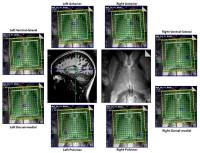
Traffic and cargo on the venous highway: distribution of venous
flow and oxygenation in the human brain.
Jill B. De Vis1, Hanzhang Lu2, Harshan
Ravi2, Jeroen Hendrikse1, and Peiying
Liu3
1Radiology, University Medical Center Utrecht,
Utrecht, Netherlands, 2Radiology,
Johns Hopkins University School of Medicine, Baltimore, MD,
United States, 3Radiology,
Johns Hopkins University Medical Center, Baltimore, MD,
United States
Arterial territory and flow have been well studied, but few
studies have been performed to investigate the venous flow
distribution. Similarly, little is known about the
oxygenation and its heterogeneity among the different venous
structures. The purpose of this study was to investigate
venous flow distribution and oxygenation.
|
| |
14:18
|
0422.
 |
Using 3D ASL to assess the change of cerebral blood flow at high
altitude: a longitudinal study
Wenjia Liu1, Bing Wu2, Dandan Zheng2,
Xin Lou1, Yulin Wang1, Li Zheng3,
Jie Liu4, and Lin Ma1
1Department of Radiology, PLA General Hospital,
Beijing, China, People's Republic of, 2GE
Healthcare, MR Research China, Beijing, Beijing, China,
People's Republic of, 3Biomedical
Engineering, Peking university, Beijing, China, People's
Republic of, 4General
Hospital of Tibetan Military Area Command, Lhasa, China,
People's Republic of
Although cerebral blood flow(CBF) at high altitude have been
researched for years, most previous studies are limited by
the use of transcranial Doppler. The conclusion of changes
in CBF depend on the assumption that the middle cerebral
arterial diameter does not alter in hypoxia, but recent
studies suggesting that this is not the case. In our study,
CBF was measured by 3D arterial spin labeling (ASL)
technique at sea level and high altitude in order to seek
the cerebrovascular response to altitude environment.
|
| |
14:30
|
0423.
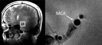 |
Imaging Changes in Cross-Sectional Area of the Middle Cerebral
Artery through the Cardiac Cycle at 7 Tesla
Esther AH Warnert1, Jasper Verbree2,
Richard G Wise1, and Matthias JP van Osch2
1Cardiff University Brain Research Imaging
Centre, Cardiff University, Cardiff, United Kingdom, 2Radiology,
Leiden University Medical Center, Leiden, Netherlands
Arterial stiffness is an important marker for
cerebrovascular health, as increased stiffness can lead to a
range of cerebrovascular pathologies. A non-invasive
assessment of cerebral arterial stiffness could therefore be
an important imaging marker for cerebrovascular health. Here
we show the feasibility of using high field MRI to
non-invasively assess cerebral arterial stiffness by
measuring the changes in cross-sectional area of the middle
cerebral artery throughout the cardiac cycle.
|
| |
14:42
|
0424.
 |
Impact of calibration method on the reproducibility of CBF
mapping using multiple post-labeling-delay PASL - Permission Withheld
Joana Pinto1, Pedro Vilela2, Michael
A. Chappell3, and Patrícia Figueiredo1
1ISR-Lisboa/LARSyS and Department of
Bioengineering, Instituto Superior Técnico – Universidade de
Lisboa, Lisbon, Portugal, 2Imaging
Department, Hospital da Luz, Lisbon, Portugal, 3Institute
of Biomedical Engineering, University of Oxford, Oxford,
United Kingdom
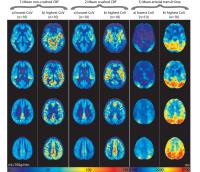
Absolute CBF quantification using ASL requires the
normalization of the control-label difference images by the
equilibrium magnetization, M0. A voxelwise calibration
method is currently recommended for single
post-labelling-delay (PLD) PCASL. However, the impact of
using an M0t map
obtained directly from the ASL data, with no need for an
extra scan, by fitting a saturation-recovery curve to the
control image time-series in multiple-PLD PASL remains to be
investigated. Here, we show that, using this type of
acquisition, voxelwise calibration significantly reduced
inter- and intra-subject variability in gray matter CBF
measurements relative to methods based on a reference
tissue.
|
| |
14:54
|
0425.
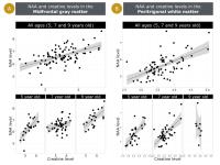 |
Regional differences in absolute metabolite level couplings in a
longitudinal study of children
Martha J Holmes1, Frances C Robertson1,
Francesca Little2, Mark F Cotton3, Els
Dobbels3, Andre JW van der Kouwe4,5,
Barbara Laughton3, and Ernesta M Meintjes1
1MRC/UCT Medical Imaging Research Unit,
Department of Human Biology, University of Cape Town, Cape
Town, South Africa, 2Department
of Statistical Sciences, University of Cape Town, Cape Town,
South Africa, 3Children’s
Infectious Diseases Clinical Research Unit, Department of
Paediatrics & Child Health, Tygerberg Children’s Hospital
and Faculty of Medicine and Health Sciences, Stellenbosch
University, Cape Town, South Africa, 4A.A.
Martinos Centre for Biomedical Imaging, Department of
Radiology, Massachusetts General Hospital, Charlestown, MA,
United States, 5Department
of Radiology, Harvard Medical School, Boston, MA, United
States
1H-MRS non-invasively quantifies metabolites that play
important roles in neurodevelopment. The physiological
functions of these metabolites, however, are still debated.
Examining the regional intercorrelations between metabolites
such as NAA, creatine, choline and glutamate provides
insight about the role of individual and coupled
biochemicals in the developing brain. We examined
correlations between pairs of metabolites in the midfrontal
gray matter (MFGM), peritrigonal white matter (PWM), basal
ganglia (BG) at 5, 7 and 9 years in a cohort of South
African children. We found significant metabolite couplings
in both the MFGM and PWM, however no significant couplings
were observed in the BG.
|
| |
15:06
|
0426.
 |
Differential effects of ketamine-propofol vs propofol
anaesthesia on cerebral perfusion in children
Ruth L O'Gorman1, Philipp Buehler2,
Carola Sabandal2, Ianina Scheer3,
Malek Makki1, Markus Weiss2, Christian
Kellenberger3, and Achim Schmitz2
1Center for MR Research, University Children's
Hospital, Zurich, Switzerland, 2Anaesthesia,
University Children's Hospital, Zurich, Switzerland, 3Radiology,
University Children's Hospital, Zurich, Switzerland
Anaesthetics such as those used for sedation in pediatric
MRI affect cerebral blood flow and hemodynamics to varying
degrees. This study examines differences in cerebral
perfusion in children undergoing elective MRI under sedation
with propofol vs. a combination of propofol and ketamine.
Children induced for sedation with ketamine demonstrated on
average 14% higher whole brain perfusion values than those
induced for sedation with propofol, confirming that ketamine
and propofol exert a differential effect on brain activity
and hemodynamics.
|
| |
15:18
|
0427.
 |
Evidencing different neurochemical profiles between thalamic
nuclei using 2D-semilaser 1H-MRSI at 7T
Maxime Donadieu1,2,3, Yann Le Fur1,2,
Sylviane Confort-gouny1,2, Arnaud Le Troter1,2,
Maxime Guye1,2, and Jean-Philippe Ranjeva1,2
1CRMBM UMR 7339, Aix Marseille Université CNRS,
Marseille, France, Metropolitan, 2CEMEREM
Pole d'Imagerie, AP-HM CHU Timone, Marseille, France,
Metropolitan, 3Siemens
Healthcare, Saint-Denis, France, Metropolitan
Using 2D-semilaser 1H-MRSI sequence centered on thalamus and
acquired at 7T in 10 healthy volunteers, we demonstrate that
the neurochemical profiles (relative NAA, Cr and Cho levels)
are different between pulvinar, ventral-lateral,
dorsal-medial and anterior nuclei. Moreover, left/right
differences in neurochemical profiles, especially for NAA
levels, showed a left NAA lateralization for the
ventral-lateral nucleus and the pulvinar and in contrast
higher right NAA levels in the anterior nucleus. These
results suggest that the various neurochemical profiles of
these thalamic nuclei may be related to their functional
specificity.
|
|