| |
13:30
 |
0695.
 |
Accurate T1 and T2 mapping by direct least-squares ellipse
fitting to phase-cycled bSSFP data 
Yulia Shcherbakova1, Cornelis A.T. van den Berg2,
Jan J.W. Lagendijk3, Chrit T.W. Moonen4,
and Lambertus W. Bartels5
1Center for Imaging Sciences/Imaging Division,
UMC Utrecht, Utrecht, Netherlands, 2Dept.
of Radiotherapy/Imaging Division, UMC Utrecht, Utrecht,
Netherlands, 3Department
of Radiotherapy/Centre for Image Sciences, UMC Utrecht,
Utrecht, Netherlands, 4Center
for Imaging Sciences/ Imaging Division, UMC Utrecht,
Utrecht, Netherlands, 5Image
Sciences Institute/Department of Radiology, UMC Utrecht,
Utrecht, Netherlands
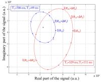
Björk et al. proposed to use the balanced steady-state free
precession (bSSFP) pulse sequence with multiple phase-cycled
acquisitions to estimate the values of T1 and
T2 using
a non-linear fitting approach. Unfortunately, they found
that this non-linear approach would face large uncertainties
for realistic SNRs. The purpose of our work was to
demonstrate that by reformulating the signal model in the
complex plane, an elliptical model can be fitted to the data
points using a linear least squares method , allowing for
more robust, accurate and simultaneous estimation of T1 and
T2 values.
|
| |
13:42
|
0696.
 |
Influence of pulse length and shape on variable flip angle T1
mapping of the human brain
Yosef Al-Abasse1 and
Gunther Helms1
1Medical Radiation Physics, Lund University,
Lund, Sweden
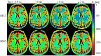
Effects of the macromolecular pool are usually neglected in
T1 mapping
using the variable flip angle (VFA) method. To demonstrate
the influence of magnetization transfer (MT) on the
estimated T1, VFA experiments were performed
using sinc and rect pulses of different pulse lengths (0.5
ms ≤ TRF ≤ 2.0 ms). Substantial variations in T1 (11-21
%) were observed. Longer rect pulses yielded lower T1 values
than those obtained by inversion recovery. This can be
explained by varying saturation of macromolecules and
inherent MT. A simplified model for the influence of TRF on
the T1 estimates
is suggested for low-power rect pulses.
|
| |
13:54
 |
0697.
 |
B0 and B1 Insensitive Robust Fat Suppression using Frequency
Offset Corrected Inversion (FOCI)
Xinzeng Wang1, Joshua S. Greer1,2,
Ivan E. Dimitrov3,4, and Ananth J. Madhuranthakam1,3
1Radiology, UT Southwestern Medical Center,
Dallas, TX, United States, 2Bioengineering,
University of Texas at Dallas, Richardson, TX, United
States, 3Advanced
Imaging Research Center, UT Southwestern Medical Center,
Dallas, TX, United States, 4Philips
Medical Systems, Cleveland, OH, United States
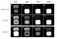
STIR uses adiabatic non-selective hyperbolic secant (HS)
inversion pulse to achieve uniform fat suppression even in
the presence of B1 inhomogeneities. However, in the regions
of increased B0 and B1 inhomogeneities, particularly at 3T
and higher field strengths, the increased bandwidth of the
HS pulse comes at the expense of higher adiabatic threshold.
In this work, we evaluated C-FOCI pulse to achieve robust
fat suppression with broader bandwidth and increased
robustness to B1 variations compared to HS pulse. We also
derived an analytical expression for the adiabatic threshold
of C-FOCI pulse and show robust performance against B0 and
B1 inhomogeneities.
|
| |
14:06
|
0698.
 |
Real-time multi-slice MRI during CPAP reveals dynamic changes in
upper airway in response to pressure change
Weiyi Chen1, Ziyue Wu2, Sally L.
Davidson Ward3, Michael C.K. Khoo4,
and Krishna S. Nayak1
1Electrical Engineering, University of Southern
California, Los Angeles, CA, United States, 2Alltech
Medical Systems USA, Solon, OH, United States, 3Children's
Hospital Los Angeles, Los Angeles, CA, United States,4Biomedical
Engineering, University of Southern California, Los Angeles,
CA, United States
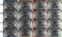
We demonstrate a novel experiment that captures the upper
airway’s instantaneous response to changes in air pressure.
We apply rapid changes in continuous positive airway
pressure (CPAP) during real-time simultaneous multi-slice
MRI. This reveals the airway area does NOT only depend on
pressure level but also different airway sections and
previous muscle tone status. This technique also enables
characterization of airway collapsibility, and is relevant
to the assessment of obstructive sleep apnea (OSA) and
treatment planning.
|
| |
14:18
|
0699.
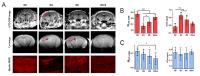 |
Imaging myelin with ultrashort-echo time (UTE) in a Multiple
Sclerosis model on a clinical 7T system
Caroline Guglielmetti1, Tanguy Boucneau2,
Peng Cao2, Annemie Van der Linden3,
Peder Larson2, and Myriam M Chaumeil1
1University of California San Francisco, San
Francisco, CA, United States, 2Department
of Radiology and Biomedical Imaging, University of
California San Francisco, San Francisco, CA, United States, 3University
of Antwerp, Antwerp, Belgium
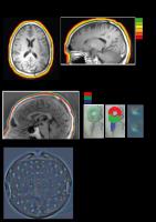
Many advances in neuroimaging have improved diagnosis and
care of Multiple Sclerosis (MS) patients. However, current
clinical methods fail to detect the majority of cortical
lesions. In this work, we used the well characterized
cuprizone mouse model for brain demyelination to evaluate
the sensitivity of in vivo ultra-short echo time (UTE)
measurements for the non-invasive detection of grey and
white matter alterations. We showed that UTE enabled the
detection of cortical lesions and the assessment of myelin
integrity in the white matter following demyelination and
spontaneous remyelination.
|
| |
14:30
|
0700.
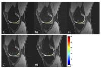 |
Simultaneous DESS imaging and T2 mapping, for knee
osteoarthritis studies
Cheng-Chieh Cheng1, Lena Franziska Schaefer1,
Jeffrey Duryea1, and Bruno Madore1
1Radiology, Brigham and Women's Hospital, Harvard
Medical School, Boston, MA, United States
The ‘dual-echo in the steady state’ (DESS) sequence is often
the method of choice for assessing cartilage damage. A
modified DESS method was developed that provides images of
similar quality to regular DESS while also providing T2 values,
a proven biomarker for the early stages of osteoarthritis.
The method exploits the fact that the two signal pathways
sampled in a DESS sequence decay differently during the TR
period, allowing T2 and
T2* to be quantified. The resulting method can
assess cartilage volume seemingly as well as regular DESS,
while also providing relevant T2 information,
without increasing scan time.
|
| |
14:42
|
0701.
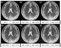 |
Quality evaluation scheme for no-reference MR images using
pre-scanned MR big data
Jinseong Jang1, Taejoon Eo1, and Dosik
Hwang1
1Electrical and Electronic Engineering, Yonsei
University, Seoul, Korea, Republic of
This study demonstrated the feasibility of no reference (NR)
image quality assessment (IQA) for magnetic resonance
imaging. Especially, this method used pre-scanned images
from other subjects. So by using prior big data, MRI can be
evaluated in no reference environments.
|
| |
14:54
|
0702.
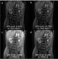 |
Multiresolution imaging using golden angle stack-of-stars and
compressed sensing
Abhishek Pandey1,2, Umit Yoruk3,
Puneet Sharma1, Diego R. Martin1,
Maria Altbach1, Ali Bilgin1,2,4, and
Manojkumar Saranathan1,4
1Department of Medical Imaging, University of
Arizona, Tucson, AZ, United States, 2Electrical
and Computer Engineering, University of Arizona, Tucson, AZ,
United States, 3Electrical
Engineering, Stanford University, Stanford, CA, United
States, 4Biomedical
Engineering, University of Arizona, Tucson, AZ, United
States
Dynamic contrast enhanced MRI requires measurement of
arterial input function with great accuracy while
maintaining high spatial resolution. Golden angle
stack-of-stars radial acquisition was used to get
reconstructions at multiple temporal resolutions. A
multiresolution reconstruction scheme is used to generate
AIFs using a very small temporal window. The accuracy of the
reconstruction method was checked on a realistic phantom and
then applied to an in vivo data. Results show that
compressed sensing reconstruction works best with high
temporal resolution (HTR) AIF giving both diagnostic image
quality and accurate GFR estimate.
|
| |
15:06
|
0703.
 |
Ultra-short echo time sequence for electrode locations in
simultaneous EEG-FMRI
Russell Butler1, Guillaume Gilbert2,
and Kevin Whittingstall3
1University of Sherbrooke, Sherbrooke, QC,
Canada, 2MR
Clinical Science - Philips Healthcare, Markham, ON, Canada, 3Diagnostic
Radiology, University of Sherbrooke, Sherbrooke, QC, Canada
Precise and accurate knowledge of EEG sensors relative to
underlying cortical tissue enhances simultaneous EEG-FMRI
studies, but to date no specialized sequence for providing
these locations exists. We propose an ultra-short echo time
sequence (UTE) to highlight the plastic casing and wiring of
a 64 channel MR compatible EEG cap. We show that the UTE
resolves electrode components up to 6mm from the surface of
the scalp, allowing to locate the precise contact point of
electrode with skin and direction of wire leading away from
the electrode in all subjects (n=8).
|
| |
15:18
|
0704.
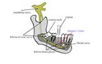 |
Can Zero TE imaging be a viable alternative to micro CT in
dentistry imaging : application in tooth implanting and
extraction ?
Yu Kang1, Bing Wu2, Shikuo Fu2,
and Nan Hong1
1Peking University people's hospital, Beijing,
China, People's Republic of, 2GE
healthcare MR Research China, Beijing, China, People's
Republic of
Micro CT is currently used for dentistry imaging. Not only
it is associated with radiation, it also offers poor
contrast of the mandible canal, whose position needs to be
precisely known during tooth implantation and extraction.
Conventional MRI fails for this case due to the short T2
time of the teeth as well as the susceptibility in the oral
cavity. Zero TE imaging, due to its technical uniqueness,
seems to be a viable solution to this. In this work, imaging
of the the jaw of a patient with both CT, conventional MR
and zTE imaging was performed.
|
|