| |
13:30
 |
0930.
 |
B0-Atlas with Field-Probe Guidance: Application in Real-Time
Field Control 
Simon Gross1, Yolanda Duerst1,
Laetitia Maëlle Vionnet1, Christoph Barmet1,2,
and Klaas Paul Pruessmann1
1Institute for Biomedical Engineering, ETH and
University of Zurich, Zurich, Switzerland, 2Skope
Magnetic Resonance Inc., Zurich, Switzerland
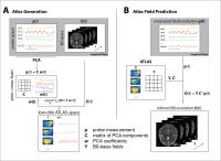
A novel model for the prediction of B0-maps from
external field measurements is presented. It is based on the
joint analysis of training data from simultaneously acquired
B0-maps and magnetic field evolution measured with NMR field
probes. A first application to real-time shim feedback is
demonstrated.
|
| |
13:42
|
0931.
 |
Model-based rapid field map prediction for dynamic shimming
applications
Yuhang Shi1, Johanna Vannesjo1, Karla
L. Miller1, and Stuart Clare1
1Nuffield Department of Clinical Neurosciences,
University of Oxford, Oxford, United Kingdom
This work presents a rapid field map prediction method based
on the individual subject's quick localizer scan and a large
brain field map database to accelerate the field map
acquisition stage for dynamic shimming applications. Our
model-based method is able to better identify the steep
change in the field associated with some slices in the lower
part of the brain, however a low-resolution field map
performs better for the rest of the brain.
|
| |
13:54
|
0932.
 |
Fast B0 first order inhomogeneity estimation using radial
acquisition
Ali Aghaeifar1,2, Alexander Loktyushin1,
Christian Mirkes1,3, Axel Thielscher1,
and Klaus Scheffler1,3
1Max Planck Institute for Biological Cybernetics,
Tübingen, Germany, 2IMPRS
for Cognitive and Systems Neuroscience, Tübingen, Germany, 3Department
of Biomedical Magnetic Resonance, University of Tübingen,
Tübingen, Germany
B0 field inhomogeneity is a major source of distortion in MR
images. Current approaches to dynamic shimming require extra
acquisition time or external hardware. We propose a method
that estimates first order shim errors by using projections
of radial acquisition. The errors can be estimated from
three projections multiple times in each measurement, which
makes the method highly robust. The proposed method is
evaluated in simulation and in vivo. Obtained results show a
strong agreement between applied and measured first order
shim errors.
|
| |
14:06
|
0933.
 |
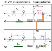
BMART: B0 Mapping using Rewind Trajectories
Corey Allan Baron1 and
Dwight G. Nishimura1
1Electrical Engineering, Stanford University,
Stanford, CA, United States
B0 inhomogeneity
leads to image artifacts and/or blurring. These issues can
be addressed by using a B0 map,
which typically requires an extra scan. In addition to the
longer total scan time required, motion occurring between
the acquisition of the imaging data and B0 map
can lead to misregistration. The proposed method utilizes
images reconstructed from rewind trajectories to construct a
B0 map.
In pulse sequences that already use gradient rewinds (e.g.,
bSSFP), a B0 map
that is inherently registered to the imaging data can be
created with no additional scan time.
|
| |
14:18
|
0934.
 |
Broadband Frequency Mapping with Balanced SSFP
Oliver Bieri1,2, Grzegorz Bauman1,2,
and Carl Ganter3
1Radiology, University Hospital Basel, Basel,
Switzerland, 2Biomedical
Engineering, University of Basel, Basel, Switzerland, 3Diagnostic
Radiology, Technical University Munich, Munich, Germany
A new method for accurate and fast broadband frequency
mapping with balanced steady state free precession is
introduced. The method mitigates the need for advanced phase
unwrapping algorithms from a matrix pencil analysis of
sequentially shifted echo times. Typically, the new method
offers a spectral resolution in the range of Hertz with a
sensitivity range in the order of several thousands of
Hertz.
|
| |
14:30
|
0935.
 |
Determination of Relative B1+ Sensitivities Using Accelerated
Simultaneous Excitation with Multiple Transmit Channels and
Controlled Aliasing
Iulius Dragonu1, Craig Buckley1, Peter
Weale1, Matthew D Robson2, and Aaron T
Hess2
1Siemens Healthcare Ltd, Frimley, Camberley,
United Kingdom, 2University
of Oxford Centre for Clinical Magnetic Resonance Research
(OCMR), John Radcliffe Hospital, Headington, United Kingdom
Radiofrequency shimming with multiple channel excitation is
a well established method to increase the transverse
magnetic field homogeneity and reduce SAR at high magnetic
field strength(≥7T). To harness the benefits of a parallel
transmit system, the magnitude and relative phase of each
transmit channel must be determined during a calibration
scan. We propose a new strategy to accelerate the
acquisition of such calibration images by simultaneously
exciting several transmit channels and reconstructing the
calibration images using the technique similar to
simultaneous multi-slice acquisitions.
|
| |
14:42
|
0936.
 |
Combining B1 mapping with TIAMO for fast and accurate
multi-channel RF shimming in 7 Tesla body MRI
Sascha Brunheim1,2, Stephan Orzada1,
Soeren Johst1, Marcel Gratz1,2,
Maximilian N. Voelker1, Oliver Kraff1,
Martina Floeser3, Andreas K. Bitz3,
Mark E. Ladd1,3, and Harald H. Quick1,2
1Erwin L. Hahn Institute for Magentic Resonance
Imaging, University Duisburg-Essen, Essen, Germany, 2High
Field and Hybrid MR Imaging, University Hospital Essen,
Essen, Germany, 3Medical
Physics in Radiology, German Cancer Research Center (DKFZ),
Heidelberg, Germany
With current methods the mitigation of transmit field
inhomogeneity at ultrahigh field by multi-channel RF
shimming with conventional methods is relatively time
consuming. This applies in particular for parallel
transmit/receive in-vivo body imaging within breath-hold and
during organ motion. Therefore, we propose a new technique
merging fast acquired relative single channel maps and the
spatial-dependent flip-angle distribution of two
complementary shims to define absolute transmit coil maps
for fast and accurate RF shim calculation. The performance
of this technique is validated against established methods
in phantom measurements and its reliability is shown in
comparison to simulation data serving as reference.
|
| |
14:54
|
0937.
 |
Silent, Free-Breathing B1+ Mapping using DREAM
Kay Nehrke1 and
Peter Börnert1,2
1Philips Research, Hamburg, Germany, 2Radiology,
LUMC, Leiden, Netherlands
To improve the workflow for B1+ calibration
on a dual transmit MRI system, the DREAM B1+ mapping
sequence has been streamlined for acoustic noise reduction
and free-breathing acquisition using a standard external
respiratory motion sensor. About 10 dB reduction in sound
pressure level were achieved by optimizing the echo order
with respect to gradient strength reduction. Feasibility was
shown in volunteer experiments on abdominal B1+ mapping.
|
| |
15:06
|
0938.
 |
DREAM Based Receive Sensitivity Correction
Wyger Brink1 and
Andrew Webb1
1Radiology, Leiden University Medical Center,
Leiden, Netherlands
Imaging methods at high fields can suffer from receive
non-uniformities from the body coil, particularly when the
body coil is used as a reference for intensity correction.
In this work we show that the DREAM B1 mapping sequence can
be used for receive uniformity correction in RF-shimmed
whole-body imaging at 3T.
|
| |
15:18
|
0939.
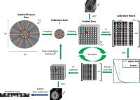 |
Simultaneous Estimation of Auto-calibration Data and Gradient
Delays in non-Cartesian Parallel MRI using Low-rank Constraints
Wenwen Jiang1, Peder E.Z Larson2, and
Michael Lustig3
1Bioengineering, UC Berkeley/ UCSF, Berkeley, CA,
United States, 2Radiology
and Biomedical Imaging, UCSF, San francisco, CA, United
States, 3Electrical
Engineering and Computer Science, UC Berkeley, Berkeley, CA,
United States
Gradient timing delay errors in non-Cartesian trajectories
often induce spurious image artifacts. More importantly,
misaligned k-space center data results in auto-calibration
errors for parallel imaging methods. We propose a general
approach that simultaneously estimates consistent
calibration data and corrects for gradient delays. We pose
the joint estimation problem as a low-rank minimization
problem, and solve it using a Gauss-Newton method. We
demonstrate the feasibility of the proposed method by
simulation and phantom experiments.
|
|