| |
10:45
 |
0016.
 |
Characterization of the macromolecular baseline with a
metabolite-cycled double-inversion recovery sequence in the
human brain at 9.4T 
Ioannis Angelos Giapitzakis1,2, Roland Kreis 3,
and Anke Henning 1,4
1Max Planck Institute for Biological Cybernetics,
Tübingen, Germany, 2IMPRS
for Cognitive and Systems Neuroscience, University of
Tuebingen, Tuebingen, Germany, 3Depts.
Radiology and Clinical Research, University of Bern, Bern,
Switzerland, 4Institute
of Biomedical Engineering, University and ETH, Zürich,
Switzerland
Macromolecular resonances (MM) overlap with
metabolites resulting in inaccurate quantification of the
metabolites due to baseline distortion. This effect becomes
even more severe in case of short echo times (TE). The
purpose of this study was the development of an adiabatic
pulse for double inversion recovery and investigation of
impact to include MM into quantification of 9.4T MRS data of
human brain. This is the first study where MC-STEAM is
combined with a double inversion technique. The results
showed the advantages of UHF and MC as well as the necessity
of the inclusion of MM baseline in the basis set.
|
| |
10:57
|
0017.
 |
Evidence for regional and spectral differences of macromolecule
signals in human brain using a crusher coil at 7 Tesla
Nicolas Geades1, Carrie Wismans2,
Mariska Damen2, Penny Gowland1, Hans
Hoogduin2, Vincent Boer2, Dennis Klomp2,
and Jannie Wijnen2
1Sir Peter Mansfield Imaging Centre, University
of Nottingham, Nottingham, United Kingdom, 2Department
of Radiology, University Medical Centre Utrecht, Utrecht,
Netherlands
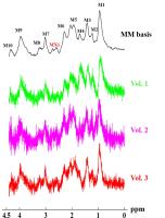
The regional, spectral and relaxation differences of
macromolecules (MM) in the human brain were investigated
using T1 mapping, metabolite nulling and high resolution
MRSI with a crusher coil at 7T. Differences between
macromolecular signal of GM and WM were observed by all
three methods. The T1 mapping showed different T1 relaxation
time of MM in GM and WM. Metabolic maps created by fitting
an averaged WM spectrum showed differences in M1 and M2. The
macromolecules in the metabolite nulled data showed a
different M4 in GM and WM. Some of these differences can be
explained by differences in T1 relaxation.
|
| |
11:09
 |
0018.
 |
Improvement of 2-hydroxyglutarate detectability using optimized
triple-refocusing difference editing at 7T in vivo
Sandeep K Ganji1, Zhongxu An1, Vivek
Tiwari1, Marco Pinho2, Edward Pan3,
Bruce Mickey4, Elizabeth Maher5, and
Changho Choi1
1Advanced Imaging Research Center, UT
Southwestern Medical Center, Dallas, TX, United States, 2Radiology,
UT Southwestern Medical Center, Dallas, TX, United States, 3Neurology
and Neurotherapeutic, UT Southwestern Medical Center,
Dallas, TX, United States, 4Neurological
Surgery, UT Southwestern Medical Center, Dallas, TX, United
States, 5Internal
Medicine, UT Southwestern Medical Center, Dallas, TX, United
States
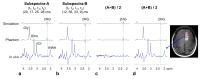
2-hydroxyglutarate (2HG) has become an important biomarker
in the diagnosis and management of glioma patients as well
as in the workup of an undiagnosed mass. The 1H MRS signals
of 2HG are extensively overlapped with other metabolite
signals. Specifically, uncertainty in 2HG evaluation arising
from the spectral overlap of the 2HG 2.25-ppm signal with
the GABA 2.29-ppm resonance may be a major obstacle when the
2HG level is relatively low. Here we report a novel
triple-refocusing difference editing that provides complete
differentiation between 2HG and GABA signals at 7T.
|
| |
11:21
|
0019.
 |
Indirectly-Detected and Spin-Amplified Heteronuclear MRS and MRI - Permission Withheld
Chencai Wang1, Chaohsiung Hsu1,
Stephanie Wolohan1, and Yung-Ya Lin1
1Department of Chemistry and Biochemistry, UCLA,
Los Angeles, CA, United States
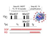
A general indirect-detection and spin-amplification scheme
has been developed to enhance the sensitivity of
heteronuclear MRS and MRI based on dynamic instability of
the solvent proton magnetization under collective feedback
fields of radiation damping and the distant dipolar field.
The heteronuclear solute spins are first detected by the
solvent proton spins through various magnetization transfer
mechanisms and serve as small “input” signals to perturb the
solvent proton magnetization, which is prepared in an
unstable state. The weakly detected signal is then amplified
through subsequent nonlinear evolution of the solvent proton
magnetization to achieve 10x SNR improvement for 13C MRS and
MRI.
|
| |
11:33
|
0020.
 |
Remodeling of energy metabolism revealed by 31P magnetization
transfer in a transgenic rat model of Huntington’s disease
Brice Tiret1,2, Maria-Angeles Carrillo-de Sauvage1,2,
Huu Phuc Nguyen3,4, Nicole El Massioui5,6,
Valérie Doyère5,6, Vincent Lebon1,2,
Emmanuel Brouillet1,2, and Julien Valette1,2
1CEA/DSV/I2BM/MIRCen, Fontenay-aux-Roses, France, 2CNRS
Université Paris-Saclay UMR 9199, Fontenay-aux-Roses,
France, 3Institute
of Medical Genetics and Applied Genomics, University of
Tuebingen, Tuebingen, Germany, 4Centre
for Rare Diseases, University of Tuebingen, Tuebingen,
Germany, 5Paris-Saclay
Institute of Neuroscience, Université Paris-Sud, UMR 9197,
Orsay, France, 6Centre
National de la Recherche Scientifique, Orsay, France
Localized 31P
MRS with progressive magnetization transfer (MT) is
performed in the BACHD transgenic rat model of Huntington’s
disease to assess energy metabolism. Localized measurements
of the ATP formation rate through creatine kinase and
oxidative phosphorylation (ATPsynthase) are performed in the
rat brain for the first time. Results show that ATPsynthase
rate is reduced by a factor 2, which is partly compensated
by higher cerebral concentrations of phosphocreatine to
generate ATP via creatine kinase.
|
| |
11:45
|
0021.
 |
Investigating machine learning approaches for quality control of
brain tumor spectra
Sreenath P Kyathanahally1, Victor Mocioiu2,
Nuno Miguel Pedrosa de Barros3, Johannes Slotboom3,
Alan J Wright4, Margarida Julià-Sapé 2,
Carles Arús2, and Roland Kreis1
1Depts. Radiology and Clinical Research,
University of Bern, Bern, Switzerland, 2Centro
de Investigación Biomédica en Red en Bioingeniería,
Biomateriales y Nanomedicina (CIBER-BBN), Universitat
Autònoma de Barcelona, Barcelona, Spain, 3DRNN,
Institute of Diagnostic and Interventional
Neuroradiology/SCAN, University Hospital Bern, Bern,
Switzerland, 4CRUK
Cambridge Institute, University of Cambridge, Cambridge,
United Kingdom
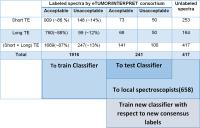
Despite many potential applications of MR spectroscopy in
the clinic, its usage is limited – and the need for human
experts to identify bad quality spectra may contribute to
this. Previous studies have shown that machine learning
methods can be developed to accept or reject a spectrum
automatically. In this study, we extend this to different
machine learning methods on 1916 spectra from the eTUMOUR
and INTERPRET databases. The RUSBoost classifier, which
handles unbalanced data, improved specificity and accuracy
compared to other classifiers, in particular in combination
with an extended feature set and multi-class labels.
|
| |
11:57
|
0022.
 |
Automatic quality assessment of short and long-TE brain tumour
MRSI data using novel Spectral Features
Nuno Miguel Pedrosa de Barros1,2, Urspeter Knecht1,
Richard McKinley1, Jonathan Giezendanner1,
Roland Wiest1, and Johannes Slotboom1
1Institute for Diagnostic and Interventional
Neuroradiology, Inselspital, Bern, Switzerland, 2University
of Bern, Bern, Switzerland
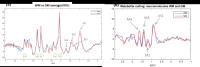
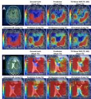
MRSI-data frequently contains bad-quality spectra which
strongly limits its clinical-use. Current clinical practice
in our institute is that these bad-quality spectra are
filtered out by an MRS-expert, at the expense of long
processing times. In this work we present a new method for
automatic quality assessment of both long and short-TE
MRSI brain tumour data. This method is based upon a novel
set of spectral features, and it is as accurate as an expert
but considerably faster (3/4 minutes vs 3seconds).
|
| |
12:09
|
0023.
 |
Fast frequency–sweep spectroscopic imaging with an ultra-low
flip angle
Junyu Guo1, Zoltan Patay1, and Wilbrun
E. Reddick1
1St Jude Children's Research Hospital, Memphis,
TN, United States
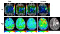
We present a novel, simple and fast MR spectroscopic imaging
technique and show its conceptual validation with
simulations and demonstrate proof-of-principle with phantom
and human studies. First, compared to the conventional
spectroscopic imaging in the time-domain, our method
acquires data in the frequency domain, allowing flexible
non-uniform sampling to speed up the acquisition. Second,
using ultra-small RF pulses offers intrinsic water and fat
suppression, greatly simplifying the scanning procedures.
Third, this new technique has hundreds of times lower energy
deposition than conventional MRI scans. We believe our
method could allow spectroscopic imaging to play a larger
role in clinical applications.
|
| |
12:21
|
0024.
 |
Parameterization of measured macromolecular background in
ultra-short acquisition delay 1H
MRSI in the brain at 7T
Michal Považan1,2, Gilbert Hangel1,
Bernhard Strasser1, Eva Heckova1,
Lukas Hingerl1, Stephan Gruber1,
Siegfried Trattnig1,2, and Wolfgang Bogner1
1High Field MR Center, Department of Biomedical
Imaging and Image-guided Therapy, Medical University Vienna,
Vienna, Austria, 2Christian
Doppler Laboratory for Clinical Molecular MR Imaging,
Vienna, Austria
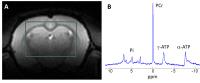
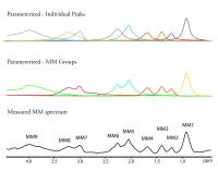
Ultra-short echo/acquisition delay MRS spectra have a strong
characteristic background consisting of macromolecule (MM)
resonances superimposed on the signal of metabolites.
Typically a single metabolite-nulled MM spectrum is included
into quantification routine to account for this. To detect
more prominent regional and pathologic changes, we replaced
this single MM spectrum by individual MM peaks. We found
that the MM peaks in a 2.3-0.5 ppm region are higher in gray
matter compared to white matter, whereas the MM peaks from
2.9 to 3.2 ppm were significantly higher in white matter of
healthy volunteers and one MS patient.
|
| |
12:33
|
0025.
 |
Stochastic excitation scheme for estimating longitudinal
relaxation and radiofrequency transmit inhomogeneity in single
voxel spectroscopy
Assaf Tal1
1Chemical Physics, Weizmann Institute of Science,
Rehovot, Israel
A stochastic excitation and corresponding dictionary
matching scheme is presented for quantifying metabolite
concentrations, longitudinal relaxation times and transmit
inhomogeneity in single voxel proton magnetic resonance
spectroscopy in the human brain.
|
|