| |
13:30
 |
0910.
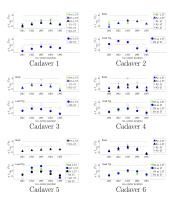 |
Evaluation of RF Induced Lead Tip Heating at 1.5T and 3T in
Cadavers with Cardiac Pacemakers or ICDs 
Volkan Acikel1, Patrick Magrath1,2,
Scott E Parker1, Holden H Wu1, Peng Hu1,
Paul J Finn1, and Daniel B Ennis1,2
1Department of Radiological Sciences, University
of California Los Angeles, Los Angeles, CA, United States, 2Department
of Bioengineering, University of California Los Angeles, Los
Angeles, CA, United States
MRI exams for patients with pacemakers and implanted
cardioverter defibrillators (ICDs) are contraindicated at
all clinical field strengths. The aim of this study was to
measure directly RF induced lead tip heating during MRI
exams of cadavers with existing devices at both 1.5T and 3T.
|
| |
13:42
 |
0911.
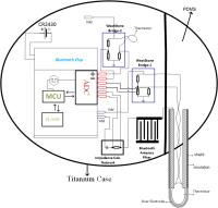 |
Subacute In-vivo RF Heating of an Active Medical Implantable
Device Under MRI Using Temperature Sensor Implant
Berk Silemek1, Oktay Algin1,2, Cagdas
Oto3, and Ergin Atalar1,4
1UMRAM, Bilkent University, Ankara, Turkey, 2Department
of Radiology, Atatürk Education and Research Hospital,
Ankara, Turkey, 3Faculty
of Veterinary Medicine, Ankara University, Ankara, Turkey, 4Electrical
and Electronics Engineering, Bilkent University, Ankara,
Turkey
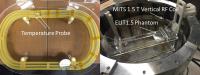
RF tissue heating of the Active Medical Implantable Devices
(AIMD) is a well-known problem. However, due to the complex
structure of the body, in vivo testing of the AIMDs’ heating
under MRI cannot be verified with phantoms completely. Acute
in vivo experiments damage the tissue and body’s
thermoregulation response changes which can affect the
measurements and investigation of the problems. Here, we
propose a Temperature Sensor Implant setup to eliminate
hyperacute effects of the surgery and enable real-time
temperature monitoring of the tip of the implant during MRI
examination
|
| |
13:54
|
0912.
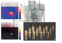 |
Experimental System for RF-Heating Characterization of Medical
Implants during MRI
Earl Zastrow1,2, Myles Capstick1,3,
and Niels Kuster1,2
1IT'IS Foundation, Zurich, Switzerland, 2Department
of Information Technology and Electrical Engineering,
ETH-Zurich, Zurich, Switzerland, 3Zurich
MedTech AG, Zurich, Switzerland
Patients with elongated conductive implants are generally
excluded from MRI diagnostics because the interaction of the
implant with MRI-induced RF fields can lead to hazardous
localized heating in surrounding tissues. Depending on the
complexity of the lead structure, numerical assessment of
implant-RF interactions may require excessive computational
overhead and may not be feasible. To overcome this
challenge, an experimental system, based on the revised Tier
3 of the ISO/IEC TS 10974, is developed and validated with
full-wave electromagnetics simulations. The experimental
system is designed for the assessment of RF-induced heating
of implants, irrespective of the complexity of the implant
structure.
|
| |
14:06
|
0913.
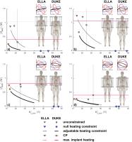 |
Convex optimization of MRI exposure for RF-heating mitigation of
leaded implants: extended coverage of clinical scenarios at 128
MHz
Earl Zastrow1,2, Juan Córcoles3, and
Niels Kuster1,2
1IT'IS Foundation, Zurich, Switzerland, 2Department
of Information Technology and Electrical Engineering,
ETH-Zurich, Zurich, Switzerland, 3Department
of Electronic and Communication Technology, Universidad
Autónoma de Madrid, Madrid, Spain
Interactions of long insulated implants with conductive
wires (e.g., cardiac pacemaker and deep-brain stimulator)
with RF during MRI can lead to excessive local heating of
tissue at the vicinity of the implant and is one of the
contraindication to MRI. We present the preliminary results
of a convex optimization method that can be used to suppress
the local deposited power in tissue in a controllable
manner. The performance of the proposed method is evaluated,
as a function of the trade-off between homogeneity of |B1+|
and the mitigated RF-induced power deposition caused by the
implant, for multiple clinical scenarios at 128 MHz.
|
| |
14:18
|
0914.
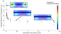 |
Simulation and Experimental Measurements of Flow Effects on
Radio Frequency Induced Heating of a Stent
David C. Gross1,2, Benjamin Scandling1,
and Orlando P. Simonetti3,4
1Biomedical Engineering, The Ohio State
University, Columbus, OH, United States, 2Dorothy
M. Davis Heart and Lung Research Institute, The Ohio State
University Wexner Medical Center, Columbus, OH, United
States, 3Radiology,
The Ohio State University Wexner Medical Center, Columbus,
OH, United States, 4Internal
Medicine, Division of Cardiovascular Medicine, The Ohio
State University Wexner Medical Center, Columbus, OH, United
States
The goal of this study was to investigate the influence of
blood flow on the temperature rise of a peripheral vascular
stent during MRI with flow phantom experiments and computer
simulations. RF heating experiments of a vascular stent are
performed during MRI at 3.0T in a flow phantom. The
temperature rise of the stent is measured with varied flow
rates. The temperature rise of the stent was over 10°C
without flow, and was reduced by 50% with a flow rate of
only 50 mL/min. Blood flow significantly reduces the
temperature rise of stents and the surrounding tissue during
RF heating.
|
| |
14:30
|
0915.
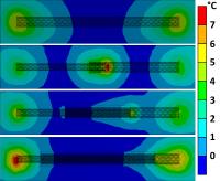 |
RF Induced Heating of Overlapped Stents
Peter Serano1,2, Maria Ida Iacono1,
Leonardo M. Angelone1, and Sunder S. Rajan1
1U.S. Food and Drug Administration, Washington,
DC, United States, 2Electrical
and Computer Engineering, University of Maryland, College
Park, MD, United States
In this study, the authors present an analysis of a
potentially overlooked clinical scenario, namely overlapped
stents separated with a layer of insulation. Electromagnetic
and thermal simulations as well as measurements were
performed to test such configurations. The results show that
implanted medical devices that include gapped conductive
structures, like overlapped stents, can affect the location
and magnitude of peak heating near the implant.
|
| |
14:42
|
0916.
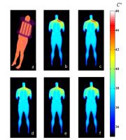 |
Extremely Rapid Temperature Predictions Considering Numerous
Physiological Phenomena
Giuseppe Carluccio1,2 and
Christopher Michael Collins1,2
1Radiology, Center for Advanced Imaging
Innovation and Research (CAI2R), New York, NY, United
States, 2Radiology,
Bernard and Irene Schwartz Center for Biomedical Imaging,
New York, NY, United States
In a patient exam, SAR may cause temperature increase
potentially leading to tissue damage or thermoregulatory
distress. Hence, development of fast and accurate
temperature computation methods could be useful for safety
assurance. We propose a method considering more factors than
ever before (including SAR, respiration, perspiration,
convection, conduction, and local perfusion rates), where
the temperature over an entire MRI exam is rapidly estimated
exploiting the linearity of the bioheat equation. Nonlinear
effects due to thermoregulatory mechanisms of the human
body, such as the variation of local blood perfusion rate,
are approximated with a fast spatial filter.
|
| |
14:54
|
0917.
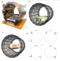 |
Incident electric field on implanted lead vs. source position
and field polarization
Elena Lucano1,2, Micaela Liberti2,
Gonzalo G Mendoza1, Tom Lloyd3,
Francesca Apollonio2, Steve Wedan3,
Wolfgang Kainz1, and Leonardo M Angelone1
1Center for Devices and Radiological Health,
Office of Science and Engineering Laboratories, U.S. Food
and Drug Administration, Silver Spring, MD, United States, 2Department
of Information Engineering, Electronics and
Telecommunications, Univerisity of Rome "Sapienza", Rome,
Italy, 3Imricor
Medical Systems, Burnsville, MN, United States
We aim to generate a quantitative method for RF-safety of
patients with partially implanted leads at 64 MHz. Within
this aim, the position of the RF feeding sources and the
orientation of the polarization is often unknown, as it is
the quantitative effect of such variables on the induced
currents on the leads. The Electric field profile was
studied by means of simulations and measurements with a coil
loaded with a phantom, and simulations with an anatomical
human model. Changes of up to 40% of E-field magnitude were
observed. Future work is needed to develop a systematic
exposure procedure.
|
| |
15:06
|
0918.
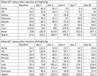 |
Biodistribution of ferumoxytol: a longitudinal MRI study - Permission Withheld
Tilman Schubert1,2, Utaroh Motosugi3,
Diego Hernando1, Camilo A Campo1,
Samir Sharma4, Scott Reeder1,4,5,6,7,
and Shane Wells1
1Radiology, University of Wisconsin Madison,
Madison, WI, United States, 2Clinic
for Radiology and Nuclear Medicine, Basel University
Hospital, Basel, Switzerland, 3Department
of Radiology, University of Yamanashi, Yamanashi, Japan, 4Medical
Physics, University of Wisconsin Madison, Madison, WI,
United States, 5Biomedical
Engineering, University of Wisconsin Madison, Madison, WI,
United States, 6Medicine,
University of Wisconsin Madison, Madison, WI, United States, 7Emergency
Medicine, University of Wisconsin Madison, Madison, WI,
United States
Ferumoxytol has gained increasing interest as a negative
MR-contrast agent due to its high r2* relaxivity. However,
limited data is available about the temporal course of the
biodistribution of ferumoxytol. This study evaluated the
biodistribution of ferumoxytol in different tissue types
using repeated MR-measurements until the 30th day after
administration. Our longitudinal MRI-study demonstrated that
tissues of the monocyte−macrophage system show different,
dose dependent R2* peaks after ferumoxytol injection. These
results could help to determine the optimal, tissue specific
imaging delay after ferumoxytol administration. Tissues not
containing monocytes/macrophages parallel the time course of
ferumoxytol in the blood pool.
|
| |
15:18
|
0919.
 |
MRI RF-Induced Pacemaker Lead Heating: Effect of Single vs
Dual-lead Systems
Shi Feng1, Shiloh Sison2, Jazmine
Garcia3, Gabriel Mouchawar3, and
Richard Williamson3
1Hardware development, St. Jude Medical, Sylmar,
CA, United States, 2St.
Jude Medical, Sunnyvale, CA, United States, 3St.
Jude Medical, Sylmar, CA, United States
Metallic leads of an implanted electronic device such as a
pacemaker may behave as antennae in the strong radio
frequency electromagnetic field of MRI. The induced current
surrounding the electrodes may heat the local tissue. The
MRI-induced tissue heating around the electrodes of a
pacemaker have only been investigated for pacemakers
employing a single lead. In this paper, we examine the
MRI-induced temperature rise (TR) of the tip electrode(s)
associated with a pacemaker system with two St. Jude Medical
Tendril 2088 STS leads, and compare it to the single result.
Both transfer function and in vitro temperature rise are
investigated.
|
|