10:45
|
0026.
 |
An illustrated comparison of background field elimination
methods for phase MRI and QSM 
Ferdinand Schweser1,2, Wei Li3, Hongfu
Sun4, Dong Zhou5, Nicola Bertolino1,
Paul Polak1, Yi Wang5, Alan H Wilman4,
Kristian Bredies6, Robert Zivadinov1,
and Simon Daniel Robinson7
1Buffalo Neuroimaging Analysis Center, Department
of Neurology, Jacobs School of Medicine and Biomedical
Sciences, The State University of New York at Buffalo,
Buffalo, NY, United States, 2MRI
Molecular and Translational Research Center, Jacobs School
of Medicine and Biomedical Sciences, The State University of
New York at Buffalo, Buffalo, NY, United States, 3Research
Imaging Institute, The University of Texas Health Science
Center, San Antonio, TX, United States, 4Department
of Biomedical Engineering, University of Alberta, Edmonton,
AB, Canada, 5Department
of Radiology, Weill Cornell Medical College, New York, NY,
United States, 6Institute
for Mathematics and Scientific Computing, University of
Graz, Graz, Austria, 7High
Field MR Center of Excellence, Department of Radiology,
Medical University of Vienna, Vienna, Austria
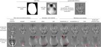
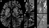
Elimination of background fields is an essential step in
phase MRI and QSM, with many different approaches proposed
over the past years. However, it is currently unclear how
the various methods perform relative to each other and what
their respective strengths and weaknesses are, because a
multi-center quantitative comparison of all techniques has
not yet been carried out. In this work we quantitatively
compare inverse Laplace filtering, SHARP , V-SHARP, iSMV ,
LBV, HARPERELLA, iHARPERELLA, PDF, and RE-SHARP in a
collaborative effort.
The background correction performance was similar with
all methods, with iSMV and LBV yielding the best results.
|
10:57
 |
0027.
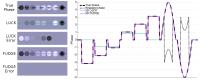 |
Fast Unwrapping using Discrete Gradient Evaluation (FUDGE): an
analytical correction to the Laplacian-based phase unwrapping
technique for discrete data.
Amanda Ching Lih Ng1, Meei Pyng Ng2,
Sonal Josan3, Shawna Farquharson4,
Claire Mulcahy4, and Roger J Ordidge1
1Dept of Anatomy & Neuroscience, The University
of Melbourne, Parkville, Australia, 2Dept
of Mathematics & Statistics, The University of Melbourne,
Parkville, Australia, 3Siemens
Healthcare, Melbourne, Australia,4Imaging, The
Florey Institute of Neuroscience and Mental Health,
Melbourne, Australia
Laplacian-based phase unwrapping is commonly used to
pre-process phase for methods such as Quantitative
Susceptibility Mapping (QSM). However, the formulation was
derived with the assumption of a continuous signal and a
continuous Fourier transform. When applied to discrete MRI
phase data, serious errors in phase can occur, resulting in
substantial errors in QSM estimates. We present a
mathematically correct Laplacian-based phase unwrapping
formula, based on the assumption of the discrete nature of
MRI phase data and processing. Our results reflect the
mathematical predictions of the old and new formulations.
|
11:09
|
0028.
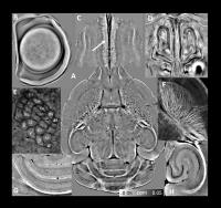 |
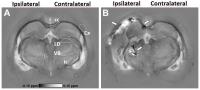
Imaging Whole Mouse Brain Cytoarchitecture by Quantitative
Susceptibility Mapping at 10-µm Resolution
Hongjiang Wei1, Luke Xie2, Russell
Dibb3, Wei Li4, Kyle Decker3,
G. Allan Johnson3,5, and Chunlei Liu1,5
1Brain Imaging and Analysis Center, Duke
University, Durham, NC, United States, 2Utah
Center for Advanced Imaging Research, Department of
Radiology, University of Utah, Salt Lake City, UT, United
States, 3Center
for In Vivo Microscopy, Duke University, Durham, NC, United
States, 4Research
Imaging Institute, University of Texas Health Science
Center, San Antonio, TX, United States, 5Department
of Radiology, School of Medicine, Duke University, Durham,
NC, United States
In this study, we demonstrate that whole brain
cytoarchitecture can be revealed by QSM at 10-μm resolution
at 9.4T. Using QSM, we are able to reveal exquisite
anatomical details such as retina layers of the eyeball,
glomeruli in olfactory bulb, barrel cortex, medium-sized
spiny neurons in striatum, cell layers of cerebellum, and
hippocampus. This ultra-high resolution QSM of the intact
mouse brain is a powerful dataset to allow analysis and
visualization of the brain cytoarchitecture in 3D.
|
11:21
|
0029.
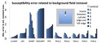 |
A Novel Method for Background Field Removal in Abdominal QSM
Debra E. Horng1,2, Samir D. Sharma1,
Scott B. Reeder1,2,3,4,5, and Diego Hernando1
1Radiology, University of Wisconsin, Madison, WI,
United States, 2Medical
Physics, University of Wisconsin, Madison, WI, United
States, 3Medicine,
University of Wisconsin, Madison, WI, United States, 4Biomedical
Engineering, University of Wisconsin, Madison, WI, United
States, 5Emergency
Medicine, University of Wisconsin, Madison, WI, United
States
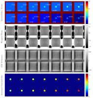
We introduce a QSM background field removal method based on
harmonic function theory. Methods based on the mean value
theorem compute the value at the center of a spherical
kernel. Conversely, a new method based on the extended
Poisson kernel can compute the value at any location in a
spherical kernel. The new kernel is evaluated for accuracy
near air/tissue interfaces, resulting in low errors compared
to existing methods. Our new method is fast (analytic) and
is designed for performance near air/tissue interfaces in
abdominal QSM.
|
11:33
|
0030.
 |
Toward Iron Distribution Mapping using Quantitative
Susceptibility Mapping (QSM): A Comparison of Histological Iron
Concentration Maps with Magnetic Susceptibility Maps
Andreas Deistung1, Verena Endmayr2,
Simon Hametner2, Hans Lassmann2,
Jürgen Rainer Reichenbach1, Simon Daniel Robinson3,
Thomas Haider4, Hannes Traxler5,
Evelin Haimburger6, Siegfried Trattnig3,
and Günther Grabner3,6
1Medical Physics Group, Institute of Diagnostic
and Interventional Radiology, Jena University Hospital –
Friedrich Schiller University Jena, Jena, Germany, 2Center
for Brain Research, Medical University of Vienna, Vienna,
Austria, 3High
Field Magnetic Resonance Centre, Department of Biomedical
Imaging and Image-guided Therapy, Medical University of
Vienna, Vienna, Austria, 4University
Clinic for Trauma Surgery, Medical University of Vienna,
Vienna, Austria, 5Center
of Anatomy and Cellbiology, Medical University of Vienna,
Vienna, Austria, 6Department
of Health Sciences and Social Work, Carinthia University of
Applied Sciences, Klagenfurt, Austria
Quantitative susceptibility mapping (QSM) provides a unique
view into cerebral iron distribution in
vivo. However, not only paramagnetic iron complexes but
also diamagnetic myelin around axons contribute to the
magnetic susceptibility. To further validate QSM for iron
mapping we present a histochemical-driven approach to
quantify iron in post mortem brain tissue and compare the
spatial distribution of iron with in
situmagnetic susceptibility maps. Direct comparison
between histological iron concentration and susceptibility
maps revealed excellent correspondence between iron
accumulations and elevated susceptibility in deep gray
matter and can improve the understanding of biophysical
origins of susceptibility variations within brain tissue.
|
11:45
|
0031.
 |
Feasibility Study of High Resolution Mapping for Myelin Water
Fraction and Frequency Shift using Tissue Susceptibility
Zhe Wu1,2, Hongjian He1,2, Ying Chen1,2,
Song Chen1,2, Hui Liu3, Yiping P. Du2,
and Jianhui Zhong1,2
1Center for Brain Imaging Science and Technology,
Zhejiang University, Hangzhou, China, People's Republic of, 2Department
of Biomedical Engineering, Zhejiang University, Hangzhou,
China, People's Republic of,3NEA MR
Collaboration, Siemens Ltd., China, Shanghai, China,
People's Republic of
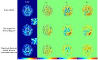
A three-step method for high resolution myelin water
fraction (MWF) and frequency shift mapping of white matter
components using tissue susceptibility is presented in this
study. Tissue susceptibility induced phase was calculated by
the simultaneously acquired QSM from the same multi-echo GRE
(mGRE) dataset, and was used as the phase part of complex
data for a subsequent fitting to a three-pool white matter
model. Benefit from the background phase removal and
magnetic dipole deconvolution procedures during QSM
calculation, the result reveals much less misfitting when
comparing with direct fitting to original mGRE data. These
generated quantitative maps can be potentially used for
quantitative studies of demyelinated diseases.
|
11:57
|
0032.
 |
Preconditioned QSM to Determine a Large Range of Susceptibility
Over The Entire Field Of View from Total Field
Zhe Liu1, Youngwook Kee2, Dong Zhou2,
Pascal Spincemaille2, and Yi Wang1,2
1Biomedical Engineering, Cornell University,
Ithaca, NY, United States, 2Radiology,
Weill Cornell Medical College, New York, NY, United States
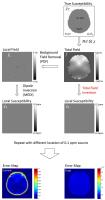
We propose a Preconditioned QSM calculating susceptibility
over the entire field of view (FOV), which eliminates the
errors associated with background field removal. The
background is regarded as part of the region with large
susceptibilities, which is determined by a preconditioned
conjugate gradient solver with enhanced convergence. Our
data demonstrate that our preconditioned QSM provides a
susceptibility map of the entire head accurately depicting
skin, bone, air filled sinuses and hemorrhages.
|
12:09
|
0033.
 |
MRI in Multiple Sclerosis: The curiosity of apparent
susceptibility increases at simultaneous iron loss
Vanessa Wiggermann1,2, Simon Hametner3,
Enedino Hernandez-Torres2,4, Verena Endmayr3,
Christian Kames5, and Alexander Rauscher2
1Physics and Astronomy, University of British
Columbia, Vancouver, BC, Canada, 2Pediatrics,
University of British Columbia, Vancouver, BC, Canada, 3Neuroimmunology,
Medical University of Vienna, Vienna, Austria,4UBC
MRI Research Centre, University of British Columbia,
Vancouver, BC, Canada, 5Engineering
Physics, University of British Columbia, Vancouver, BC,
Canada
Quantitative Susceptibility Mapping has shown great
potential to be used for clinical diagnoses due to its high
sensitivity to change and high spatial resolution. Notably,
the ability to quantify damage has been appealing. However,
attributing susceptibility increases or decreases to certain
mechanisms has been challenging. In particular,
interpretation of MR signal changes during multiple
sclerosis lesion formation is lacking consistency and
histological validation. Here, we investigated the
hypothesis that apparent changes of the lesion tissue may be
in fact due to changes in the lesions vicinity and caution
is required when interpreting the quantitative
susceptibility signal in multiple sclerosis lesions.
|
12:21
|
0034.
 |
Quantitative susceptibility mapping of the rat brain after
traumatic brain injury
Karthik Chary1, Mikko J. Nissi2,3,
Ramón I. Rey4, Eppu Manninen1, Karin
Shmueli5, Alejandra Sierra1, and Olli
Gröhn1
1Department of Neurobiology, A.I. Virtanen
Institute for Molecular Sciences, University of Eastern
Finland, Kuopio, Finland, 2Department
of Applied Physics, University of Eastern Finland, Kuopio,
Finland, 3Finland
Diagnostic Imaging Center, Kuopio University Hospital,
Kuopio, Finland, 4Department
of Neurology, Clinical Neurosciences Research Laboratory,
Hospital Clínico Universitario, Health Research Institute of
Santiago de Compostela (IDIS), University of Santiago de
Compostela, Santiago de Compostela, Spain, 5Department
of Medical Physics & Biomedical Engineering, University
College London, London, United Kingdom
Our aim was to test the sensitivity of QSM to demyelination,
iron and calcifications in a rat model of TBI. Ex
vivo QSM
data were obtained from five injured and four sham control
rats, six months after TBI. Our results showed
susceptibility changes in white matter areas consistent with
myelin staining. Perilesional cortex became more diamagnetic
after TBI. Thalamic nuclei showed variable responses as
diamagnetic calcification and paramagnetic iron accumulation
occurred in the same brain areas. Overall, QSM showed
sensitivity to TBI changes. However, further studies are
required to better understand the influence of potentially
counteracting pathological processes.
|
12:33
|
0035.
 |
Suitable reference tissues for quantitative susceptibility
mapping of the brain
Sina Straub1, Till Schneider2,3,
Martin T. Freitag3, Christian H. Ziener3,
Heinz-Peter Schlemmer3, Mark E. Ladd1,
and Frederik B. Laun1
1Department of Medical Physics in Radiology,
German Cancer Research Center (DKFZ), Heidelberg, Germany, 2Department
of Neuroradiology, University of Heidelberg, Heidelberg,
Germany, 3Department
of Radiology, German Cancer Research Center (DKFZ),
Heidelberg, Germany
Since QSM is only able to quantify magnetic susceptibility
relative to a reference value, a suitable reference tissue
must be available to be able to compare different subjects
and stages of disease. To find such a suitable reference
tissue for QSM of the brain, melanoma patients with and
without brain lesions were measured. 12 reference tissues
were chosen and assessed in multiple measurements of the
same patient and amongst different patients. The posterior
limb of the internal capsule and a cerebrospinal fluid
volume in the atrium of the lateral ventricles appeared to
be most suitable reference tissues.
|
|