| |
13:30
|
0705.
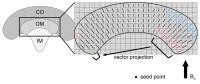 |
Frequency tensor imaging (FTI) at a single orientation by vector
projection 
Luke Xie1, Russell Dibb2, Chunlei Liu2,
and Vivian S. Lee1
1Utah Center for Advanced Imaging Research,
Radiology, University of Utah, Salt Lake City, UT, United
States, 2Brain
Imaging Analysis Center, Radiology, Duke University Medical
Center, Durham, NC, United States
STI is sensitive to tissue microstructure and can detect
subtle changes in disease states. However, STI remains a
challenging protocol due to the physical reorientation with
respect to the magnetic field. Current studies of the heart
and kidney are limited to ex-vivo imaging.
In this study, we present frequency tensor imaging (FTI) at
a single image acquisition without rotating the object. FTI
takes advantage of tissue structure already pointing in
multiple directions with respect to the magnetic field in a
single orientation dataset. This technique offers the
potential for susceptibility-based tensor imaging of the
abdomen in the clinic.
|
| |
13:42
|
0706.
 |
Automatic Renal Cortex Segmentation Using Machine Learning for
MR Urography
Umit Yoruk1,2, Brian Hargreaves2, and
Shreyas Vasanawala2
1Electrical Engineering, Stanford University,
Stanford, CA, United States, 2Radiology,
Stanford University, Stanford, CA, United States
Glomerular filtration rate (GFR) estimation can be achieved
using dynamic contrast enhanced MRI (DCE-MRI) and
pharmacokinetic models. The segmentation of kidneys is
essential for obtaining the time intensity curves needed by
these models. Manual segmentation of kidneys is one of the
most time consuming and labor-intensive steps of GFR
analysis as it can take several hours and require trained
personnel. Here, we introduce a novel method for automatic
renal segmentation based on morphological segmentation and
machine learning, and assess the performance of the method.
|
| |
13:54
|
0707.
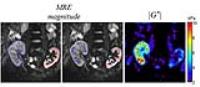 |
Magnetic Resonance Elastography (MRE) for the assessment of
renal allograft function
Jing Guo1, Stephan Marticorena1,
Florian Dittmann1, Andreas Fehlner1,
Sebastian Hirsch1, Thomas Fischer1,
Jürgen Braun2, and Ingolf Sack1
1Radiology, Charité - Universitätsmedizin Berlin,
Berlin, Germany, 2Department
of Medical Informatics, Charité - Universitätsmedizin
Berlin, Berlin, Germany
In vivo assessment of the renal allograft function post
kidney transplantation is challenging. We here demonstrate
that multifrequency MR elastography (MMRE) can detect renal
allograft dysfunction with good diagnostic accuracy
(AUROC:0.91 [95% CI 0.80-1.02; p < 0.001]). Renal stiffness
is significantly lower in dysfunctional transplant kidney
and correlates moderately with glomerular filtration rate
and resistive index. MMRE may serve as a non-invasive
imaging maker to detect renal allograft dysfunction in an
early stage and to monitor renal allograft function
longitudinally.
|
| |
14:06
|
0708.
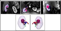 |
3D Printed Renal Cancer Models Derived from MRI data:
Application in Pre-surgical Planning
Nicole Wake1, Temitope Rude2, William
C Huang2, Michael D Stifelman2, James
F Borin2, Daniel K Sodickson1, and
Hersh Chandarana1
1Bernard and Irene Schwartz Center for Biomedical
Imaging, Center for Advanced Imaging Innovation and
Research, Department of Radiology, New York University
School of Medicine, New York, NY, United States, 2Department
of Urology, New York University School of Medicine, New
York, NY, United States
The objective of this study was to determine how
patient-specific 3D printed renal tumor models derived from
MRI data can influence pre-surgical planning. These 3D
printed models may alter the surgical plan, especially for
trainees and young surgeons. Future, outcome based studies
may help to determine the impact of these 3D printed models
on surgical outcomes and patient care.
|
| |
14:18
|
0709.
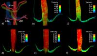 |
Diurnal Variation of Renal Blood Flow using 4D Flow MRI
Sylvana García-Rodríguez1, Alejandro
Roldán-Alzate1,2, Camilo A. Campo1,
Scott B. Reeder1, Oliver Wieben1,3,
and Christopher J. François1
1Radiology, University of Wisconsin-Madison,
Madison, WI, United States, 2Mechanical
Engineering, University of Wisconsin-Madison, Madison, WI,
United States, 3Medical
Physics, Univerisity of Wisconsin-Madison, Madison, WI,
United States
This study investigated the diurnal changes in renal blood
flow in healthy volunteers using 4D flow MRI, to determine
the optimal time of day to perform renal blood flow
measurements. Five 4D flow MRI acquisitions were performed
throughout the day in seven healthy subjects to mimic
potential imaging scheduling time points. Significant
differences in renal blood flow were observed depending upon
time of day and prandial status. This study confirms the
importance of timing of renal MRI studies assessing kidney
function.
|
| |
14:30
|
0710.
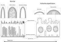 |
Reduced susceptibility anisotropy in ischemia reperfusion
kidneys: evidence of cellular organization as a source of
contrast
Luke Xie1, Vivian Lee1, Russell Dibb2,
Yi Qi2, Nian Wang3, G. Allan Johnson2,
and Chunlei Liu3
1Radiology, University of Utah, Salt Lake City,
UT, United States, 2Center
for In Vivo Microscopy, Radiology, Duke University Medical
Center, Durham, NC, United States, 3Brain
Imaging Analysis Center, Radiology, Duke University Medical
Center, Durham, NC, United States
Diffusion tensor imaging (DTI) and susceptibility tensor
imaging (STI) can assess the integrity of the nephron where
STI provides additional molecular information. STI has
demonstrated sensitivity to changes in kidney disease
models. The source of susceptibility anisotropy is
hypothesized to be the organized tubules, basement membrane,
and the organized lipids. Ischemia reperfusion is one
particular disease model with well known cellular
disorganization in specific nephron segments. In the present
study, we applied STI in a model of ischemia perfusion to
demonstrate changes in susceptibility anisotropy and to
provide additional evidence that the cellular organization
is a major contributor.
|
| |
14:42
|
0711.
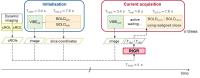 |
Prospective Image Alignment for Time-Resolved Renal BOLD MRI
Inge Manuela Kalis1, David Pilutti1,
Axel Joachim Krafft1,2,3, and Michael Bock1
1Dept. of Radiology - Medical Physics, University
Medical Center Freiburg, Freiburg, Germany, 2German
Cancer Consortium (DKTK), Heidelberg, Germany, 3German
Cancer Research Center (DKFZ), Heidelberg, Germany
Renal function can be analyzed by time-resolved BOLD MRI
before, during and after a functional challenge.
Inconsistent kidney positions from one measurement to
another hamper the analysis of renal parenchyma and medulla
over time. Here, a new method, Kidney ALIgnment for BOLD
Renal Imaging (KALIBRI), with prospective rigid image
registration of each kidney is proposed.
|
| |
14:54
|
0712.
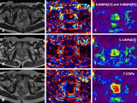 |
MR Elastography of The Prostate with A Mode-Conversion
Endourethral Driver: Feasibility at 3.0 T
Jin Wang1, Kevin J. Glaser2, Bingjun
He1, Tianhui Zhang1, Jun Pang3,
Ziying Yin2, Zhuang Kang1, Qungang
Shan1, Meng Yin2, Forghanian-Arani
Arvin2, and Richard L. Ehman2
1Department of Radiology, The Third Affiliated
Hospital of Sun Yat-Sen University, Guangzhou, China,
People's Republic of, 2Department
of Radiology, Mayo Clinic, Rochester, MN, United States, 3Department
of Urology, The Third Affiliated Hospital of Sun Yat-Sen
University, Guangzhou, China, People's Republic of
Prostate cancer(PCa) is one of the leading causes of
cancer-related deaths in men. Detection of clinically
significant PCa is a major challenge. We evaluated the
feasibility of a novel approach for quantitatively imaging
the stiffness of prostate gland, using a conventional
urinary catheter as a source of shear waves for MR
elastography. Results in 19 examinations showed that the
approach, which uses conventional commercially-available MRE
drivers can generate shear wave fields in the prostate that
are suitable processing. Measurements of regional prostate
stiffness in patients with benign prostatic hypertrophy and
PCa reveal trends that provide motivation for further
evaluation of prostatic MRE.
|
| |
15:06
|
0713.
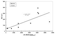 |
Adrenal gland iron measurement using MRI-R2* in patients with
iron overload: a feasibility Study
Sarah Keller1, Bjoern Schoennagel1,
Zhiyue Jerry Wang2, Hendrick Kooijman3,
Gerhard Adam1, Roland Fischer4,5, and
Jin Yamamura1
1Diagnostic and Interventional Radiology,
University Medical Center Hamburg-Eppendorf, Hamburg,
Germany, 2Radiology,
University of Texas Southwestern Medical Center, Dallas, TX,
United States, 3Philips
Medical Care, Hamburg, Germany, 4Radiology,
Children’s Hospital & Research Center Oakland, Oakland, CA,
United States, 5Biochemistry,
University Medical Center Hamburg-Eppendorf, Hamburg,
Germany
In recent years, hepatic, cardiac, and even pancreatic iron
deposition has been studied in detail. However, the presence
and incidence of iron disposition of normal-sized adrenal
glands has not been adequately reported. The purpose of this
study was to evaluate the levels of iron deposition in the
adrenal glands in patients with iron overload.
|
| |
15:18
|
0714.
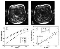 |
Improving the accuracy of renal perfusion measurements from ASL
by using multiple TIs: Validation with DCE MRI
Christopher Charles Conlin1,2, Yangyang Zhao2,
and Jeff Lei Zhang1,3
1Utah Center for Advanced Imaging Research,
University of Utah, Salt Lake City, UT, United States, 2Bioengineering,
University of Utah, Salt Lake City, UT, United States, 3Radiology,
University of Utah School of Medicine, Salt Lake City, UT,
United States
This study presents an approach for measuring renal
perfusion from multi-TI ASL data and examines the impact of
TI-sampling density on perfusion estimation. Our approach
incorporates a tracer-kinetic model of the ASL difference
signal and a correction for inversion-efficiency artifacts.
It was used to measure renal perfusion in human subjects
from ASL data sampled at different numbers of TIs and
validated against an established DCE-MRI technique. For ASL
data sampled at more than two TIs, our approach showed good
agreement and correlation with DCE-MRI, demonstrating robust
modeling of the ASL difference signal and accurate
measurement of renal perfusion.
|
|