| |
13:30
|
0969.
 |
Magnetic Resonance Imaging quantification of venous return in
pregnant women: A comparison between supine and left lateral
tilt position. 
Emer J Hughes1, Anthony N Price2,
Laura McCabe1, Kelly Pegoretti Baruteau1,
Jana Hutter2, Olivia Carney1, Andreia
S Gaspar2, Joseph V Hajnal2, and Mary
Rutherford1
1Perinatal Imaging and Health, Kings College
London, London, United Kingdom, 2Biomedical
Engineering, Kings College London, London, United Kingdom
In-vivo imaging of the fetus is commonly undertaken in the
left-lateral position to prevent compression of the inferior
vena cava (IVC) and hence a vasovagal episode. Studies have
shown that the IVC has collateral pathways, such as the
lumbar venous plexus and the lumbar veins that provide
collateral venous return. Here, we use phase contrast
imaging to assess the venous return pathways in pregnant
women lying supine and left lateral tilt in the MRI scanner.
We found that the spinal venous plexus and the ascending
lumbar veins act as a complimentary venous return system to
maintain vascular homeostasis in pregnant women lying
supine. This supports the proposition that it is feasible to
scan pregnant women safely in the supine position.
|
| |
13:42
 |
0970.
 |
In vivo localization and timing of oxygen delivery in human
placenta based on BOLD MRI
Jie Luo1,2, Esra Abaci Turk1,2, Polina
Golland3,4, Borjan Gagoski1, Carolina
Bibbo5, Drucilla J Roberts6, Norberto
Malpica7, Julian N Robinson5, Patricia
Ellen Grant1, and Elfar Adalsteinsson2,3,8
1Fetal-Neonatal Neuroimaging & Developmental
Science Center, Boston Children's Hospital, Harvard Medical
School, Boston, MA, United States, 2Madrid-MIT
M+Vision Consortium in RLE, Massachusetts Institute of
Technology, Cambridge, MA, United States, 3Department
of Electrical Engineering and Computer Science,
Massachusetts Institute of Technology, Cambridge, MA, United
States, 4Computer
Science and Artificial Intelligence Laboratory (CSAIL),
Massachusetts Institute of Technology, Cambridge, MA, United
States, 5Maternal
and Fetal Medicine, Brigham and Women's Hospital, Boston,
MA, United States,6Obstetric and Perinatal
Pathology, Massachusetts General Hospital, Boston, MA,
United States, 7Medical
Image Analysis and Biometry Laboratory, Universidad Rey Juan
Carlos, Madrid, Spain, 8Harvard-
MIT Health Sciences and Technology, Massachusetts Institute
of Technology, Cambridge, MA, United States
Clinically there is no direct measurement of oxygen delivery
in placenta. In this study, we propose a method to map the
timing of oxygen delivery in the human placenta in vivo.
Healthy placentae show p?????n? that agree with normal
perfusion timing in response to maternal hyperoxygenation.
Pathological placentas exhibit a more dispersed timing
across the placenta. Better understanding of the timing in
different type of pathology may be achieved by spatial
correlation between placental pathology and in vivo placenta
images in both healthy and pathological placenta based on
BOLD signal change in response to maternal hyperoxygenation.
|
| |
13:54
|
0971.
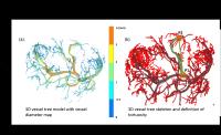 |
Placental vascularization quantification using ex-vivo magnetic
resonance angiography
Bailiang Chen1,2,3, Jie Duan2,3,4,
Jacques Felblinger1,2,3,5, Olivier Morel2,3,4,
and Marine Beaumont 1,3,5
1CHRU Nancy, CIC-IT 1433, Inserm,
Vandoeuvre-lès-Nancy, France, 2Imagerie
Adaptative Diagnostique et Interventionnelle, Université de
Lorraine, Nancy, France, 3U947,
Inserm, Nancy, France, 4Service
d’obstétrique et médecine fœtale, Pôle de
Gynécologie-Obstétrique, CHRU Nancy, Nancy, France, 5Pôle
S2R, CHRU Nancy, Nancy, France
Abnormal uteroplacental vascurlarization can cause major
obstetric complications such as intra-uterine growth
restriction or abnormally invasive placenta. Clinical 3D
ultrasound imaging cannot discriminate maternal and fetal
flow in utero-placental unit, thus blocking a better
understanding of the pathology. Conventional ex-vivo
vascularization quantification relies on 2D histological
slices using samples dissected from placenta. Micro-CT was
applied to fixed small animal placenta but with complicated
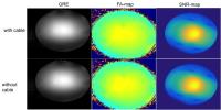
and long preparation. Here we presented the flexibility of a
comprehensive 3D vascularization characterization of a fresh
healthy human placenta using ex-vivo MRA. A quantification
framework is proposed with defined systematic metrics to
characterize the vascularization.
|
| |
14:06
|
0972.
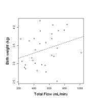 |
MRI Quantification of uterine blood flow in third trimester
human pregnancy and relation to birthweight - Permission Withheld
David J LOMAS1, Rebecca HAWKES1,
Andrew N PRIEST1, Nicholas HILLIARD1,
Andrew PATTERSON1, Pat SET1, and
Martin J GRAVES1
1Radiology, University of Cambridge &
Addenbrooke's Hospital, Cambridge, United Kingdom
Non-invasive measurement of uterine blood flow (UBF) during
pregnancy is desirable to assess fetal well-being but
difficult using Doppler ultrasound. This work demonstrates
an MR method of identifying the uterine arteries in 31 early
3rd trimester normal human pregnancies and quantifying
absolute UBF using cine phase contrast. Results are
comparable with other methods for quantifying flow and
demonstrate a correlation with actual birthweight. The
method has potential for future UBF monitoring during
pregnancy.
|
| |
14:18
|
0973.
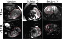 |
Three-Dimensional Placental Perfusion Imaging Using
Velocity-Selective Arterial Spin Labeling
Zungho Zun1, Ajit Shankaranarayanan2,
Nickie Niforatos-Andescavage1, Samantha Bauer1,
Diane Lanham1, Dorothy Bulas1, Adre J
Du Plessis1, and Catherine Limperopoulos1
1Children’s National Medical Center, Washington,
DC, United States, 2GE
Healthcare, Menlo Park, CA, United States
Pregnancies complicated by placental insufficiency such as
fetal growth restriction and preeclampsia are characterized
by reduced placental perfusion. Conventional MR perfusion
imaging involves the use of gadolinium-based contrast
agents, which are contraindicated in pregnancy. In this
study we demonstrate the utility of non-invasive placental
perfusion imaging using velocity-selective arterial spin
labeling and 3D image acquisition with whole placenta
coverage, and present global and regional placental
perfusion in high and low-risk pregnancies. Global placental
perfusion matched ranges of previously reported values.
However, perfusion was heterogeneous and regional placental
perfusion measured within the placental lobules reached
levels two-fold higher than the global placental perfusion
measurement.
|
| |
14:30
|
0974.
 |
Fetal cardiac MRI and flow measurement using Optimized Doppler
Ultrasound Sensor (DUS) gating
Jin Yamamura1, Bjoern Schoennagel1,
Manuele Tavares de Sousa2, Christian Ruprecht1,
Gerhard Adam1, and Fabian Kording1
1Diagnostic and Interventional Radiology,
University Medical Center Hamburg-Eppendorf, Hamburg,
Germany, 2Obstetrics
and Reproductive Medicine, University Medical Center
Hamburg-Eppendorf, Hamburg, Germany
The commonly used method to evaluate the fetal heart is
echocardiography (ECG). However, the detection of congenital
heart diseases by ECG varies from 45% to 74% and an
alternative imaging modality would be desirable. Fetal
cardiac MRI has the potential to visualize anatomy and to
assess functional parameters of the fetal heart. External
fetal cardiac gating using a newly developed Doppler
ultrasound sensor (DUS) has been introduced in previous
studies. The purpose of this study was to perform fetal
cardiac MRI as well as MR flow measurement within great
vessels using for external fetal cardiac gating in human
fetus and to optimize the device.
|
| |
14:42
|
0975.
 |
Comparative Study of Modelling DW-MRI Data From High-grade
Serous Carcinomas and Clear Cell Carcinomas
Feng Wang1, Jianyu Liu1, Yan Zhou1,
and Lizhi Xie2
1Radiology Department of Peking University Third
Hospital, Beijing, China, People's Republic of, 2GE
Healthcare, MR Research China, Beijing, Beijing, China,
People's Republic of
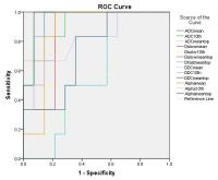
The aim of this study was to assess if the histogram
analysis of mono-exponential, bi-exponential and stretched
exponential models of diffusion-weighted MRI (DW-MRI)
parameters can differentiate two common subtypes of ovarian
epithelial cancer: high-grade serous carcinomas (HGSCs) and
clear cell carcinomas(CCCs). Based on an entire-tumour
measurement, the following histogram parameters were derived
from ADC, D, D*, F, DDC and α maps, respectively: the mean
of the whole tumor, the 10th percentile and the mean of the
top 10 percent. We concluded that ADC, D, F, DDC and α have
showed good diagnostic performance by analyzing these data.
|
| |
14:54
|
0976.
 |
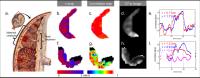
Diffusion-weighted MR Imaging (DW-MRI) in advanced epithelial
ovarian and primary peritoneal cancer: anatomic site-specific
changes following neoadjuvant chemotherapy for detecting
residual viable tumor
Jennifer C Wakefield1,2, Jessica M Winfield1,2,
Gordon Stamp3, Alison MacDonald2,
Charlotte Hodgkin4, Ayoma Attygalle2,
Desmond Barton2, Robin Crawford4,
Susan Freeman4, and Nandita M deSouza1,2
1Division of Radiotherapy and Imaging, Cancer
Research UK Cancer Imaging Centre, The Institute of Cancer
Research, London, United Kingdom, 2The
Royal Marsden Hospital, Sutton, United Kingdom,3Department
of Medicine, Centre for Pathology, Imperial College London,
London, United Kingdom, 4Departments
of Gynaecological Oncology and Radiology, Addenbrooke’s
Hospital, Cambridge University Hospitals NHS Foundation
Trust, Cambridge, United Kingdom
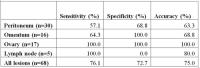
An understanding of the apparent diffusion coefficient (ADC)
changes following neoadjuvant chemotherapy at different
metastatic sites in advanced ovarian and primary peritoneal
cancer is essential to establish the utility of ADC as a
biomarker in site-specific response assessment in this
disease. In this study, we found that there was variability
in the detection accuracy of DW-MRI between different
disease sites and the ADC shows utility as an adjunct to
morphological imaging for the detection of viable tumor.
Further studies with larger numbers of lesions are needed to
interrogate differences between microscopic and non-viable
and residual macroscopic tumor fully.
|
| |
15:06
|
0977.
 |
Diagnostic Performance of Endovaginal Zoom EPI Images for
Detecting Cervix Cancer after Distortion Correction using
Gradient Reversal
Nandita deSouza1, Matthew Orton1, Kate
Downey1, Veronica Morgan1, David
Collins1, Sharon Giles1, and Geoffrey
Payne1
1CRUK/EPSRC Cancer Imaging Centre, The Institute
of Cancer Research and The Royal Marsden NHS Foundation
Trust, Sutton, United Kingdom
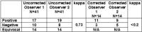
Diffusion-weighted MRI (DW-MRI) suffers from distortion
induced by susceptibility variation and eddy-currents. To
correct this for endovaginal imaging of the uterine cervix,
we implemented the forward and reversed gradient technique
proposed by Chang and Fitzpatrick in the phase-encode
direction and assessed clinical utility of the technique.
This required acquisition of two images of the cervix under
the same conditions. Correction of distortions
significantly improved diagnostic performance for an
experienced observer when images were viewed with the T2-W
images. Correction allowed definitive diagnosis in a third
of cases with tumour volumes of <0.2cm3 classified as
equivocal on uncorrected images.
|
| |
15:18
|
0978.
 |
Quantitative DCE-MRI as predictors of immediate ablation
efficiency in MR-HIFU treatment of uterine fibroids based on
reference region model and entire-tumor histogram analysis
Chenxia Li1,2, Chao Jin1, Ting Liang1,2,
Gang Niu1, Yitong Bian1, Keserci
Bilgin3, and Jian Yang1,2
1Department of Radiology, The First Affiliated
Hospital of Xi’an Jiaotong University, Xi'an, China,
People's Republic of, 2Department
of Biomedical Engineering, School of Life Science and
Technology of Xi’an Jiaotong University, Xi'an, China,
People's Republic of, 3Philips
Healthcare, Seoul, Korea, Republic of
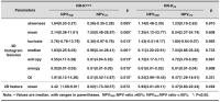
The aim is to investigate whether quantitative DCE-MRI could
be a predictor of immediate ablation efficiency in MR-HIFU
of uterine fibroids. 24 eligible female underwent DCE-MRI
during screening procedure and immediately after MR-HIFU
therapy.They were divided into high non-perfused volume
(NPV) ratio (>60%) and low NPV ratio (<60%) group. The
reference region model was used for 3D histogram
analysis.All histogram metrics of RR-Ktrans showed
significant difference between two groups. The correlation
of RR-Ktrans and
NPV ratio was significantly negative (r=-0.6). It indicated
that the 3D histogram metrics of RR-Ktrans might
be a sensitive predictor used for patients selection in
MR-HIFU.
|
|