| |
16:00
|
0730.
 |
Complex congenital heart defects in infants produce lasting
decreases in functional network segregation 
Vincent Jerome Schmithorst1, Jodie Votava-Smith2,
Vince Lee1, Vidya Rajagopalan2,
Shaheda Suleiman1, Lisa Paquette2, and
Ashok Panigrahy1
1Radiology, Children's Hospital of Pittsburgh of
UPMC, Pittsburgh, PA, United States, 2Children's
Hospital of Los Angeles, Los Angeles, CA, United States
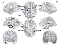
We used functional connectivity MRI and graph analysis to
investigate the impact of congenital heart disease (CHD) on
functional network topology in neonates. Cost-dependent and
cost-independent analyses both showed decreases in global
segregation (transitivity). The cost-dependent analysis
showed a decrease in clustering coefficient (reflective of
nodal changes) while the cost-independent analysis showed a
decrease in modularity and an increase in participation
coefficient (reflective of changed community structure).
Minimal differences were seen for CHD patients scanned
post-operatively compared to those scanned pre-operatively.
Results indicate complex CHD results in lasting changes to
functional network topology not ameliorated by the effects
of surgery.
|
| |
16:12
 |
0731.
 |
Altered Cortical and Subcortical Structures and Structural
Connectivity in Perinatally HIV-infected Children
Santosh Kumar Yadav1, Rakesh Kumar Gupta2,
Ravindra Kumar Garg3, Vimala Venkatesh4,
Ena Wang1, Francesco M Marincola1, and
Mohammad Haris1
1Division of Translational Medicine, Sidra
Medical and Research Center, Doha, Qatar, 2Department
of Radiology, Fortis Memorial Research Institute, Gurgaon,
India, 3Department
of Neurology, King George Medical University, Lucknow,
India, 4Department
of Microbiology, King George Medical University, Lucknow,
India
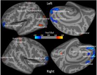
Cortical thickness, subcortical volumes and structural brain
connectivity changes in HIV-seropositive children were
evaluated in comparison to HIV-seronegative children. HIV-seropositive
children showed altered cortical thicknesses, subcortical
volumes and structural connectivity compared to those of
HIV-seronegative children. In addition, changes in cortical
and subcortical structures were significantly correlated
with CD4+ counts and neuropsychological scores in HIV-seropositive
children. We suggest that neuronal injury due to
HIV-infection and inflammation might be possible reasons for
the altered cortical thickness, subcortical volumes and
connectivity in these patients.
|
| |
16:24
 |
0732.
 |
Mapping longitudinal white matter changes in extremely preterm
born infants
Eliza Orasanu1, Andrew Melbourne1,
Marc Modat1, Marco Lorenzi1, Herve
Lombaert2, Zach Eaton-Rosen1, Nicola
Robertson3, Giles Kendall4, Neil
Marlow5, and Sebastien Ourselin1
1Translational Imaging Group, Centre for Medical
Image Computing, University College London, London, United
Kingdom, 2INRIA,
Palaiseau, France, 3Academic
Neonatology, Institute for Women's Health, University
College London, London, United Kingdom, 4Academic
Neonatology, Institute for Women's Health, University
College Hospital, London, United Kingdom, 5Institute
for Women's Health, University College London, London,
United Kingdom
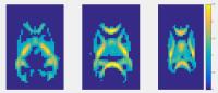
During the preterm period, the brain undergoes changes in
volume, structure and cortical folding, which can be
connected with cognitive abilities in preterm born infants.
Diffusion MRI allows us to investigate microstructural
changes during this period. In this study we registered the
longitudinal diffusion tensor images of six extremely
preterm born infants and looked at white matter changes. The
corpus callosum and internal capsule exhibits the most
microstructural changes during this crucial period and we
hypothesis that this can affect the neurodevelopment in
these infants.
|
| |
16:36
|
0733.
 |
Combining lesion burden with cortical malformation morphology
strongly predicts motor outcomes in children with cerebral palsy
Alex Pagnozzi1, Nicholas Dowson1,
James Doecke1, Simona Fiori2, Andrea
Guzzetta3, Roslyn N Boyd4, and Stephen
Rose1
1The Australian e-Health Research Centre, CSIRO
Health & Biosecurity, Brisbane, Australia, 2Stella
Maris Institute, Pisa, Italy, 3Stella
Maris institute, Pisa, Italy, 4The
University of Queensland, Queensland Cerebral Palsy and
Rehabilitation Research Centre, Brisbane, Australia
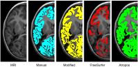
Magnetic Resonance Imaging (MRI) is the clinical standard
for assessing developmental brain injury in children with
Cerebral Palsy (CP). We propose an automated process that
segments the spectrum of white and grey matter injury,
including tissue lesions and malformations of the cortex,
and correlates biomarkers of injury with the Assisting Hand
Assessment (AHA), a clinical score quantifying hand
function. The proposed method is shown to perform accurate
tissue and injury segmentation using T1 and T2 MRI compared
to the manual classification of injury, and was
significantly correlated with AHA (p<0.001).
|
| |
16:48
|
0734.
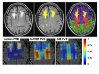 |
Quantitative Spectroscopic Imaging in Metachromatic
Leukodystrophy: value in prognosis and treatment monitoring.
Diane van Rappard1, Antoine Klauser2,
Marjan Steenweg1, Marjo van der Knaap1,
Nicole Wolf1, and Petra Pouwels3
1Child Neurology, VU University Medical Center,
Amsterdam, Netherlands, 2Centre
d'Imagerie BioMédicale, Geneva University, Geneva,
Switzerland, 3Physics
& Medical Technology, VU University Medical Center,
Amsterdam, Netherlands
Currently, hematopoietic stem cell transplantation (HSCT) is
the only treatment option for patients with metachromatic
leukodystrophy (MLD). This study in MLD patients and
controls investigated the possible additional prognostic
value of quantitative MRSI. In WM (consisting of lesions and
NAWM), ratios of Cho/NAA and Ins/NAA were significantly
higher in patients who were considered non-eligible for HSCT
than in eligible patients. Follow-up of successfully treated
patients showed partial normalization of concentrations and
ratios. This study suggests that quantitative MRS can
support the decision whom to treat, especially when
neurological and cognitive examinations are ambiguous.
|
| |
17:00
|
0735.
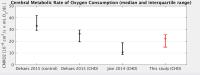 |
Optimizing unanesthetized cerebral oxygen consumption measures:
comparison of MRI and near-infrared spectroscopy (NIRS)
approaches in neonates with congenital heart disease
Jeffrey N Stout1, Silvina Ferradal2,
Borjan Gagoski2, Lilla Zollei3, Divya
S Bolar3,4, Alex Lin5, Henry H Cheng6,
Elfar Adalsteinsson1,7,8, and Patricia Ellen
Grant2
1Harvard-MIT Health Sciences and Technology,
Massachusetts Institute of Technology, Cambridge, MA, United
States, 2Fetal-Neonatal
Neuroimaging and Developmental Science Center, Boston
Children’s Hospital, Boston, MA, United States, 3Martinos
Center for Biomedical Imaging, MGH/Harvard Medical School,
Boston, MA, United States, 4Department
of Radiology, Massachusetts General Hospital, Boston, MA,
United States, 5Department
of Radiology, Brigham and Women's Hospital, Boston, MA,
United States, 6Department
of Cardiology, Boston Children’s Hospital, Boston, MA,
United States, 7Department
of Electrical Engineering and Computer Science,
Massachusetts Institute of Technology, Cambridge, MA, United
States, 8Institute
for Medical Engineering and Science, Cambridge, MA, United
States
Concern for cerebral perfusion in neonates with congenital
heart disease (CHD) has driven investigations into cerebral
hemodynamics. MRI in combination with bedside NIRS has the
potential to provide complementary measures of hemodynamics
to guide surgical timing and assess response to surgery. We
compare MRI and NIRS measures of cerebral hemodynamics.
Modality results compare well to literature studies, but
intermodality correlation is limited. Before combining
modalities additional studies are needed to better
understand why cerebral blood flow and CMRO2 measures
in MRI and NIRS differ.
|
| |
17:12
|
0736.
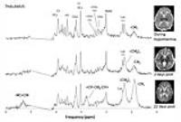 |
Quantitating polyunsaturated fatty acids in neonates with
hypoxic-ischemic brain injury
Jessica Lee Wisnowski1,2, Aaron J Reitman3,4,
Tai-Wei Lee Wu3, Eugenia Ho5, Claire
McLean6, Douglas Lee Vanderbilt6,
Marvin D Nelson1, Ashok Panigrahy7,
Philippe Lee Friedlich3,8, and Stefan Lee Bluml1
1Radiology, Children's Hospital Los Angeles, Los
Angeles, CA, United States, 2Rudi
Schulte Research Institute, Santa Barbara, CA, United
States, 3Center
for Fetal and Neonatal Medicine, Children's Hospital Los
Angeles, Los Angeles, CA, United States, 4Division
of Neonatal Medicine, LAC + USC Medical Center, Los Angeles,
CA, United States, 5Division
of Child Neurology, Children's Hospital Los Angeles, Los
Angeles, CA, United States, 6Children's
Hospital Los Angeles, Los Angeles, CA, United States, 7Radiology,
Children's Hospital of Pittsburgh of UPMC, Pittsburgh, PA,
United States, 8Division
of Neonatal Medicine, University of Southern California, Los
Angeles, CA, United States
Polyunsaturated fatty acids (PUFA) are endogenous components
of cellular membranes and a potential biomarker for
apoptosis following hypoxic-ischemic (HI) brain injury.
Prior studies have applied 1H-MRS
techniques for quantifying PUFA in human carcinomas. Here,
using a retrospective dataset of 1,046 neonatal 1H-MRS
spectra, we demonstrate that PUFA can be routinely
characterized in newborns using a modified LCModel
(Provencher, Inc) pipeline.
|
| |
17:24
|
0737.
 |
Tract-specific analysis of white matter fasciculi in a large
cohort of preterm infants
Diliana Pecheva1, Hui Zhang2, Gareth
Ball1, Mary Rutherford1, Nigel Kennea 3,
Joseph V. Hajnal1, Daniel Alexander2,
A. David Edwards1, and Serena J. Counsell1
1Centre for the Developing Brain, King's College
London, London, United Kingdom, 2Department
of Computer Science & Centre for Medical Image Computing,
University College London, London, United Kingdom,3Neonatal
Unit, St Georges Hospital NHS Trust, London, United Kingdom
Preterm birth adversely affects brain development and
diffuse white matter (WM) injury is often observed in
preterm infants. Diffusion tensor imaging (DTI) allows us to
study these effects in vivo. In this study tract-specific
analysis, a novel method for large infant cohort analyses,
was used to study the effects of age at scan and prematurity
at birth on major WM tracts in 384 preterm infants. Our
results show that age at scan is associated with widespread
changes in DTI metrics across WM tracts, while the impact of
prematurity at birth is more localized.
|
| |
17:36
|
0738.
 |
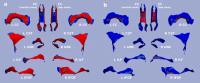
Brain Reorganization in Young Children with Epilepsy Surgery:
Longitudinal Tractography-Based Connectome Study - Permission Withheld
Jeong-Won Jeong1,2, Eishi Asano1,
Csaba Juhasz1,2, and Harry T. Chugani1,2
1Pediatrics and Neurology, Wayne State
University, Detroit, MI, United States, 2Translational
Imaging Laboratory, Children's Hospital of Michigan,
Detroit, MI, United States
Both ictal and interictal epileptic activities can lead to
progressive deterioration of affected brain structure and
function with an additional indirect impairment of
functional reorganization (or compensation) in no-epileptic
areas. This study applies whole brain connectome analysis
for children with intractable focal epilepsy in order to
investigate the potential effect of epilepsy surgery and
surgical outcome on the pattern of axonal plasticity in the
contralateral hemisphere. We found that post-operative
seizures are associated with increased connectivity, most
pronounced in the temporal pole region of the contralateral
hemisphere. Such increased connectivity may be an imaging
marker of recurrent epilepsy after focal cortical resection.
|
| |
17:48
|
0739.
 |
Disrupted Development and Integrity of Frontal White Matter in
Patients Treated for Pediatric Posterior Fossa Tumors
John O Glass1, Robert J Ogg1, Jung W
Hyun2, Julie H Harreld1, Yimei Li2,
Amar Gajjar3, and Wilburn E Reddick1
1Diagnostic Imaging, St. Jude Children's Research
Hospital, Memphis, TN, United States, 2Biostatistics,
St. Jude Children's Research Hospital, Memphis, TN, United
States, 3Oncology,
St. Jude Children's Research Hospital, Memphis, TN, United
States
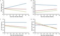
This study assessed the longitudinal white matter (WM)
microstructure of 129 patients and 72 normal healthy
age-similar controls. WM volume, fractional anisotropy (FA),
and radial (RAD) and axial (AX) diffusivity trajectories
were examined. After surgery but before any additional
therapy, frontal WM volume in patients was similar to
controls, while FA and AX were reduced in patients,
suggestive of acute, indirect microstructural/axonal injury
caused by disease and/or surgical excision. Over the next
three years, AX, RAD, and WM volume decreased in patients,
which would be consistent with possible resolution of axonal
swelling combined with chronic demyelination.
|
|