13:30
|
|
Introduction |
13:42
 |
0950.
 |
Bayesian Exponential Random Graph Modeling of Whole-Brain
Structural Networks across Lifespan 
Michel R.T. Sinke1, Willem M. Otte1,2,
Alberto Caimo3, Cornelis J. Stam4, and
Rick M. Dijkhuizen1
1Biomedical MR Imaging and Spectroscopy Group,
Center for Image Sciences, University Medical Center
Utrecht, Utrecht, Netherlands, 2Department
of Pediatric Neurology, Brain Center Rudolf Magnus,
University Medical Center Utrecht, Utrecht, Netherlands, 3Social
Network Analysis Research Centre, Interdisciplinary
Institute of Data Science, University of Lugano, Lugano,
Switzerland, 4Department
of Clinical Neurophysiology, Neuroscience Campus Amsterdam,
VU University Medical Center, Amsterdam, Netherlands
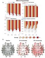
Comparison of brain networks that differ in size or edge
density may be inadequate with frequently applied
descriptive graph analysis methods. To resolve this, we
propose an alternative framework based on Bayesian
generative modeling, allowing unbiased assessment of local
substructures that shape the global network topology.
Structural networks were derived from DTI-based whole-brain
tractography of 382 healthy subjects (age: 20-86 years), and
successfully simulated. Despite clear effects of age and hub
damage on network topologies, relative contributions of
local substructures did not change significantly. The use of
generative models may shed new light on the complex (re)organization
of the brain.
|
13:54
|
0951.
 |
Resting state fMRI of spinal cord is keeping synchronistic with
brain - Permission Withheld
Jinsong Zhang1, Lingzhi Wang2, and Jun
Li2
1Radiology department,Xijing Hospital, MRI room,
Xi'an, China, People's Republic of, 2School
of Life Science and Technology, Xidian University, Xi'an,
China, People's Republic of
The spinal cord and brain form central nervous system and
sensory and motor signals are relayed by spinal cord and
processed by brain. Studies have suggested that resting
state functional connectivity (rsFC) are fundamental, common
feature of the entire nervous system. However, it still
remain unknown the correlation between rsFCs within spinal
cord and brain. The present study discovered dorsal and
ventral resting state networks (RSNs) within spinal cord and
sensory-motor RSN within brain. Further, correlation
analysis suggest that dorsal and ventral RSNs connected to
sensory and motor RSNs respectively.
|
14:06
 |
0952.
 |
Functional connectivity self-regulation of cerebellum and
primary motor area with fMRI-Brain Computer Interfaces. Pilot
results.
Patricia Andrea Vargas1,2, Ranganatha Sitaram1,3,4,5,
Pradyumna Sepúlveda2,6, Cristian Montalba2,
Mohit Rana1, Cristián Tejos2,6, and
Sergio Ruiz1,3
1Department of Psychiatry, Faculty of Medicine,
Interdisciplinary Center for Neuroscience, Pontificia
Universidad Católica de Chile, Santiago, Chile, 2Biomedical
Imaging Center, Pontificia Universidad Católica de Chile,
Santiago, Chile, 3Institute
of Medical Psychology and Behavioral Neurobiology,
University of Tübingen, Tübingen, Germany, 4Sree
Chitra Tirunal Institute of Medical Sciences and Technology,
Trivandrum, India, 5Institute
for Biological and Medical Engineering, Pontificia
Universidad Católica de Chile, Santiago, Chile, 6Department
of Electrical Engineering, Pontificia Universidad Católica
de Chile, Santiago, Chile
In recent years there is a growing interest in the potential
application of Brain-Computer interfaces (BCI) for
psychiatric and neurological disorders. After stroke, if the
primary motor cortex (M1) is affected, it is common to find
a “deactivation” of the contralateral cerebellum. The aim
of this study was to evaluate the feasibility of achieving
volitional control of M1-cerebellum functional connectivity,
in healthy subjects with an fMRI-BCI system.
The results indicate that volitional self-regulation of
cerebellum-M1 connectivity is feasible with fMRI-BCI. The
data also suggests that cerebellum is more easily recruited
than M1.
|
14:18
|
0953.
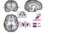 |
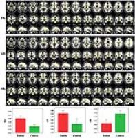
Connectivity-based parcellation of nucleus accumbens into
putative core and shell guiding for stereotactic target
localization and alterations in each NAc subdivision in mTLE
patients
Xixi Zhao1, Junling Wang1, Xiangliang
Tan1, Xiang Xiao1, Zeyu Zheng1,
Yingjie Mei2, Queenie Chan3, Yikai Xu1,
Ru Yang4, and Qianjin Feng4
1Department of Medical Imaging Center, Nanfang
Hospital, Southern Medical University, Guangzhou, China,
People's Republic of, 2Philips
Healthcare, Guangzhou, China, People's Republic of, 3Philips
Healthcare, HongKong, China, People's Republic of, 42School
of Biomedical Engineering and Guangdong Provincial Key
Laboratory of Medical Image Processing, Southern Medical
University, Guangzhou, China, People's Republic of
NAc was supposed be involved in epileptogenesis, especially
shell portion. The exact parcellation within the NAc and
structural alterations in vivo of NAc subdivisions in EP
patients remains unclear. We used diffusion probabilistic
tractography to subdivide NAc into putative core shell
subdivisions in individual mTLE patients for guiding NAc
shell stereotactic target localization. Our results revealed
that both left and right mTLE patients exhibited decreased
FA and increased MD in shell portion of bilateral NAc, which
may reflect neuronal degeneration and damage caused by
seizure mainly in shell portions, and suggest a possible
role of the NAc shell in epileptogenesis
|
14:30
|
0954.
 |
Brain White Matter Plasticity and Functional Reorganization
Underlying the Central Pathogenesis of Idiopathic Trigeminal
Neuralgia
Linying Guo1, Tian Tian1, and Wenzhen
Zhu1
1Department of Radiology, Tongji Hospital, Tongji
Medical College, Huazhong University of Science and
Technology, Hubei, China, People's Republic of
Previous studies on trigeminal neuralgia (TN) have mainly
focused on peripheral nerve damage, but little is known
about the structural and functional changes in central
nervous system (CNS) that can occur following trigeminal
nerve dysfunction. In this study, we used diffusion kurtosis
imaging (DKI) and functional connectivity density (FCD)
mapping in TN patients to investigate both structural and
functional changes in CNS. We found TN patients have
correlated white matter and FCD reorganization that may
contribute to pathologic algogenic system. Our findings may
be helpful guidance for systematic therapeutics in both
peripheral and central nerves.
|
14:42
|
0955.
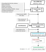 |
Connectivity Domain Analysis of the Default Mode Network in Mild
Traumatic Brain Injury at The Acute Stage
Armin Iraji1, Natalie Wiseman2, Robert
Welch3, Brian O'Neil3, E. Mark Haacke1,4,
and Zhifeng Kou1,4
1Department of Biomedical Engineering, Wayne
State University, Detroit, MI, United States, 2Department
of Psychiatry and Behavioral Neurosciences, Wayne State
University, Detroit, MI, United States,3Department
of Emergency Medicine, Wayne State University, Detroit, MI,
United States, 4Department
of Radiology, Wayne State University, Detroit, MI, United
States
Most functional and structural MRI studies in mild traumatic
brain injury (mTBI) are performed at the group level.
Recently, there is concern regarding the validity of
group-level analyses findings in mTBI due to the
heterogeneity of TBI. However, while group-level analysis
cannot demonstrate a complete view of impairments, we
hypothesize that there are similar patterns in group-level
and subject-level findings, especially in higher order brain
activities and networks. We evaluated this in the DMN using
a new framework known as the connectivity domain. This is
the first study of utilizing the connectivity domain to
investigate changes after a brain disorder.
|
14:54
|
0956.
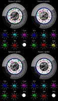 |
Brain connectivity of glioblastoma patients using MR-PET and DTI
data
Ana Carina Mendes1, Ana-Maria Oros-Peusquens2,
André Santos Ribeiro1,3, Karl-Josef Langen2,
Carolin Weiß Lucas4, Nadim Jon Shah2,
and Hugo Alexandre Ferreira1
1Institute of Biophysics and Biomedical
Engineering, Faculty of Sciences of the University of
Lisbon, Lisbon, Portugal, 2Forschungszentrum
Juelich GmbH, Institute of Neurosciences and Medicine-INM4,
Juelich, Germany, 3Centre
for Neuropsychopharmacology, Division of Brain Sciences,
Department of Medicine, Imperial College London, London,
United Kingdom, 4Center
of Neurosurgery, University of Cologne, Cologne, Germany
Methods capable of mapping brain connectivity pathways may
prove useful by providing valuable information in order to
prevent sequelae following a surgical intervention. This
study presents an approach for the whole-brain connectivity
evaluation of nine patients with lateralized gliobastoma,
using the Multimodal Imaging Brain Connectivity Analysis
(MIBCA) toolbox to process MR and PET data. Results show
changes in connectivity metrics across both hemispheres for
all patients accompanied by an increased number of fibres
which may result from reorganization of connectivity
pathways caused by the disruption of the original ones by
the tumour.
|
15:06
|
0957.
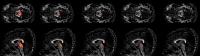 |
Thresholding to Improve the Specificity of High Spatial and
Angular Resolution In Vivo Diffusion-Weighted Tractography to
Estimate Brain Stem Connectivity.
Matthew Hey1, Luis Colon-Perez2,
William Triplett3, David Fitzgerald4,
and Thomas Mareci5
1University of Florida, Gainesville, FL, United
States, 2Department
of Psychiatry, University of Florida, Gainesville, FL,
United States, 3Department
of Physical Therapy, University of Florida, Gainesville, FL,
United States, 4Department
of Neurology, University of Florida College of Medicine,
Gainesville, FL, United States, 5Department
of Biochemistry and Molecular Biology, University of Florida
College of Medicine, Gainesville, FL, United States
The spatial resolution of diffusion-weighted (DWI) images
limits the white matter streamline fiber tracks, which can
be followed in the brain stem. To address this issue, we
introduce a high spatial resolution protocol and the use of
a threshold to limit the false positive in streamline track
density maps by requiring that a minimum amount of fibers
pass through a voxel. This provides increased accuracy in
the visualization of streamlines connecting specific regions
of the brain stem and may allow the recognition of
structural abnormalities due to neurological diseases.
|
15:18
|
0958.
 |
Influence of repetitive transcranial magnetic stimulation on
functional connectivity and hemodynamics in the rat brain
Julia Boonzaier1, Geralda A. F. van Tilborg1,
Mark J.R.J. Bouts2,3,4, Petar P.I. Petrov5,
Caroline L. van Heijningen1, Gerard van Vliet1,
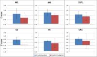
Annette van der Toorn1, Sebastiaan F.W. Neggers5,
and Rick M. Dijkhuizen1
1Center for Image Sciences, University Medical
Center Utrecht, Utrecht, Netherlands, 2Institute
of Psychology, Leiden University, Leiden, Netherlands, 3Department
of Radiology, Leiden University Medical Center, Leiden,
Netherlands, 4Leiden
Institute for Brain and Cognition (LIBC), Leiden University,
Leiden, Netherlands, 5Department
of Psychiatry, Brain Center Rudolf Magnus, University
Medical Center Utrecht, Utrecht, Netherlands
Repetitive transcranial magnetic stimulation (rTMS) is a
non-invasive neuromodulation technique
with the ability to change cortical excitability, however
its precise mechanism of action is not completely
understood. Therefore, by acquiring resting-state fMRI and
perfusion MRI data
we assessed
the influence of unilateral low-frequency (inhibitory) rTMS on
functional connectivity and hemodynamics instimulated cortical
tissue in rats. After four consecutive days of rTMS we
measured reduced interhemispheric functional connectivity
between homotopic sensorimotor regions,
while cerebral blood flow remained largely unaffected. This
reduction in interhemispheric functional connectivity may be
due to the inhibitory effect of low-frequency rTMS on
cortical excitability.
|
|