| |
10:00
|
0327.
 |
UK Biobank: Brain imaging protocols and first data release 
Karla L Miller1, Neal K Bangerter2,
Fidel Alfaro Almagro1, David L Thomas3,
Essa Yacoub4, Junqian Xu5, Andreas J
Bartsch1,6, Saad Jbabdi1, Stamatios N
Sotiropoulos1, Mark Jenkinson1, Jesper
Andersson1, Ludovica Griffanti1, Peter
Weale7, Iulius Dragonu7, Steve Garratt8,
Sarah Hudson8, Rory Collins8,9, Paul M
Matthews10, and Stephen M Smith1
1FMRIB Centre, University of Oxford, Oxford,
United Kingdom, 2Electrical
and Computer Engineering, Brigham Young University, Provo,
UT, United States, 3Department
of Brain Repair and Rehabilitation, UCL Institute of
Neurology, University College London, London, United
Kingdom, 4Center
for Magnetic Resonance Research, University of Minnesota,
Minneapolis, MN, United States, 5Icahn
School of Medicine at Mount Sinai, New York, NY, United
States, 6Department
of Neuroradiology, University of Heidelberg, Heidelberg,
Germany, 7Siemens
Healthcare (UK), London, United Kingdom, 8UK
Biobank Ltd, Stockport, United Kingdom, 9Nuffield
Department of Population Health, University of Oxford,
Oxford, United Kingdom, 10Department
of Medicine, Imperial College London, London, United Kingdom
UK Biobank is a prospective epidemiological study of 500,000
participants consisting of extensive questionnaires,
physical measures and biological samples, linking to
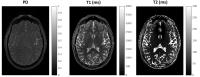
long-term health outcomes. The imaging extension for the UK
Biobank ultimately aims to image 100,000 subjects from this
cohort, including brain, cardiac and body MRI, bone scans
and carotid ultrasound. We overview the brain imaging
component, which includes structural, functional and
diffusion MRI. The value of this open resource arises not
only from multi-modal/multi-organ imaging, but also from the
depth of other demographic, phenotypic and exposure data,
and will increase over time as clinical outcomes are
realized in the population.
|
| |
10:12
 |
0328.
 |
On the Relationship between Cellular and Hemodynamic Properties
of the Human Brain Cortex over Adult Lifespan
Yue Zhao1, Jie Wen2, Anne Cross3,
and Dmitriy Yablonskiy2
1Chemistry, Washington University in St. Louis,
St. Louis, MO, United States, 2Radiology,
Washington University in St. Louis, St. Louis, MO, United
States, 3Neurology,
Washington University in St. Louis, St. Louis, MO, United
States
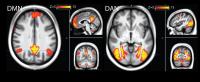
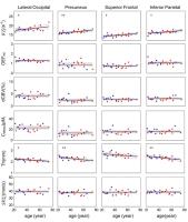
Establishing baseline MRI biomarkers for normal brain aging
is significant and valuable. In this study, we use
previously developed approach to measure tissue-specific
transverse relaxation rate constant (R2*t) and
BOLD contributions to GRE signal, thus providing information
on tissue cellular and hemodynamic properties. The VSF
approach is applied for background gradient correction
together with navigator echo to minimize artifacts from
physiological fluctuations. Our results show age-related R2*t increases
in most cortical regions and age-independent behavior of
most hemodynamic parameters. We hypothesize that R2*t could
serve as a biomarker of the cortical “cellular packing
density”, which mostly reflects the neuronal density.
|
| |
10:24
|
0329.
 |
Venous metrics in a large cohort of healthy elderly individuals
from susceptibility-weighted images and quantitative
susceptibility maps
Phillip G. D. Ward1,2, Parnesh Raniga1,
Nicholas J. Ferris1,3, David G. Barnes2,4,
David L. Dowe2, Elsdon Storey5, Robyn
L. Woods6, and Gary F. Egan1,7
1Monash Biomedical Imaging, Monash University,
Clayton, Australia, 2Faculty
of Information Technology, Monash University, Clayton,
Australia, 3Monash
Imaging, Monash Health, Clayton, Australia, 4Monash
eResearch Centre, Monash University, Clayton, Australia, 5Department
of Neurology, Monash University, Clayton, Australia, 6Department
of Epidemiology & Preventative Medicine, Monash University,
Melbourne, Australia, 7ARC
Centre of Excellence for Integrative Brain Function,
Melbourne, Australia
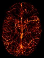
In this study we examine venous characteristics of elderly
individuals in a large healthy population. Venograms were
generated from susceptibility-weighted images and
quantitative susceptibility maps using state-of-the-art
automated venography. Venous density and oxygen extraction
fraction were calculated in different brain regions. The
pattern of metabolic demand (oxygen extraction fraction) is
found to be consistent with rest and passive observation.
Additionally, our results suggest that venous density may be
a potential biomarker.
|
| |
10:36
|
0330.
 |
In Vivo Characterization of Brain Ultrashort-T2 Components
Tanguy Boucneau1,2, Shuyu Tang1,3,
Misung Han1, Roland G Henry1,4, Duan
Xu1,3, and Peder Eric Zufall Larson1,3
1Radiology and Biomedical Imaging, University of
California - San Francisco, San Francisco, CA, United
States, 2Physics,
Ecole Normale Supérieure de Cachan, Cachan, France, 3UC
Berkeley-UCSF Graduate Program in Bioengineering, University
of California, Berkeley and University of California, San
Francisco, San Francisco, CA, United States, 4Neurology,
University of California - San Francisco, San Francisco, CA,
United States
It has recently been shown that myelin contains ultrashort
T2 components with sub-millisecond relaxation times that are
not observed with conventional pulse sequences and maybe
associated with bound protons in the myelin phospholipid
membranes.We performed ultrashort T2* relaxometry in vivo to
characterize these components with a 3D ultrashort echo time
(UTE) pulse sequence at 7T.We observed an ultrashort T2
component (T2* $$$\approx 100 \mu s$$$) as well as a short
T2 component (T2* $$$\approx 1.5 ms$$$) that had a distinct
frequency shift corresponding to the methylene proton
chemical shift, which to our knowledge has never been
observed in vivo.These components were validated in an ex
vivo post-mortem brain specimen, and may provide valuable
new biomarkers of myelin density, structure, and integrity.
|
| |
10:48
|
0331.
 |
Multi-parameter mapping, fat/water separation and functional
imaging with a two-sequence brain morphometry protocol
Andre Jan Willem van der Kouwe1, Fikret Isik
Karahanoglu1, Matthew Dylan Tisdall1,
Paul Wighton1, Himanshu Bhat2, Thomas
Benner3, and Jonathan R Polimeni1
1Athinoula A. Martinos Center, Department of
Radiology, Massachusetts General Hospital, Charlestown, MA,
United States, 2Siemens
Healthcare, Charlestown, MA, United States, 3Siemens
Healthcare, Erlangen, Germany
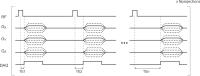
We present an efficient two-sequence protocol for
quantifying multiple parameters in a 1 mm isotropic brain
morphometry examination. The protocol comprises a multiple
gradient echo (TE), multiple inversion (TI) time MPRAGE
(MEMPxRAGE) and a two-flip-angle balanced SSFP (TrueFISP)
sequence. Proton density and T1 maps
are estimated from the MEMPxRAGE data using the multi-TI
data and a Bloch simulation. With the T1 map
and TrueFISP data, the T2 map
is estimated using DESPOT2. Fat, water and B0 maps are
obtained from the multi-TE data using the IDEAL algorithm.
The MEMPxRAGE scan includes embedded 3D EPI-based
navigators encoding low resolution functional information.
|
| |
11:00
|
0332.
 |
Reproducibility of fast three-dimensional macromolecular proton
fraction mapping of the human brain: global tissue
characterization and volume measurements
Vasily L. Yarnykh1,2
1Radiology, University of Washington, Seattle,
WA, United States, 2Research
Institute of Biology and Biophysics, Tomsk State University,
Tomsk, Russian Federation
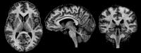
A new method for fast high-resolution whole-brain
three-dimensional (3D) mapping of the macromolecular proton
fraction (MPF) based on three source images has been
recently proposed. In this study, reproducibility of
repeated MPF measurements in white and gray matter with
simultaneous estimation of tissue volumes using automated
segmentation of 3D MPF maps obtained with isotropic
resolution of 1.25 mm was assessed. MPF measurements in
brain tissues are highly reproducible with coefficients of
variation <1.5%. 3D MPF mapping provides “all-in-one”
solution for simultaneous characterization of myelination
and volumetric changes in brain tissues.
|
| |
11:12
|
0333.
|
Automated Measurements of Brain Morphometry Derived from
T1-weighted Magnetic Resonance Imaging Fluctuate from Morning to
Afternoon - Permission Withheld
Aaron Trefler1, Neda Sadeghi2, Adam
Thomas1, Carlo Pierpaoli2, Chris Baker1,
and Cibu Thomas3
1National Institute of Mental Health, Bethesda,
MD, United States, 2National
Institute of Child Health and Human Development, Bethesda,
MD, United States, 3Center
for Neuroscience and Regenerative Medicine, Bethesda, MD,
United States
Automated measures of brain morphometry derived from
T1-weighted (T1W) images are typically used as proxy
measures to investigate the relation between brain structure
and behavior. However, the computation of T1W morphometric
measures can be influenced by subject-related factors such
as head motion1 and
level of hydration2. Here, we provide a
comprehensive assessment of the impact of time-of-day (TOD)
on widely used measures of brain morphometry in healthy
young adults. Our results show that the apparent volume of
all major tissue compartments as well as measures of brain
morphometry such as cortical thickness and gray matter
density are significantly influenced by TOD.
|
| |
11:24
 |
0334.
 |
Optimized Inversion-Time Schedules For High-Resolution
Multi-Inversion EPI Quantitative Measurements of T1 - Permission Withheld
Ouri Cohen1,2, Ville Renvall3, and
Jonathan Polimeni1,2
1Athinoula A. Martinos Center, Charlestown, MA,
United States, 2Radiology,
Massachusetts General Hospital, Boston, MA, United States, 3Department
of Neuroscience and Biomedical Engineering, Aalto University
School of Science, Espoo, Finland
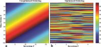
A novel optimized method for high-resolution quantitative
EPI measurements of T1 is
introduced and validated on a 3T clinical scanner in a
phantom and a healthy volunteer. The method offers a 5-fold
acceleration in scan time over previous techniques allowing
fully quantitative 1.2 mm3 isotropic T1 maps in
less than 30 seconds.
|
| |
11:36
|
0335.
 |
Cerebral gray matter volume changes caused by exposure to
hypobaric environment: a preliminary study
Dandan Zheng1, Wenjia Liu2, Li Zheng3,
and Lin Ma2
1MR Research China, GE Healthcare, Beijing,
China, People's Republic of, 2Radiology
Department, Beijing Military General Hospital, Beijing,
China, People's Republic of, 3Department
of Biomedical Engineering, College of Engineering, Peking
University, Beijing, China, People's Republic of
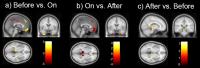
Acute mountain sickness is a series of pathologic reactions
during rapid exposing to low pressure hypoxic high altitude
environment, which is a widespread illness among
un-acclimatized individuals in plateau. Human always stay in
plain will display some common physiological and
pathological changes of brain, such as change of cerebral
blood flow, cerebral pressure and brain volume. The aim of
the present study was to investigate whether there was
different change of gray matter volume in some brain regions
related to AMS development before, during and after exposing
to the real high altitude environment.
|
| |
11:48
|
0336.
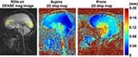 |
Regional Brain Motion Varies with Subject Positioning: A Study
Using Displacement Encoding with Stimulated Echoes (DENSE)
Xiaodong Zhong1, Zihan Ye2, Tucker
Lancaster3, Deqiang Qiu3, Brian M.
Dale4, Amit Saindane3, and John N.
Oshinski2,3
1MR R&D Collaborations, Siemens Healthcare,
Atlanta, GA, United States, 2Biomedical
Engineering, Georgia Institute of Technology, Atlanta, GA,
United States, 3Department
of Radiology and Imaging Sciences, Emory University,
Atlanta, GA, United States, 4MR
R&D Collaborations, Siemens Healthcare, Cary, NC, United
States
Displacement encoding with stimulated echoes (DENSE) with
high motion sensitivity was used to investigate the
influence of subject position (prone versus supine) on
regional brain motion. Preliminary results in 9 volunteers
demonstrated that there is a significant difference in
displacement with a change in position. Displacements were
significantly increased in the frontal lobe going from the
prone to the supine position and significantly increased in
the occipital lobe going from the supine to the prone
position.
|
|