| |
10:30
|
0876.
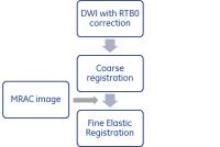 |
Improvement in Alignment & Signal Uniformity via Realtime B0
Correction and Image Registration in Multi-station PET/MR Whole
body Diffusion Imaging - Video Not
Available
Maggie Mei Kei Fung1, Abhishek Sharma2,
Justin Lahrman3, Lloyd Estkowski4, and
Ersin Bayram5
1MR Apps & Workflow, GE Healthcare, New York, NY,
United States, 2MR
Engineering, GE Healthcare, Bangalore, India, 3MR
Apps & Workflow, GE Healthcare, Waukesha, WI, United States, 4MR
Apps & Workflow, GE Healthcare, Menlo Park, CA, United
States, 5MR
Apps & Workflow, GE Healthcare, Houston, TX, United States
In a PET/MR imaging, anatomical alignment between PET & MR
images and good visualization of spine & lymph node are
critical in the clinical interpretation of diseases. In
whole body multi-station diffusion weighted imaging (DWI),
it is common to observe signal drop off and spatial
misalignment due to B0 inhomogeneity. In this study, we
proposed a two-prong approach in improving the signal
uniformity & spatial alignment by combining a real-time
slice-by-slice B0 correction technique and an image
registration technique. We have validated the approach in 18
volunteers with various physical attributes.
|
| |
10:42
|
0877.
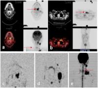 |
Diagnostic ability of Whole-Body Diffusion-Weighted Imaging in
malignant tumors compared with PET-CT 
Xiaoyi Wang1, Ning Wu1, Yanfeng Zhao1,
Han Ouyang1, Lizhi Xie2, Jin Zhang1,
Li Liu1, Wenjie Zhang1, Rong Zheng1,
Ying Liang1, and Ying Liu1
1Department of Diagnostic Imaging, PET-CT Center,
Cancer Hospital, Chinese Academy of Medical Sciences, Peking
Union Medical College, Beijing,China, Beijing, China,
People's Republic of, 2GE
Healthcare China, Beijing, China, Beijing, China, People's
Republic of
Because of its convenience in whole body examination, whole
body MRI is growing popular, especially in the tumor
diagnosis. In the present work, the diagnostic ability of
whole-body diffusion-weighted imaging in malignant lesions
is compared with that obtained with 18F-FDG
PET-CT. We found that WBDWI was an effective method for
screening bone metastasis, especially suitable for
radiation-vonuerable population, and it is better than
PET-CT in detecting low grade malignant tumor. In summary,
WBDWI can be used as a potential alternative to PET/CT in
addition to conventional MR examination.
|
| |
10:54
|
0878.
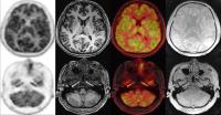 |
PET/MR attenuation correction using Zero Echo Time imaging in
15O-water study - Permission Withheld
Mohammad Mehdi Khalighi1, Gaspar Delso2,
Praveen K. Gulaka3, Audrey Peiwen Fan3,
Bin Shen4, Aileen Hoehne4, Prachi
Singh3, Jun-Hyung Park4, Dawn Holley3,
Frederick T. Chin3,4, and Greg Zaharchuk3,4
1Applied Science Lab, GE Healthcare, Menlo Park,
CA, United States, 2Applied
Science Lab, GE Healthcare, Zurich, Switzerland, 3Radiology
Department, Stanford University, Stanford, CA, United
States, 4Molecular
Imaging Program, Stanford University, Stanford, CA, United
States
Accurate identification of bone tissue is important to
generate attenuation correction maps on a PET/MR scanner for
quantification of tracer activity in PET images. Head
atlas-based attenuation correction and a new zero echo time
technique (ZTE) for attenuation correction are compared in
an 15O-water
brain study. The comparison shows that ZTE-based attenuation
correction provides more accurate identification of bone
tissue and thus of the tracer activity. Any mismatch in bone
identification will affect the tracer activity, especially
in voxels close to the bone.
|
| |
11:06
|
0879.
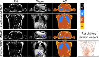 |
Respiratory Resolved Attenuation Correction Maps for Motion
Compensated PET-MR using Dixon-GRPE
Christoph Kolbitsch1,2, Radhouene Neji3,
Matthias Fenchel4, and Tobias Schaeffter1,2
1Division of Imaging Sciences and Biomedical
Engineering, King's College London, London, United Kingdom, 2Physikalisch-Technische
Bundesanstalt (PTB), Braunschweig and Berlin, Germany, 3MR
Research Collaborations, Siemens Healthcare, Frimley, United
Kingdom, 4MR
Oncology Application Development, Siemens Healthcare,
Erlangen, Germany
Quantitative PET requires accurate attenuation correction
(AC) information. For simultaneous PET-MR acquisitions in
the thorax or abdomen these MRAC images are obtained in a
single breathhold which can lead to misregistration errors
between breathhold MRAC and free-breathing PET data. Here we
present a method which obtains accurate AC information (Dice
coefficient higher than 0.85) during free-breathing and
yields additional respiratory motion fields which can be
utilised in motion-compensated MR and PET reconstructions.
The proposed Dixon-GRPE method led to improvements of up to
50% in sharpness (FWHM) and a 33% improvement in the
quantification of the specific uptake value (SUV).
|
| |
11:18
|
0880.
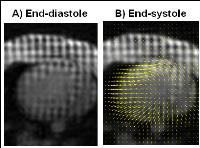 |
Impact of MR-based PET motion correction on the quantification
of myocardial blood flow: an in-vivo simultaneous MR/PET study
Yoann Petibon1, Behzad Ebrahimi1,
Timothy G Reese1,2, Nicolas Guehl1,
Marc D Normandin1, Nathaniel M Alpert1,
Georges El Fakhri1, and Jinsong Ouyang1
1Center for Advanced Medical Imaging Sciences,
Radiology, Massachusetts General Hospital and Harvard
Medical School, Boston, MA, United States, 2Athinoula
A. Martinos Center, Radiology, Massachusetts General
Hospital and Harvard Medical School, Boston, MA, United
States
Dynamic PET imaging enables absolute quantification of
myocardial blood flow (MBF). However, motion of the heart
during imaging deteriorates the accuracy of PET MBF
measurements. Simultaneous MR/PET makes it possible to
compensate PET images for motion by incorporating MR-based
motion information inside the PET reconstruction process. In
this study, we propose and assess the impact of a tagged-MRI
based PET motion-correction technique for improved PET MBF
quantification using an in-vivo simultaneous
MR/PET study.
|
| |
11:30
|
0881.
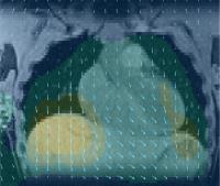 |
Efficient 5D imaging of thorax and abdomen for MR-guided PET
motion correction
Christian Würslin1, Dominik Fleischmann2,
and Roland Bammer1
1Radiological Sciences Laboratory, Stanford
University, Stanford, CA, United States, 2Cardiovascular
Imaging Section, Department of Radiology, Stanford
University, Stanford, CA, United States
Cardiac imaging under free breathing is a desirable tool for
clinical routine, which can provide improved patient comfort
and shorter examination times. Furthermore, it can be used
in the context of MR-guided PET motion correction in
simultaneous PET-MRI. Here, we propose a radial acquisition
and reconstruction framework for the acquisition of these
images. A piecewise rigid respiration motion model enables a
highly efficient use of the acquired data to either achieve
higher image quality or shorter examination times than
standard, dual-gated techniques.
|
| |
11:42
|
0882.
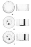 |
MR-PET simultaneous acquisitions with attenuation correction
using LSO background radiation.
Liliana Lourenco Caldeira1, Theodoros Kaltsas1,
Jürgen Scheins1, Elena Rota Kops1,
Lutz Tellmann1, Uwe Pietrzyk1,
Christoph Lerche1, and N. Jon Shah1,2
1Institute of Neuroscience and Medicine,
Forschungszentrum Jülich, Jülich, Germany, 2Department
of Neurology, Faculty of Medicine, JARA, RWTH Aachen
University, Aachen, Germany
In this work, the goal is to perform attenuation correction
(AC) for MR-PET scanners using the background activity from
LSO (Cerium-doped Lutetium Oxyorthosilicate) scintillator
used in PET scanners. This approach has the advantage of
obtaining a geometrically aligned AC map with the PET
emission scans, which can be useful for coil AC maps. We
demonstrate our approach for the Siemens 3T MR-BrainPET with
a Tx/Rx 8-channel head coil and a 3-rod phantom.
|
| |
11:54
 |
0883.
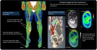 |
[18F]FDG PET/MRI Of Patients With Chronic Pain Alters
Management: Early Experience.
Daehyun Yoon1, Deepak Behera1, Dawn
Holley1, Pamela Gallant1, Ma Agnes
Martinez Ith2, Ian Carroll3, Matthew
Smuck2, Brian Hargreaves1, and Sandip
Biswal1
1Radiology, Stanford University, Palo Alto, CA,
United States, 2Orthopaedic
Surgery, Stanford University, Palo Alto, CA, United States, 3Anesthesia,
Stanford University, Palo Alto, CA, United States
The chronic pain sufferer is currently faced with a lack of
objective tools to identify the source of their pain.
Increased inflammation of the nervous system, vessels,
muscles, and other tissues in chronic pain sufferers and
[18F]fluorodeoxyglucose positron emission
tomography/magnetic resonance imaging ([18F]FDG PET/MRI) has
emerged as a sensitive clinical tool to identify increased
inflammation. We plan to develop clinical [18F]FDG PET/MRI
method to more accurately localize sites of hypermetabolic
foci as it relates to pain generators. Early clinical
results suggest that [18F]FDG PET/MRI can identify
abnormalities in chronic pain patients and can immediately
affect their management.
|
| |
12:06
|
0884.
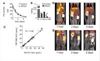 |
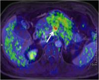
Distribution and metabolism of 89Zr-labeled HDL nanoparticles in
atherosclerotic rabbits: in vivo, longitudinal imaging with
PET/MRI
Claudia Calcagno1,2, Carlos Perez-Medina1,2,
Tina Binderup3, Mark E Lobatto4, Seigo
Ishino1,2, Mootaz Eldib1,2, Philip
Robson1,2, Sarayu Ramachandran1,2,
Thomas Reiner5, Edward Fisher6, Zahi A
Fayad1,2, and Willem JM Mulder1,2
1Department of Radiology, Icahn School of
Medicine at Mount Sinai, New York, NY, United States, 2Translational
and Molecular Imaging Institute, Icahn School of Medicine at
Mount Sinai, New York, NY, United States, 3University
of Copenaghen, Copenaghen, Denmark, 4Academisch
Medisch Centrum, Amsterdam, Netherlands, 5Memorial
Sloan Kettering Cancer Center, New York, NY, United States, 6New
York University School of Medicine, New York, NY, United
States
Abundant, active inflammatory cells are a hallmark of
high-risk atherosclerotic plaques. High-density lipoprotein
(HDL) is a natural nanoparticle composed of phospholipids,
cholesterol and apolipoprotein A-I (APOA1), which has been
shown to have atheroprotective properties. . The recent
development of combined PET/MRI scanners and new advances in
radio-labeling technology gives the opportunity to
investigate theese properties in vivo. Using a unique set-up
combining PET/CT and PET/MRI, we non-invasively assess the
pharmacokinetics, distribution, metabolism and turnover of 89Zr-HDL’s
in a rabbit model of atherosclerosis.
|
| |
12:18
|
0885.
 |
PET/MRI in Pancreatic and Periampullary Cancer: Correlating
Diffusion-weighted Imaging, MR spectroscopy, and Glucose
Metabolic Activity With Clinical Stage
Bang-Bin Chen1, Yu-Wen Tien2, Ming-Chu
Chang3, Mei-Fang Cheng4, Yu-Ting Chang3,
Chih-Horng Wu1, Xin-Jia Chen1,
Ting-Chun Kuo2, Shih-Hung Yang5, I-Lun
Shih1, Hong-Shiee Lai2, and Tiffany
Ting-Fang Shih1
1Medical Imaging and Radiology, National Taiwan
University Medical School and Hospital, Taipei, Taiwan, 2Surgery,
National Taiwan University Medical School and Hospital,
Taipei, Taiwan, 3Internal
Medicine, National Taiwan University Medical School and
Hospital, Taipei, Taiwan, 4Nuclear
Medicine, National Taiwan University Medical School and
Hospital, Taipei, Taiwan, 5Oncology,
National Taiwan University Medical School and Hospital,
Taipei, Taiwan
We demonstrated that PET/MRI provides numerous useful
imaging biomarkers for clinical staging and pathological
grading in patients with pancreatic cancer or periampullary
cancer. ADCmin was lower in tumors with N1 and an advanced
TNM stage. Choline levels were higher in T4 and poorly
differentiated tumors. Tumors with high glucose metabolic
activity, as reflected by MTV and TLG, were at a more
advanced T stage, exhibited lymph node and distant
metastasis, and were at an advanced TNM stage. Moreover,
compared with MTV or ADCmin alone, the MTV/ADCmin ratio
demonstrated the highest predictive ability for determining
the clinical TNM stage. Thus, integrated PET/MRI could
provide complementary information on tumor characteristics,
and these combined data could have stronger clinical or
pathological implications than MRI or PET alone.
|
|