| |
14:15
|
0107.
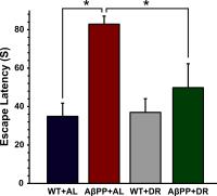 |
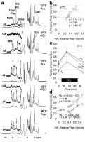
Dietary Restriction Improved Memory and Neuronal Metabolism in
AßPP-PS1 Mouse Model of Alzheimer’s Disease: A 1H-[13C]-NMR
Study 
Anant Bahadur Patel1 and
Kamal Saba1
1NMR Microimaging and Spectroscopy, CSIR-Centre
for Cellular and Molecular Biology, Hyderabad, India
Alzheimer's disease (AD) is the most common
neurodegenerative disorders. Currently no effective
treatment available for AD. Dietary restriction (DR) has
been shown to improve longevity in rodents. In the present
study, we evaluated the effects of DR on memory and brain
energy metabolism in AβPP-PS1 mouse model of AD using 1H-[13C]-NMR
spectroscopy in conjunction with infusion of [1,6-13C2]glucose.
Our findings suggest that DR intervention had improved the
memory and the neuro-metabolic activity in the AD mice.
|
| |
14:27
 |
0108.
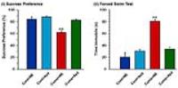 |
A 1H-[13C]-NMR
Study for Understanding Antidepressant Action of Lanicemine in
Chronic Unpredictable Mild Stress Model of Depression
Pravin K Mishra1 and
Anant Bahadur Patel1
1NMR Microimaging and Spectroscopy, CSIR-Centre
for Cellular and Molecular Biology, Hyderabad, India
Though, ketamine possess rapid antidepressant properties,
its use is limited due to addictive and psychotomimetic
properties. In the current study, we have evaluated the
antidepressant activity of lanicemine in CUMS model of
depression by 1H-[13C]-NMR
spectroscopy together with infusion of [1,6-13C2]glucose.
Exposure of lanicemine restored behavioral phenotype and
activity of excitatory and inhibitory neurons in depression.
|
| |
14:39
|
0109.
 |
Brain Glycogen Supercompensation: A Role in the Development of
Hypoglycemia Unawareness?
Gulin Oz1, Mauro DiNuzzo2, Anjali
Kumar3, Amir Moheet3, Kristine
Kubisiak4, Lynn E. Eberly4, and
Elizabeth R. Seaquist3
1Radiology, Center for Magnetic Resonance
Research, University of Minnesota, Minneapolis, MN, United
States, 2Museo
storico della fisica e Centro di studi e ricerche Enrico
Fermi, Rome, Italy, 3Medicine,
University of Minnesota, Minneapolis, MN, United States, 4Biostatistics,
School of Public Health, University of Minnesota,
Minneapolis, MN, United States
Supercompensated brain glycogen levels may contribute to the
development of hypoglycemia associated autonomic failure
(HAAF) following recurrent hypoglycemia (RH) by providing
energy for the brain during subsequent periods of
hypoglycemia. To assess the role of glycogen
supercompensation in the generation of HAAF, we estimated
the level of brain glycogen supercompensation following RH
using 13C
MRS and compared it to that following acute hypoglycemia
(AH). Glycogen levels were found to increase after both AH
and RH, but to a lesser extent after RH. These data suggest
that glycogen supercompensation may be an epiphenomenon of
HAAF.
|
| |
14:51
 |
0110.
 |
In vivo detection of hypothalamic glucose metabolism in HFD and
regular fed mice
Blanca Lizarbe1, Antonie Cherix1,
Lijing Xin2, Hongxia Lei2,3, and Rolf
Gruetter1,3,4
1Laboratory for Functional and Metabolic Imaging
(LIFMET), Ecole Polytechnique Fédérale de Lausanne,
Lausanne, Switzerland, 2Animal
imaging and technology core (AIT), Center for Biomedical
Imaging (CIBM), Ecole Polytechnique Fédérale de Lausanne,
Lausanne, Switzerland, Lausanne, Switzerland, 3Department
of Radiology, University of Geneva, Geneva, Switzerland, 4Department
of Radiology, University of Lausanne, Lausanne, Switzerland
Obesity is a pandemic syndrome that leads to reduced life
expectancy, increasing the risk of heart disease, type-2
diabetes and some type of cancers. Noteworthy, to
understand the mechanisms of obesity onset and development,
several animal models, such as administration high fat
diets, have been developed. We used 1H-[13C]
MRS methods in regular and in high fat diet fed mice to
investigate the effects of high caloric diets and obesity in
the hypothalamus, its effects in glucose metabolism and
metabolic fluxes in neurons and glia. We found differences
that suggest impaired glucose metabolism in the hypothalamus
of obese mice.
|
| |
15:03
|
0111.
 |
Amide proton signals as pH indicator for in vivo MRS and MRI of
the brain – Responses to hypercapnia and hypothermia
Takashi Watanabe1, Jens Frahm1, and
Thomas Michaelis1
1Biomedizinische NMR Forschungs GmbH,
Max-Planck-Institut für biophysikalische Chemie, Göttingen,
Germany
Using proton MRS/MRI of mouse brain at 9.4 T, this work
provides the first in
vivo evidence
of concurrent pH-dependent changes of amide signals and
related metabolic responses to hypercapnia and hypothermia.
During hypercapnia, amide MRS signals of glutamine and of
unspecific compounds increase by ≥50% at 37°C and 22°C. They
are strongly correlated with intracellular pH determined
from a shift in creatine phosphokinase equilibrium. In MRI,
saturation transfer to water protons alters signal
intensities in dependence on pH and temperature. Irradiation
of aliphatic compounds at -3.5 ppm frequency offset from
water predominantly saturates lipids and water associated
with myelin.
|
| |
15:15
|
0112.
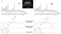 |
Assessing metabolic and structural alterations of brain cells in
the APP/PS1/tauP301L mouse model of Alzheimer’s disease using
MRS and diffusion-weighted MRS in vivo
Clemence Ligneul1,2, Marco Palombo1,2,
Juliette Le Douce1,2, Pierrick Jego1,2,
Martine Guillermier1,2, Gilles Bonvento1,2,
and Julien Valette1,2
1CEA/DSV/I2BM/MIRCen, Fontenay-aux-Roses, France, 2CNRS
Université Paris-Saclay UMR 9199, Fontenay-aux-Roses, France
In this work we use in vivo MRS and diffusion-weighted MRS
to detect alterations in cellular metabolism and structure
in a triple transgenic APP/PS1/tauP301L mouse model of
Alzheimer’s disease. We are able to detect massive
remodeling of metabolic content in the hippocampus, as well
as subtle but significant variations in diffusion properties
of astrocytic metabolites. These results are essentially
consistent with the metabolic and structural signature of
activated astrocyte, a cell status represented around
amyloid plaques.
|
| |
15:27
|
0113.
 |
Brain Sodium MRI depicts upper motor neuron involvement in
Amyotrophic Lateral Sclerosis patients
Aude-Marie Grapperon1, Adil Maarouf2,3,
Annie Verschueren1, Amandine Sevy1,
Elisabeth Soulier2, Sylviane Confort-Gouny2,
Patrick Viout2, Jean-Philippe Ranjeva2,
Maxime Guye2,3, Sharham Attarian1, and
Wafaa Zaaraoui2
1APHM, Hôpital Timone, Pôle Neurosciences,
Marseille, France, 2CRMBM
- CNRS - Aix-Marseille Université, Marseille, France, 3APHM,
Hôpital Timone, CEMEREM, Marseille, France
Amyotrophic lateral sclerosis (ALS) is a lethal
neurodegenerative disease that involves the death of upper
(in brain) and lower (in spine) motor neurons. As
conventional MRI failed to show brain motor neurons
impairment in ALS, advanced techniques are needed to improve
the diagnosis of the disease and monitor its progression. 23Na
brain MRI was performed to 4 ALS patients and showed
accumulation of sodium in the primary motor areas in the 3
patients presenting with clinical brain motor neuron signs.
Besides, more patients were clinically affected, more the
sodium accumulation was extended. In conclusion, sodium
accumulation, which is an indicator of neuronal injury,
could be a marker of ALS diagnosis and disease progression.
|
| |
15:39
|
0114.
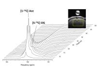 |
Modulations of cerebral TCA cycle activity studied by
hyperpolarized Acetate 13C MRS - Permission Withheld
Elise Vinckenbosch1, Mor Mishkovsky1,
Arnaud Comment2, and Rolf Gruetter1,3
1Laboratory of functional and metabolic imaging,
EPFL, Lausanne, Switzerland, 2Institute
of Physics of Biological Systems, EPFL, Lausanne,
Switzerland, 3Department
of Radiology, University of Lausanne and Geneva, Lausanne,
Switzerland
Hyperpolarized [1-13C] acetate enables for in
vivo detection
of 2-oxoglutarate, a tricarboxylic acid (TCA) cycle
intermediate, in intact brain at high field. The aim of this
study is to examine saturation substrate dose conditions and
to compare it with a partially inhibited TCA cycle model. We
conclude that 2-oxoglutarate production rate can be
calculated as a function of varying substrate concentrations
and is affected as well as the cerebral acetate kinetics by
TCA cycle activity modulations.
|
| |
15:51
|
0115.
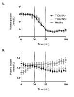 |
Brain lactate concentration falls in response to hypoglycemia in
type 1 diabetes patients with impaired awareness of hypoglycemia
Evita Wiegers1, Hanne Rooijackers2,
Cees Tack2, Arend Heerschap1, Bastiaan
de Galan2, and Marinette van der Graaf1,3
1Radiology and Nuclear Medicine, Radboud umc,
Nijmegen, Netherlands, 2Internal
Medicine, Radboud umc, Nijmegen, Netherlands, 3Pediatrics,
Radboud umc, Nijmegen, Netherlands
TThe effect of hypoglycemia on cerebral lactate
concentration was assessed in patients with type 1 diabetes
(T1DM) and impaired awareness of hypoglycemia (IAH),
patients with normal awareness of hypoglycemia (NAH) and in
healthy subjects. Brain lactate concentrations were
determined during stable euglycemic and stable hypoglycemic
conditions using a J-editing semi-LASER 1H-MRS
sequence at 3T. We found a 20% decrease in brain lactate
concentration in T1DM patients with IAH in response to
hypoglycemia, which may reflect increased lactate oxidation.
No changes in cerebral lactate concentrations were observed
in the other two groups.
|
| |
16:03
|
0116.
 |
Differential Metabolic Profiles in Rat Retrosplenial Cortex,
Cingulate Cortex and Medial Prefrontal Cortex: Relationship with
Cytoarchitecture and Functional Implications
Hui Zhang1 and
Hao Lei1
1National Center of Magnetic Resonance in Wuhan,
State Key Laboratory of Magnetic Resonance and Atomic and
Molecular Physics, Wuhan Institute of Physics and
Mathematics, Chinese Academy of Sciences, Wuhan, China,
People's Republic of
In this study, we measured regional neurochemical variations
in rat prelimbic cortex (PrL)/infralimbic cortex (IL),
cingulate cortex (Cg) and retrosplenial cortex (RSC) with
in vivo 1H-MRS at 7T. It was found that the regional
metabolic variations follow
cytoarchitectural/receptor-architectonical organization in
these brain regions.
|
|