| |
08:00
 |
1128.
 |
Towards accurate spinal cord morphometry with in situ grid
phantom calibrated gradient non-linearity correction 
Joseph Allan Borrello1,2,3, Joo-won Kim2,4,
Mootaz Eldib2,4, and Junqian Xu2,4,5
1Graduate School of Biomedical Sciences, Icahn
School of Medicine at Mount Sinai, New York, NY, United
States, 2Translational
and Molecular Imaging Institute, Icahn School of Medicine at
Mount Sinai, New York, NY, United States, 3Mount
Sinai Institute of Technology, Icahn School of Medicine at
Mount Sinai, New York, NY, United States, 4Department
of Radiology, Icahn School of Medicine at Mount Sinai, New
York, NY, United States, 5Department
of Neuroscience, Icahn School of Medicine at Mount Sinai,
New York, NY, United States
Spinal cord cross sectional area (SCCSA) holds promise as a
biomarker of neurological disorders. However, the large FOVs
required to obtain SCCSA from a large portions of the spinal
cord are accompanied by significant spatial distortions due
to gradient nonlinearity. While MRI vendors supply spatial
unwarping algorithms, site-specific variations in the
gradient linearity are present, which affects the
reproducibility of longitudinal and multi-site studies. We
have fabricated an in situ phantom designed to provide a
spatial point of reference, in conjunction with numerically
optimizing the unwarping with measurements at two table
positions, to provide scanner-specific gradient
non-linearity unwarping.
|
| |
08:12
|
1129.
 |
Fully-integrated T1, T2, T2*, white and gray matter atlases of
the spinal cord
Benjamin De Leener1, Manuel Taso2,3,
Vladimir Fonov4, Arnaud Le Troter2,3,
Nikola Stikov1,5, Louis Collins4,
Virginie Callot2,3, and Julien Cohen-Adad1,6
1Institute of Biomedical Engineering,
Polytechnique Montreal, Montreal, QC, Canada, 2Centre
de Résonance Magnétique Biologique et Médicale (CRMBM), UMR
7339, Aix-Marseille Université (AMU), CNRS, Marseille,
France, 3Centre
d'Exploration Métabolique par Résonance Magnétique (CEMEREM),
Hôpital de la Timone, AP-HM, Marseille, France, 4Montreal
Neurological Institute (MNI), McGill University, Montreal,
QC, Canada, 5Montreal
Heart Institute, Montreal, QC, Canada, 6Functional
Neuroimaging Unit, CRIUGM, Universite´ de Montre´al,
Montreal, QC, Canada
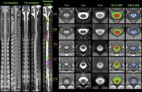
The spinal cord MRI community currently lacks a standard
reference template covering the entire cord, therefore
hindering the feasibility of large multi-center studies.
Here, we propose the MNI-Poly-AMU50, the first MRI template
of the entire spinal cord and brainstem, based on 50
subjects, available for multiple contrasts (T1-,
T2- and T2*-weighted), and integrating
probabilistic atlases of the white and gray matter. These
templates provide a common framework for co-registering
multi-parametric data. All developments are freely available
as part of the Spinal Cord Toolbox.
|
| |
08:24
|
1130.
 |
High-resolution quantitative magnetic resonance imaging of the
human cervical spinal cord at 7T
Aurélien Massire1,2,3, Manuel Taso1,2,3,4,
Maxime Guye1,2, Jean-Philippe Ranjeva1,2,3,
and Virginie Callot1,2,3
1Centre de Résonance Magnétique Biologique et
Médicale (CRMBM), UMR 7339, CNRS, Aix-Marseille Université,
Marseille, France, 2Centre
d'Exploration Métabolique par Résonance Magnétique (CEMEREM),
Hôpital de la Timone, Pôle d’imagerie médicale, AP-HM,
Marseille, France, 3iLab-Spine
- Laboratoire international - Imagerie et Biomécanique du
rachis, Marseille, France, 4LBA,
UMR T24, Aix-Marseille Université, IFSTTAR, Marseille,
France
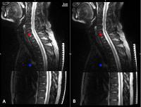
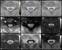
A high-resolution multi-parametric MRI protocol dedicated to
7T cervical spinal cord (SC) investigation using a
commercial prototype transceiver radiofrequency coil array
is proposed. This work pushes forward SC quantitative MRI by
reporting T1/T2/T2* relaxation
times mapping as well as diffusion tensor imaging metrics at
the C3 cervical level on a cohort of ten healthy volunteers.
Automatic segmentation and registration of these
multi-parametric acquisitions to SC templates enable group
studies with quantitative evaluation within regional WM
tracts and GM horns never reported so far at 7T. This study
lays the groundwork for improved characterization of
degenerative SC pathologies at ultra-high field.
|
| |
08:36
|
1131.
 |
Validating Myelin Water Imaging with Electron Microscopy in Rat
Spinal Cord
Henry Szu-Meng Chen1, Nathan Holmes2,
Wolfram Tetzlaff2,3, and Piotr Kozlowski4,5
1Physics and Astronomy, University of British
Columbia, Vancouver, BC, Canada, 2Zoology,
University of British Columbia, Vancouver, BC, Canada, 3ICORD,
Vancouver, BC, Canada, 4UBC
MRI Research Centre, Vancouver, BC, Canada, 5Radiology,
University of British Columbia, Vancouver, BC, Canada
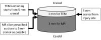
Quantitative T2 based myelin water imaging measures myelin
content by probing the properties of the water trapped in
myelin and therefore depends on its morphology. We compared
MR myelin water fraction (MWF) to electron microscopy
derived myelin content using a rat injury model and found
that MWF correlates strongly with the amount of myelin lipid
bilayers in both intact myelin and myelin debris and that
myelin debris appears to consist of areas of either normally
spaced myelin or large vacuous spaces. No significant
differences were found in myelin spacing among normal, 3
week, and 8 weeks post injury time points.
|
| |
08:48
|
1132.
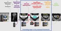 |
Fully-integrated framework for registration of spinal cord white
and gray matter
Sara Dupont1, Benjamin De Leener1,
Manuel Taso2,3, Nikola Stikov1,4,
Virginie Callot2,3, and Julien Cohen-Adad1,5
1Neuroimaging Research Laboratory (NeuroPoly),
Institute of Biomedical Engineering, École Polytechnique de
Montréal, Montréal, QC, Canada, 2Centre
de Résonance Magnétique Biologique et Médicale (CRMBM), UMR
7339, CNRS, Aix-Marseille Université, Marseille, France, 3Centre
d'Exploration Métabolique par Résonance Magnétique
(CEMEREM), Hôpital de la Timone, AP-HM, Marseille, France, 4Montreal
Heart Institute (MHI), Montréal, QC, Canada, 5Functional
Neuroimaging Unit, CRIUGM, Université de Montréal, Montréal,
QC, Canada
The spinal cord (SC) white and gray matter can be affected
by a large number of pathologies. Being able to segment
precisely the SC internal structure would be useful to
better understand SC diseases, improve diagnosis and assess
treatment efficiency. We introduce a complete framework for (i) multi-atlas
automatic segmentation of the gray-matter, (ii) accurate
registration to the MNI-Poly-AMU template and (iii)extraction
of quantitative metric using partial volume information.
Results showed improved accuracy of template registration
when adding prior automatic gray-matter segmentation. The
proposed method is freely available and provides an unbiased
framework for quantitative analysis of SC MRI data.
|
| |
09:00
 |
1133.
 |
Fully automated grey and white matter segmentation of the
cervical cord in vivo
Ferran Prados1,2, Manuel Jorge Cardoso1,
Marios C Yiannakas2, Luke R Hoy2,
Elisa Tebaldi2, Hugh Kearney2, Martina
D Liechti2, David H Miller2, Olga
Ciccarelli2, Claudia Angela Michela Gandini
Wheeler-Kingshott2,3, and Sebastien Ourselin1
1Translational Imaging Group, Medical Physics and
Biomedical Engineering, University College London, London,
United Kingdom, 2NMR
Research Unit, Queen Square MS Centre, Department of
Neuroinflammation, UCL Institute of Neurology, University
College London, London, United Kingdom, 3Brain
Connectivity Center, C. Mondino National Neurological
Institute, Pavia, Italy
We propose and validate a new fully automated spinal cord
(SC) segmentation technique that incorporates two different
multi-atlas segmentation propagation and fusion techniques:
Optimized PatchMatch Label fusion (OPAL) and Similarity and
Truth Estimation for Propagated Segmentations (STEPS). We
collaboratively join the advantages of each method to obtain
the most accurate SC segmentation. The new method reaches
the inter-rater variability, providing automatic
segmentations equivalents to inter-rater segmentations in
terms of DSC 0.97 for whole cord for any subject.
|
| |
09:12
|
1134.
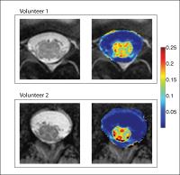 |
High-Resolution Single-Point qMT of the Lumbar Cord
Alex K. Smith1,2, Richard D. Dortch1,2,3,
Samantha By1,2, Robert L. Barry2,
Chris R. Thompson2, Kristen George-Durrett2,
Bailey D. Lyttle2, and Seth A. Smith1,2,3
1Department of Biomedical Engineering, Vanderbilt
University, Nashville, TN, United States, 2Vanderbilt
University Institute of Imaging Science, Vanderbilt
University, Nashville, TN, United States, 3Department
of Radiology and Radiological Sciences, Vanderbilt
University, Nashville, TN, United States
The spinal cord is responsible for mediating neurologic
function, and in particular, the lumbar cord is integral to
lower extremity function. However, lumbar cord quantitative
MRI studies have been limited due to its size, location, and
composition. A single-point quantitative magnetization
transfer was recently developed, but has not been applied to
the lumbar cord. Therefore, we have implemented an
assessment of qMT at the thoracolumbar bulge to characterize
the MT effect in the thoracolumbar cord in healthy
volunteers.
|
| |
09:24
|
1135.
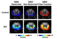 |
White Matter Swelling Masked Axonal Loss Detected by Diffusion
Basis Spectrum Imaging (DBSI)
Tsen-Hsuan Lin1, Mitchell Hallman1,2,
Fay Hwang1, Yong Wang1,3,4,5,
Sheng-Kwei Song1,4,5, and Peng Sun1
1Radiology, Washington University School of
Medicine, St. Louis, MO, United States, 2Perelman
School of Medicine at the University of Pennsylvania,
Philadelphia, PA, United States, 3Obstertic
and Gynecology, Washington University School of Medicine,
St. Louis, MO, United States, 4The
Hope Center for Neurological Disorders, Washington
University School of Medicine, St. Louis, MO, United States, 5Biomedical
Engineering, Washington University in St. Louis, St. Louis,
MO, United States
The extent of axonal loss plays a significant role in
irreversible neurological impairment in spinal cord injury
(SCI). We detected a 15% axonal loss in SCI mice using
diffusion basis spectrum imaging (DBSI) that was masked by
injury induced white matter swelling.
|
| |
09:36
|
1136.
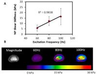 |
In-vivo Characterization of Human Lumbar Intervertebral Discs by
Magnetic Resonance Elastography: Diurnal Changes in Shear
Stiffness and Relationship with Degeneration
Benjamin A Walter1,2, Prasath Mageswaran1,3,
Hazem Mashaly1,4, William Thoman 1,4,
Daniel Boulter5, Luciano Prevedello 5,
Xuan Nguyen 5,
Mo Xiaokui 6,
Ehud Mendel 1,4,
William Marras1,3, and Arunark Kolipaka1,2,5,7
1Spine Research Institute, The Ohio State
University, Columbus, OH, United States, 2Biomedical
Engineering, The Ohio State University, Columbus, OH, United
States, 3Integrated
Systems Engineering, The Ohio State University, Columbus,
OH, United States, 4Neurological
Surgery, The Ohio State University Wexner Medical Center,
Columbus, OH, United States, 5Radiology,
The Ohio State University Wexner Medical Center, Columbus,
OH, United States, 6Biomedical
Informatics, The Ohio State University, Columbus, OH, United
States, 7Cardiovascular
Medicine, The Ohio State University Wexner Medical Center,
Columbus, OH, United States
Magnetic resonance elastography (MRE) was used to assess
intervertebral disc (IVD) shear properties in order to
develop an objective biomarker for the IVD degeneration
process. This study characterized the frequency response
and repeatability of MRE assessment of IVD shear stiffness
and how the shear stiffness of the nucleus pulposus (NP)
region of the IVD changes during degeneration. Results
suggest that MRE derived NP shear stiffness is a repeatable
technique that can provide a relative and objective
measurement of IVD degeneration that is independent of age.
|
| |
09:48
|
1137.
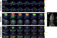 |
Developing In Vivo Perfusion Imaging Methods for Spinal Cord
Using Hyperpolarized [13C]t-Butanol and [13C, 15N2]Urea
Ilwoo Park1, Jeremy Gordon1, Sarah
Nelson1,2, and Jason Talbott1,3
1Radiology and Biomedical Imaging, University of
California San Francisco, San Francisco, CA, United States, 2Bioengineering
and Therapeutic Sciences, University of California San
Francisco, San Francisco, CA, United States, 3Brain
and Spine Injury Center (BASIC), University of California
San Francisco, San Francisco, CA, United States
This study has demonstrated the feasibility of using
hyperpolarized 13C
MRI with [13C]t-butanol and [13C,15N2]urea
for assessing in vivo perfusion in the cervical spinal cord.
T-butanol rapidly crossed the blood-brain-barrier and
diffused into spine and brain tissue, while urea
predominantly remained in vasculature. The results from this
study suggest that this technique may provide unique
non-invasive imaging tracers that are able to directly
monitor hemodynamic processes in the normal and injured
spinal cord.
|
|