| |
16:00
|
|
Introduction |
| |
16:15
|
0509.
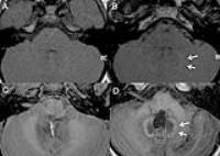 |
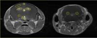
Deep brain nuclei T1 shortening after gadolinium in children:
influence of radiation and chemotherapy 
Sonja Kinner1,2, Tilman B Schubert1,3,
Susan Rebsamen1, Richard Bruce1, Scott
B Reeder1,4,5,6,7, and Howard A Rowley1
1Department of Radiology, University of Wisconsin
School of Medicine and Public Health, Madison, WI, United
States, 2Department
of Diagnostic and Interventional Radiology and
Neuroradiology, University Hospital Essen, Essen, Germany, 3Clinic
for Radiology and Nuclear Medicine, Basel University
Hospital, Basel, Switzerland, 4Department
of Medical Physics, University of Wisconsin School of
Medicine and Public Health, Madison, WI, United States, 5Department
of Emergency Medicine, University of Wisconsin School of
Medicine and Public Health, Madison, WI, United States, 6Department
of Medicine, University of Wisconsin School of Medicine and
Public Health, Madison, WI, United States, 7Department
of Biomedical Engineering, University of Wisconsin School of
Medicine and Public Health, Madison, WI, United States
Recent studies report intrinsic T1 hyperintense signal in
deep brain nuclei on MRI after multiple doses of
gadolinium-based contrast agents in adults. We investigated
whether similar T1 shortening was also found in children,
and furthermore evaluated the influence of radiochemotherapy
(RCTX) on its appearance. Signal increases were found in
2/60 children without RCTX and in 12/16 children with RCTX.
Signal ratio changes were significantly different between
the two groups and appeared with fewer doses in children
with RCTX.
|
| |
16:30
|
0510.
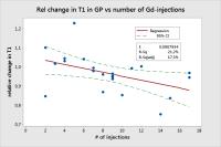 |
T1 relaxometry indicate cerebral gadolinium retention after
multiple administration of a macrocyclic Gd-based contrast
agent: A Retrospective Study in 27 patients with Glioblastoma
Multiforme
Svein Are Vatnehol1, Inge Rasmus Groote1,
Christopher Larsson1, Magne Kleppestø1,
Jonas Vardal1, and Atle Bjørnerud1,2
1The Intervention Center, Oslo University
Hospital, Oslo, Norway, 2Department
of Physics, University of Oslo, Oslo, Norway
Recent publications have shown an increase in signal
intensity on non-enhanced T1w-images for the Dentate Nucleus
and Globus Pallidus. This effect seems to be linked to
multiple administrations of linear gadolinium chelate. In
this retrospective study we have analyzed the quantitative
T1 values (qT1) and the normalized native T1 signal
intensity (nSI) for the Globus Pallidus and the nSI for the
Dentate Nucleus in patients with multiple injections of
gadobutrol (Gadovist™). Our analysis suggest a significant
change in the qT1 and nSI for the Globus Pallidus as well as
in the nSI for the Dentate Nucleus
|
| |
16:45
|
0511.
 |
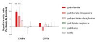
Gadolinium Deposition in the brain: Pre-clinical Investigation
of differences in concentration, Distribution and histology in
animals after repeated Administrations of linear and macrocyclic
GBCAs - Permission Withheld
Hubertus Pietsch1, Thomas Frenzel1,
Anna-Lena Frisk1, Diana Constanze Lenhard2,
Gregor Jost1, Martin Andrew Sieber1,
Astrid Zimmermann3, Volker Nischwitz3,
and Jessica Lohrke1
1Bayer Healthcare, Berlin, Germany, 2Charité,
Humboldt University Berlin, Berlin, Germany, 3Forschungszentrum
Jülich, Jülich, Germany
Recent publications reported increased T1-weighted signal
intensities in the dentate nucleus of patients who received
multiple contrast-enhanced MRI scans. In this animal study
histopathological changes and gadolinium retention in the
skin and brain of rats after twenty intravenous injections
of linear and macrocyclic GBCAs at high doses (2.5mmol/kgbw)
were systematically investigated. The Gd brain
concentrations of linear GBCAs (gadodiamide, gadopentetate
dimeglumine) were significantly higher than those of
macrocyclic agents (gadobutrol, gadoteridol). Since no
morphological changes could be detected by routine H&E
microscopic examination, immunohistochemistry and special
stains, these findings are considered be of no toxicological
relevance in rats.
|
| |
17:00
|
0512.
 |
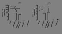
T1-weighted signal increase in the rat brain after multiple,
high-dose administrations of gadolinium based contrast agents:
Comparison of linear and macrocyclic agents - Permission Withheld
Gregor Jost1, Diana Lenhard2, Jessica
Lohrke1, Thomas Frenzel1, and Hubertus
Pietsch1
1MR and CT Contrast Media Research, Bayer
Healthcare, Berlin, Germany, 2Institute
of Vegetative Physiology, Charité, Berlin, Germany
Recent publications reported increased T1-weighted (T1w)
signal intensities (SI) in the dentate nucleus and globus
pallidus after repeated administrations of gadolinium based
contrast agents (GBCAs). In the present animal study the T1w
SI of three linear and two macrocyclic GBCAs were
systematically evaluated after ten administrations each with
a dose of 2.5 mmol/kg. Increased cerebellar nuclei to pons
SI ratios were found after administration of linear GBCAs
(significantly increased for gadodiamide and gadobenate
dimeglumine, and non-significantly increased for
gadopentetate dimeglumine). In contrast no elevated SI
ratios were observed after administration of the macrocyclic
GBCAs gadobutrol and gadoterate meglumine or saline.
|
| |
17:15
|
0513.
 |
Regional uptake and clearance of Gd(III) DTPA in the healthy
adult mouse brain
Daniel Calle1, Irene Guadilla1, Pilar
López-Larrubia1, and Sebastián Cerdán1
1Instituto de Investigaciones Biomédicas "Alberto
Sols", CSIC, Madrid, Spain
We report on the kinetics of uptake and clearance of
Gd(III)DTPA from different brain structures to healthy mice.
We fitted a biexponential model to cerebral time courses of
increase and decrease of T1w MRI
signal intensity, calculating rate constants for the uptake
(kabs) and elimination (kel). kabs showed
the rapid absorption in the ventricles and hypothalamus,
slowing down significantly in the cortex, globus pallidus
and dentante nucleus. These latter structures required 617
h. (cortex), 245 h. (globus pallidus) and approximately 100h
(hypothalamus and dentate nucleus), to remove 99% of the
administered agent, revealing very high cerebral residence
times of Magnevist.
|
| |
17:30
|
0514.
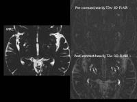 |
Contrast enhancement of perivascular spaces in the basal ganglia
Shinji Naganawa1 and
Toshiaki Taoka1
1Department of Radiology, Nagoya University
Graduate School of Medicine, Nagoya, Japan
Perivascular spaces (PVS) have been described as
non-enhancing structures with a fluid signal. In this study,
we confirmed that PVS signals are enhanced in images
obtained 4 hours after intravenous administration of
gadolinium based contrast agent (GBCA) in human subjects
without renal insufficiency. Contrast enhancement of CSF was
also observed. It is possible that GBCA in the blood vessels
might have permeated into the CSF space and PVS. This could
be the route by which GBCA is distributed to brain
parenchyma through the glymphatic
system in
subjects with a normal blood brain barrier.
|
| |
17:45
|
0515.
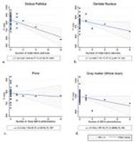 |
Regional and global assessment on relaxometric quantitative MRI
in patients with previous administration of a linear
gadolinium-based contrast agent
Hirofumi Kuno1, Hernan Jara1, Karen
Buch1, Andrew Mills1, Muhammad Mustafa
Quresh1, Neil Thayil 1,
Margaret N Chapman1, and Osamu Sakai1
1Radiology, Boston University, Boston Medical
Center, Boston, MA, United States
To assess potential regional and global correlations between
brain relaxation times and the number of prior
administrations of linear gadolinium-based contrast agents
(GBCA) using quantitative MRI. The subjects consisted of 40
patients (7 patients with multiple prior linear GBCA
exposures and 33 patients with no prior GBCA exposures) with
brain MRI using the mixed turbo spin-echo pulse sequence. T1
and T2 relaxation times were assessed in selected regions of
brain parenchyma (GP, DN, thalamus, and pons) and the whole
brain, and were demonstrated to be associated with the
number of gadolinium administrations. A stronger
relationship was demonstrated in gray matter.
|
|