| |
14:15
|
0117.
 |
U-fiber Quantification in Non-Lesional Epilepsy
Rafael O'Halloran1, Rebecca Feldman1,
Madeline Fields1, Laura Marcuse1, and
Priti Balchandani1
1Icahn School of Medicine at Mount Sinai, New
York, NY, United States
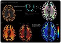
A method for the quantification of cortical-to-cortical
U-fiber fraction based on 7T MRI is presented and used to
demonstrate group differences in the the U-fiber fractions
in non-lesional and lesional epilepsy patients compared to
healthy controls. Non-lesional epilepsy patients had the
lowest u-fiber fractions followed by healthy control
subjects, and then by lesional epilepsy subjects with the
highest u-fiber fractions.
|
| |
14:27
|
0118.
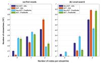 |
The influence of node assignment strategies and track
termination criteria on diffusion MRI-based structural
connectomics
Chun-Hung Yeh1, Robert Elton Smith1,
Thijs Dhollander1, Fernando Calamante1,
and Alan Connelly1
1The Florey Institute of Neuroscience and Mental
Health, Melbourne, Australia
This study highlights the issue of using the common strategy
for assigning individual streamlines to an atlas-based brain
parcellation. This process is non-trivial and can introduce
ambiguity into connectome quantification. In many fibre-tracking
algorithms, track termination criteria can cause premature
termination of streamlines within WM or CSF, which can
result in up to ~50–80% of streamlines failing in
identifying pairwise connections between nodes from
streamline endpoints. Our results demonstrate that such
issue can be largely ameliorated through the combination of
biologically meaningful track terminations and an
appropriate node assignment mechanism. This could therefore
be advantageous to structural connectome construction.
|
| |
14:39
|
0119.
 |
Behavioral response time as explained by a fiber-based analysis
of generalized fractional anisotropy measured using diffusion
spectrum imaging
Kayako Matsuo1, Yung-Chin Hsu2, Yasuo
Takehara3, Wen-Yih Isaac Tseng2, and
Norio Mori1
1Dept. Psychiatry, Hamamatsu University School of
Medicine, Hamamatsu, Japan, 2Institute
of Medical Devices and Imaging System, National Taiwan
University College of Medicine, Taipei, Taiwan, 3Dept.
Radiology, Hamamatsu University School of Medicine,
Hamamatsu, Japan
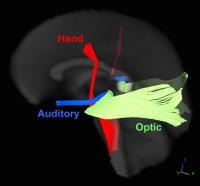
DSI on a GE 3T was conducted for 22 normal controls to
examine the neural basis of the response time (RT). RT was
measured outside the scanner using button pressing by left
or right hand in response to visual or auditory stimulation.
Faster RT was associated with greater GFA of portions near
the cortical hand area in the corticospinal tract (CST).
Left and right hand specializations were found in the deeper
CST. Greater GFA in portions near the cortex in the left
auditory radiation was associated with faster RT by visual
stimulations, suggesting an influence of language processing
speed.
|
| |
14:51
|
0120.
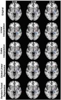 |
Image quality transfer benefits tractography of low-resolution
data
Daniel C. Alexander1, Aurobrata Ghosh1,
Samuel A. Hurley2, and Stamatios N. Sotiropoulos2
1Computer Science, UCL, London, United Kingdom, 2FMRIB,
Oxford University, Oxford, United Kingdom
We show benefits of image quality transfer to tractography.
Diffusion MRI super-resolution through image quality
transfer enables recovery of thin tracts in a dataset with
low spatial resolution (2.5mm isotropic). Specifically, we
reconstruct four pathways arising from the motor area that
have been distinguished before when using high (1.25mm)
resolution HCP data. Quantitative results confirm that image
quality transfer enhances tractography more than standard
interpolation. The results highlight the major potential of
image quality transfer in learning information from bespoke
high quality data sets to enhance the specificity of
information derived from more modest but readily available
data.
|
| |
15:03
|
0121.
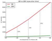 |
QuickBundlesX: Sequential clustering of millions of streamlines
in multiple levels of detail at record execution time
Eleftherios Garyfallidis1, Marc-Alexandre Côté1,
François Rheault1, and Maxime Descoteaux1
1Computer Science, Université de Sherbrooke,
Sherbrooke, QC, Canada
QuickBundlesX shows a remarkable 20+X speedup over it’s
predecessor who was until today the fastest clustering
algorithm for streamlines. In addition, it returns a useful
tree of clusters at different resolutions which allows to
query streamlines and easily process millions of streamlines
by comparing only with their neighbours.
|
| |
15:15
|
0122.
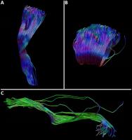 |
Structural Fingerprinting of the Human Brain: How unique is
tract shape to the individual?
Greg D Parker1, George J.A. Evans2,
and Derek K Jones1,3
1CUBRIC, School of Psychology, Cardiff
University, Cardiff, United Kingdom, 2School
of Medicine, Newcastle University, Newcastle, United
Kingdom, 3Neuroscience
and Mental Health Research Institute (NMHRI), School of
Medicine, Cardiff University, Cardiff, United Kingdom
Even amongst healthy subjects, brain function and structure
is known to be highly variable across individuals1,2.
Recently3 it
was shown that inter-subject variation in functional
connectivity is sufficient to allow robust and reliable
identification of individuals across different sessions and
tasks. Here we demonstrate for the first time that the same
is true of white matter structure; using the shape of an
individual's white matter tracts we generate fingerprints
that uniquely identify individuals across different scan
sessions.
|
| |
15:27
|
0123.
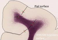 |
Fibers crossing the white/gray matter boundary: a semi-global,
histology-informed dMRI model
Michiel Cottaar1, Matteo Bastiani1,
Charles Chen2, Krikor Dikranian2,
David C. Van Essen2, Timothy E. Behrens1,
Stamatios N. Sotiropoulos1, and Saad Jbabdi1
1FMRIB, Oxford University, Oxford, United
Kingdom, 2Washington
University School of Medicine, Saint Louis, MO, United
States
Close to the cortical white/gray matter boundary surface
fiber orientations sharply transition from being nearly
tangential to the surface in the white matter to mostly
radial in the gray matter. We propose a geometric model that
describes this transition at sub-voxel resolution based on
high-resolution histology data and fit this model to lower
resolution diffusion MRI data. We assess its performance
using qualitative comparisons with histology and test the
reproducibility of the estimated parameters across multiple
diffusion MRI resolutions. This model allows the in-vivo
estimation of fiber orientations across the white/gray
matter boundary, which may improve tracking to the cortex.
|
| |
15:39
|
0124.
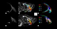 |
Microscopic DTI for quantitative tractography of MAP6-KO mice:
validation by fluorescent microscopy on cleared brains
Ulysse Gimenez1, Franck Mauconduit1,
Benoit Boulan2, Eric Denarier2,
Jacques Brocard2, Sylvie Gory-Fauré2,
Annie Andrieux2, Jean Christophe Deloulme2,
and Hana Lahrech1
1Clinatec Lab U1205, INSERM, Grenoble, France, 2Grenoble
Institute of Neurosciences, INSERM, La Tronche, France
High spatial resolution 3D DTI was developed and used for
white matter tractography to quantify neuronal tract
alterations on the MAP6-KO mouse. In this model, the
microtubule-associated protein 6 (MAP6) which is involved in
the neuromorphogenesis is deleted leading to a model
characterized by severe behavior impairments, similar to the
clinical features of schizophrenia. As 3D DTI tractography
and fluorescent microscopy on cleared brains both show a
deficiency of the post-commissural fornix, in accordance
with our previous 2D DTI results, the 3D DTI tractography
imaging is validated. Using 3D DTI tractography, new major
alterations in different neuronal tracts are detected.
|
| |
15:51
|
0125.
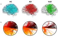 |
Network integration and segregation differentiate between
Alzheimer Disease and Vascular Dementia
Fulvia Palesi1,2, Andrea De Rinaldis2,3,
Letizia Casiraghi2,4, Gloria Castellazzi2,3,
Paolo Vitali5, Nicoletta Anzalone6,
Federica Denaro7, Elena Sinforiani8,
Giuseppe Micieli7, Egidio D'Angelo2,4,
and Claudia Angela Michela Gandini Wheeler-Kingshott2,9
1Department of Physics, University of Pavia,
Pavia, Italy, 2Brain
Connectivity Center, C. Mondino National Neurological
Institute, Pavia, Italy, 3Department
of Electrical, Computer and Biomedical Engineering,
University of Pavia, Pavia, Italy, 4Department
of Brain and Behavioral Sciences, University of Pavia,
Pavia, Italy, 5Brain
MRI 3T Mondino Research Center, C. Mondino National
Neurological Institute, Pavia, Italy,6Scientific
Institute H. S. Raffaele, Milan, Italy, 7Department
of Emergency Neurology, C. Mondino National Neurological
Institute, Pavia, Italy, 8Alzheimer's
Disease Assessment Unit, Laboratory of Neuropsychology, C.
Mondino National Neurological Institute, Pavia, Italy, 9NMR
Research Unit, Queen Square MS Centre, Department of
Neuroinflammation, UCL Institute of Neurology, University
College London, London, United Kingdom
Dementia is the most common disorder in elderly people and
comprises Alzheimer’s disease (AD) and vascular disease
(VaD). In this work graph theoretical approach was applied
to a cohort of AD, VaD and healthy controls (HC) aimed at
investigating the presence of a disease-specific pattern of
alterations. Brain structural networks were built using the
Cohen functional atlas (nodes) and advanced probabilistic
tractography (edges). Our main finding was that VaD patients
showed severe impairment in the large-scale brain networks
while AD patients mainly showed inefficiency of short-range
connections emphasizing the fact that alterations are
restricted to specific brain regions.
|
| |
16:03
|
0126.
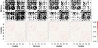 |
Estimating Network Topology in Weighted and Dense Connectomes
Luis Manuel Colon-Perez1, Michelle Couret2,
William Triplett3, Catherine Price3,
and Thomas H Mareci3
1Psychiatry, University of Florida, Gainesville,
FL, United States, 2Medicine,
Columbia University, New York, NY, United States, 3University
of Florida, Gainesville, FL, United States
Brain networks are organized in a heterogeneous range of
white-matter tract sizes suggesting that the brain is
organized in broad range of white matter connection
strengths. Studies of brain structure with a binary
connection model have shown a small-world network
topological organization of the brain. We developed a
generalized framework to estimate the topological properties
of brain networks using weighted connections, which offers a
more realistic model of the brain compared to the binary
connection model. In addition, this model reduces the need
for thresholding to obtain topological properties in dense
and weighted connectomes.
|
|