| |
14:15
 |
0157.
 |
MR elastography and DCE-MRI of the liver and spleen for
non-invasive prediction of portal pressure - Permission Withheld
Stefanie Hectors1, Mathilde Wagner1,
Octavia Bane1, Aaron Fischman2, Thomas
Schiano3, and Bachir Taouli1,4
1Translational and Molecular Imaging Institute,
Icahn School of Medicine at Mount Sinai, New York, NY,
United States, 2Department
of Interventional Radiology, Icahn School of Medicine at
Mount SInai, New York, NY, United States, 3Department
of Internal Medicine, Icahn School of Medicine at Mount
SInai, New York, NY, United States, 4Department
of Radiology, Icahn School of Medicine at Mount Sinai, New
York, NY, United States
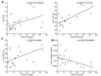
The goal of this study was to assess whether DCE-MRI
parameters and MR elastography-derived stiffness in liver
and spleen can predict portal pressure. Liver time-to-peak (TTP),
mean transit time (MTT), upslope and stiffness (LS) all
significantly correlated with hepatic venous pressure
gradient (HVPG) measurement. Sensitivity-specificity of LS
for detection of HVPG≥5mmHg and HVPG≥10mmHg were 64%-91% and
71%-89% respectively, while combined LS and spleen TTP
yielded the highest sensitivity-specificity (92%-86% for
HVPG≥5mmHg, 100%-92% for HVPG≥10mmHg). These results
indicate that combination of liver and spleen perfusion and
stiffness metrics into a multiparametric analysis maximizes
diagnostic performance for the prediction of portal
pressure.
|
| |
14:27
|
0158.
 |
Longitudinal assessment of structural and haemodynamic
parameters in compensated cirrhosis using Quantitative Magnetic
Resonance Imaging 
Chris Bradley1, Eleanor F Cox1, David
Harman2, Martin W James2, Guru P
Aithal2, I Neil Guha2, and Susan T
Francis1
1Physics and Astronomy, University of Nottingham,
Nottingham, United Kingdom, 2NIHR
Biomedical Research Unit in Gastrointestinal and Liver
Diseases, University of Nottingham, Nottingham, United
Kingdom
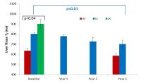
We perform a longitudinal 3 year study to assess progression
of disease in compensated cirrhosis (CC) using annual
haemodynamic and structural MR measures, and compare with a
healthy volunteer group. Longitudinal relaxation time (T1)
correlates with liver disease severity, and shows a small
variance across years in stable, compensated cirrhosis. In
contrast a large variance is shown for liver stiffness
measures using Fibroscan®. MR measures correlate well with
Enhanced Liver Fibrosis (ELF) scores. This study suggests
that MR provides a sensitive technique to assess changes in
pathophysiology of CC.
|
| |
14:39
|
0159.
 |
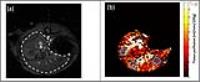
Hemodynamic Changes in the Portal Circulation in Living Related
Liver Donors, Assessed by 4D flow MRI - Permission Withheld
Alejandro Roldán-Alzate1,2, Luis A Fernandez3,
Oliver Wieben2,4, and Scott B Reeder2,4
1Mechanical Engineering, University of Wisconsin
- Madison, Madison, WI, United States, 2Radiology,
University of Wisconsin - Madison, Madison, WI, United
States, 3Surgery,
University of Wisconsin - Madison, Madison, WI, United
States, 4Medical
Physics, University of Wisconsin - Madison, Madison, WI,
United States
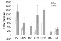
The purpose of this study was to evaluate hemodynamic
changes in the mesenteric and portal circulation of LDLT
donors in response to surgical liver resection. Four living
related liver donors were studied. Subjects were imaged
using 4D Flow MRI before and after liver resection surgery.
Highly patient-specific responses to each surgical procedure
were found. The ability to quantify hemodynamic changes in
the portal and mesenteric circulation non-invasively
demonstrates that 4D flow MRI may be a suitable tool for
both surgical planning of LDLT, and for improved
understanding of the hemodynamic changes that occur in the
liver remnant of the donor.
|
| |
14:51
|
0160.
 |
Free-Breathing 3D Liver Perfusion Quantification Using a
Dual-Input Two-Compartment Model
Satyam Ghodasara1, Vikas Gulani2, and
Yong Chen2
1Case Western Reserve University School of
Medicine, Cleveland, OH, United States, 2Radiology,
Case Western Reserve University, Cleveland, OH, United
States
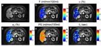
The dual-input two-compartment model was applied to liver
perfusion data, and significant differences in perfusion
parameters were found between normal hepatic parenchyma and
focal lesions, and also between HCC and metastatic lesions.
These findings support the possibility of using a
two-compartment model with 3D free-breathing acquisitions,
for lesion characterization.
|
| |
15:03
|
0161.
 |
Acceleration of Image Analysis for Liver Perfusion
Quantification Using Parallel Computational Techniques
Satyam Ghodasara1, Yong Chen2, Mark
Griswold2, Nicole Seiberlich3, and
Vikas Gulani2
1Case Western Reserve University School of
Medicine, Cleveland, OH, United States, 2Radiology,
Case Western Reserve University, Cleveland, OH, United
States, 3Biomedical
Engineering, Case Western Reserve University, Cleveland, OH,
United States
To make free-breathing liver perfusion quantification
feasible for a clinical timescale, acceleration of both
non-Cartesian parallel imaging reconstruction and non-rigid
image registration was performed with parallel computing
techniques. Our results show massively increased speed (12
minutes compared to >22.5 hours for standard computations)
with extremely minor differences in both image quality and
perfusion quantification.
|
| |
15:15
|
0162.
 |
Measurement of bulk liver perfusion: Assessment of agreement
between ASL and caval subtraction phase-contrast MRI at 9.4T
Manil Chouhan1, Rajiv Ramasawmy2, Alan
Bainbridge3, Adrienne Campbell-Washburn2,
Jack Wells2, Shonit Punwani1,
Rajeshwar Mookerjee4, Simon Walker-Samuel2,
Mark Lythgoe2, and Stuart Taylor1
1UCL Centre for Medical Imaging, University
College London, London, United Kingdom, 2UCL
Centre for Advanced Biomedical Imaging, University College
London, London, United Kingdom, 3Department
of Medical Physics, University College London Hospitals NHS
Trust, London, United Kingdom, 4UCL
Institute for Liver and Digestive Health, University College
London, London, United Kingdom
Non-invasive preclinical liver perfusion measurements could
be used to develop biomarkers and assess new treatments for
liver disease and primary/secondary malignant liver lesions.
ASL can provide regional hepatic perfusion maps, and in
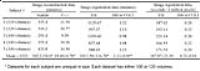
this study we compare FAIR ASL tissue perfusion measurements
with caval subtraction phase-contrast MRI, a validated
method for measuring total liver blood flow, to demonstrate
ASL overestimation but encouraging agreement between both
methods.
|
| |
15:27
|
0163.
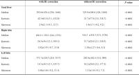 |
Quantitative Liver Function Analysis using Volumetric T1 Mapping
with Fast Multi-Slice B1 Correction on Hepatocyte-specific
Contrast Enhanced Liver Magnetic Resonance Imaging - Permission Withheld
Jeong Hee Yoon1, Jeong Min Lee1, Eun
Ju Kim2, Tomoyuki Okuaki3, and Joon
Koo Han1
1Radiology, Seoul National University Hospital,
Seoul, Korea, Republic of, 2Philips
Healthcare, Seoul, Korea, Republic of, 3Philips
Healthcare, Tokyo, Japan
Liver signal intensity on hepatobiliary phase at gadoxetic
acid-enhanced liver MRI has been reported to be useful to
estimate global and regional liver function quantitatively.
However, simple MR signal measurement is often suffering
from its sensitivity of MR field inhomogeneity and
non-linear relationship with contrast medium concentration.
Herein, we investigated of B1 correction effect on T1 map
and compared its diagnostic performance to assess liver
function according to Child-Pugh classification. In
addition, we attempted to investigate risk assessment
capability of B1 corrected T1 map for long-term clinical
outcome in patients with cirrhosis.
|
| |
15:39
|
0164.
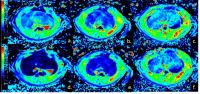 |
Gd-EOB-DTPA-enhanced MRI: evaluation of liver function by
multiple hepatocyte-phase images and T1 mapping in rats
Jia Xu1, Xuan Wang1, Yan You2,
Qin Wang1, Hui Liu3, Jing Lei1,
Huadan Xue1, and Zhengyu Jin1
1Department of Radiology, Peking Union Medical
College Hospital, Beijing, China, People's Republic of, 2Department
of Pathology, Peking Union Medical College Hospital,
Beijing, China, People's Republic of,3Siemens
Ltd. China, Shanghai, China, People's Republic of
To evaluate regional liver function preoperatively is of
great value in planning surgical management. Our Aim is to
investigate the potential of Gd-EOB-DTPA enhanced MRI in
evaluating hepatic function in rats with liver fibrosis.
Parameters calculated from Gd-EOB-DTPA enhanced MRI
exhibited moderate to high correlation with plasma
indocyanine green retention rate at 15 minutes after
intravenous injection of ICG (ICG R15) in rats with liver
fibrosis, indicating its potential in liver function
evaluation.
|
| |
15:51
|
0165.
 |
Comparison of the Hepatocyte Fraction and Conventional Image
Based Methods for the Estimation of Liver Function
Tomoyuki Okuaki1, Kosuke Morita2,
Tomohiro Namimoto3, Morikatsu Yoshida3,
Shinya Shiraishi3, Masanori Komi2,
Yasuyuki Yamashita3, and Marc Van Cauteren1
1Philips Healthcare, Tokyo, Japan, 2Department
of Central Radiology, Kumamoto University Hospital,
Kumamoto, Japan, 3Department
of Diagnostic Radiology, Faculty of Life Sciences, Kumamoto
University, Kumamoto, Japan
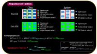
The hepatocyte fraction
(HeF) is based on simple pharmacokinetics, and can
quantitatively estimate the fraction of hepatocytes.
In this study, the HeF, liver-spleen contrast ratio and
delta T1 value were compared to the results of 99mTc-GSA scintigraphy using
the blood clearance index (HH15) and receptor index (LHL15).
The correlation coefficients of the HH15 were 0.602, 0.544
and 0.773, respectively, and of the LHL15 were 0.612, 0.670
and 0.762, respectively. The HeF quantification showed the
highest correlation with the 99mTc-GSA,
proving it to be useful for a robust evaluation of liver
function, compared to conventional imaging based
quantitative methods.
|
| |
16:03
|
0166.
 |
The change and interrelation of quantitative hepatic MR imaging
biomarkers in the course of chronic hepatitis.
Akira Yamada1, Yasunari Fujinaga1,
Yoshihiro Kitoh2, Takeshi Suzuki1,
Daisuke Komatsu1, Aya Shiobara2, Yasuo
Adachi2, Atsushi Nozaki3, Yuji Iwadate3,
Kazuhiko Ueda1, and Masumi Kadoya1
1Department of Radiology, Shinshu University
School of Medicine, Matsumoto, Japan, 2Division
of Radiology, Shinshu University Hospital, Matsumoto, Japan, 3GE
Healthcare Japan, Hino, Japan
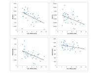
Variable quantitative hepatic imaging biomarkers including
pharmacokinetic parameters of hemodynamics and
hepatocellular uptake function, R2* and fat fraction,
apparent diffusion coefficient (ADC), liver stiffness were
obtained from the patients with chronic hepatitis using MR
imaging. The change and interrelation of these imaging
biomarkers in the course of chronic hepatitis were evaluated
quantitatively. Portal venous inflow and hepatocellular
uptake function correlated well with liver stiffness,
meanwhile, ADC showed weak correlation. Arterial
compensation, decreased blood flow speed and volume were
observed in the patients with decreased portal venous
inflow. No significant correlation was observed between
liver stiffness and R2* or fat fraction.
|
|