| |
16:00
|
0740.
 |
Abdominal MRF at Ultra-High-Field Strengths 
Martijn A Cloos1,2, Bei Zhang1,2, and
Daniel K Sodickson1,2
1Bernard and Irene Schwartz Center for Biomedical
Imaging, New York University School of Medicine, New York,
NY, United States, 2Center
for Advanced Imaging Innovation and Research (CAI2R), New
York University School of Medicine, New York, NY, United
States
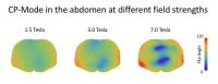
Like other magnetic resonance (MR) techniques before it,
magnetic resonance fingerprinting (MRF) was developed and
applied in the traditional context of a precisely calibrated
and uniform radiofrequency excitation field. Plug & Play
Parallel Transmission (PnP-PTX), on the other hand, was
designed to liberate MRF from these constraints. We evaluate
the impact of excitation field non-uniformities on abdominal
MRF experiments at different field strengths, and show that
PnP-PTX has the potential to alleviate these challenges, and
thereby opens opens up a new route towards robust,
quantitative, whole-body MRI for ultra-high-field systems.
|
| |
16:12
 |
0741.
 |
Spiral Acquisition for High-Speed Anatomical Imaging at 7T
Lars Kasper1,2, Christoph Barmet1,3,
Maria Engel1, Maximilian Haeberlin1,
Bertram J Wilm1, Benjamin E Dietrich1,
Thomas Schmid1, David O Brunner1,
Klaas E Stephan2,4,5, and Klaas P Pruessmann1
1Institute for Biomedical Engineering, University
of Zurich and ETH Zurich, Zuerich, Switzerland, 2Translational
Neuromodeling Unit, IBT, University of Zurich and ETH
Zurich, Zuerich, Switzerland, 3Skope
Magnetic Resonance Technologies, Zurich, Switzerland, 4Wellcome
Trust Centre for Neuroimaging, University College London,
London, United Kingdom, 5Max
Planck Institute for Metabolism Research, Cologne, Germany
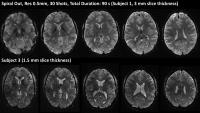
We present whole-brain, high-resolution (0.5mm) spiral
imaging with proton-density and T2* contrast at 7T in less
than a minute. Owing to a comprehensive characterization of
the imaging process, artifact-free image reconstruction from
long-readout spiral shots (20 ms) becomes feasible via an
iterative SENSE algorithm. In particular, trajectory
imperfections as well as dynamic off-resonance changes are
captured via concurrent field monitoring, while static
off-resonance as well as coil sensitivities are mapped in a
multi-echo reference scan and augment image reconstruction.
The resulting images exhibit the same geometric fidelity as
spin-warp images at a fraction of the total acquisition
duration.
|
| |
16:24
|
0742.
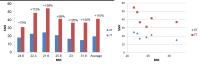 |
First proof of more than two-fold increase in intrinsic SNR for
prostate imaging at 7 tesla in comparison with 3 tesla.
Mariska P. Luttje1, Ingmar J. Voogt1,
Marco van Vulpen1, Peter R. Luijten1,
Dennis W.J. Klomp1, and Alexander J.E.
Raaijmakers1
1Imaging Division, University Medical Center
Utrecht, Utrecht, Netherlands
In this study, we demonstrate that a dipole transceive
antenna array with a loop coil receive array at 7T
substantially outperforms state of the art 3T MRI of the
prostate. Using this setup we demonstrated for the first
time the intrinsic SNR benefits of using the higher field
strength of 7 tesla for prostate MR imaging compared to a
clinically used prostate imaging setup at 3 tesla: an
overall gain in SNR of 2.1 fold as obtained in 6 subjects.
|
| |
16:36
|
0743.
 |
Utilizing the improved receive sensitivity from high
permittivity materials for SNR-challenged applications of
ultrahigh b-factor diffusion-weighted spectroscopy at 7 Tesla
Carson Ingo1, Wyger M. Brink1, Andrew
G. Webb1, and Itamar Ronen1
1C.J. Gorter Center for High Field MRI,
Department of Radiology, Leiden University Medical Center,
Leiden, Netherlands
Diffusion-weighted 7T MR spectroscopy in white matter
regions of the brain using ultrahigh b-factors have
established that intracellular metabolites exhibit
non-Gaussian diffusion. Such measurements using b-factors
well above 10,000 s/mm2 have
inherently low SNR, and so it is crucial to optimize B1 sensitivity
to ensure reliable results. Here we show that a single high
permittivity pad can increase the receive sensitivity by
~30%, resulting in potential reductions in data acquisition
time of ~70%.
|
| |
16:48
|
0744.
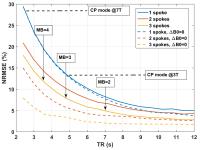 |
High resolution whole-brain diffusion MRI at 7 Tesla using
parallel RF transmission: how fast can we go?
Xiaoping Wu1, Nicolas Boulant2,
Vincent Gras2, Jinfeng Tian1,
Sebastian Schmitter1, Pierre-Francois Van de
Moortele1, and Kamil Ugurbil1
1CMRR, Radiology, University of Minnesota,
Minneapolis, MN, United States, 2CEA/NeuroSpin,
Saclay, France
The Human Connectome Project (HCP) in the WU-Minn consortium
aims to acquire multiband (MB)-accelerated whole brain
diffusion MRI (dMRI). Although shown advantageous over 3T
dMRI in inferring connectivity, the 7T acquisition suffers
from transmit B1 inhomogeneity and SAR, the latter currently
limiting the slice acceleration to an MB factor of 2 (MB=2).
In this study, we investigated numerically the highest
possible slice acceleration for 7T HCP-type dMRI acquisition
with ~1-mm isotropic resolutions. Our results suggest that
parallel RF transmission can be used to enable MB=4 while
improving flip angle homogeneity across the whole brain as
compared to a CP mode.
|
| |
17:00
|
0745.
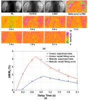 |
Quantitative Single Breath-Hold Renal ASL Perfusion Imaging at
7T
Xiufeng Li1, Pierre-Francois Van de Moortele1,
Kamil Ugurbil1, and Gregory J. Metzger1
1Center for Magnetic Resonance Research,
University of Minnesota, Minneapolis, MN, United States
In contrast to studies at 3T, where the whole body
coil is used for RF transmission, studies at 7T use local
transcieve coils, which have limited B1+ coverage
producing smaller temporal bolus widths that need to be
estimated in order to achieve proper renal blood flow (RBF)
quantification. To estimate the temporal bolus width and to
quantify RBF at 7T, single breath-hold renal perfusion
studies were performed using the FAIR ss-FSE method with
varied delay times. Based on the results form multi-delay
perfusion study, quantitative renal perfusion imaging was
further achieved by using a single-subtraction approach.
|
| |
17:12
|
0746.
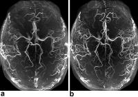 |
Prospective motion correction for ultra-high resolution Time of
Flight angiography at 7T under SAR constraints
Hendrik Mattern1, Alessandro Sciarra1,
Frank Godenschweger1, Daniel Stucht1,
Falk Lüsebrink1, and Oliver Speck1,2,3,4
1Department of Biomedical Magnetic Resonance,
Otto-von-Guericke-University Magdeburg, Magdeburg, Germany, 2Leibniz
Institute for Neurobiology, Magdeburg, Germany, 3Center
for Behavioral Brain Sciences, Magdeburg, Germany, 4German
Center for Neurodegenerative Disease, Magdeburg, Germany
At 7T, venous saturation and magnetization transfer for Time
of Flight (ToF) angiography cannot be applied directly due
to the increased specific absorption rate. Additionally,
motion artifacts can degrade the image quality. A sequence
with prospective motion correction (PMC) and sparse
saturation was implemented to overcome these challenges. In
vivo ultra-high resolution ToF angiograms were acquired,
providing dramatically improved level of detail and image
quality if PMC and sparse saturation is used. Thus, the
proposed sequence unleashes the full potential of ToF
angiography at 7T.
|
| |
17:24
|
0747.
 |
On-resonant balanced Steady-State Free Precession imaging at
9.4T
Damien Nguyen1,2, Tom Hilbert3,4,5,
Philipp Ehses6,7, Klaus Scheffler6,7,
Jean-Philippe Thiran4,5, Oliver Bieri1,2,
and Tobias Kober3,4,5
1Radiological Physics, Dep. of Radiology,
University of Basel Hospital, Basel, Switzerland, 2Department
of Biomedical Engineering, University of Basel, Basel,
Switzerland, 3Advanced
Clinical Imaging Technology (HC CMEA SUI DI BM PI), Siemens
Healthcare AG, Lausanne, Switzerland, 4Department
of Radiology, University Hospital Lausanne (CHUV), Lausanne,
Switzerland, 5LTS5,
École Polytechnique Fédérale de Lausanne, Lausanne,
Switzerland, 6High-Field
MR Center, Max Planck Institute for Biological Cybernetics,
Tübingen, Germany, 7Department
for Biomedical Magnetic Resonance, University of Tübingen,
Tübingen, Germany
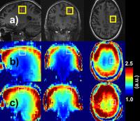
In this work, we explore the possibility of using the
recently proposed highly undersampled 3D phase-cycled
balanced Steady-State Free Precession (bSSFP) sequence
trueCISS to generate on-resonant band-free bSSFP images at
9.4T. By applying the forward signal model, it is also
possible to synthetically generate bSSFP images at higher
flip angles, which would otherwise be impossible to acquire
due to SAR limitations. Lastly, we show a maximum bSSFP
signal intensity image of the brain using the trueCISS
estimated parameter maps.
|
| |
17:36
|
0748.
 |
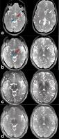
Magnetic resonance imaging of low-grade and high-grade gliomas
at 7 Tesla - Permission Withheld
Bixia Chen1,2, Philipp Dammann1,2,
Stefan Maderwald1, Soeren Johst1,
Tobias Schoemberg1,2, Lale Umutlu1,3,
Harald H. Quick1,4, Mark Edward Ladd1,5,
Ulrich Sure2, and Karsten Henning Wrede1,2
1Erwin L. Hahn Institute for MRI, University of
Duisburg-Essen, Essen, Germany, 2Department
of Neurosurgery, University Hospital Essen, University of
Duisburg-Essen, Essen, Germany, 3Institute
of Diagnostic and Interventional Radiology and
Neuroradiology, University of Duisburg-Essen, Essen,
Germany, 4High
Field and Hybrid MR Imaging, University Hospital Essen,
University of Duisburg-Essen, Essen, Germany,5Medical
Physics in Radiology, German Cancer Research Center (DKFZ),
Heidelberg, Germany
Magnetic resonance imaging (MRI) plays a major role in
diagnosis, multimodal treatment planning, and follow-up of
low-grade and high-grade gliomas. In this prospective study,
24 patients were intra-individually examined at 3 Tesla (T)
and 7T utilizing MPRAGE, T2 TSE,
T2 FLAIR,
and SWI sequences. Image evaluation had special focus on
intra-tumoral structures, vascularization, intra-lesional
hemorrhages, and contrast uptake. At 7T, intra-tumoral
structures were depicted in excellent image quality.
Especially SWI was superior at 7T compared to 3T and
revealed microhemorrhages and vascularization patterns
correlating with histopathology, possibly providing an
additional imaging predictor for future grading of malignant
gliomas.
|
| |
17:48
|
0749.
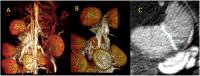 |
High-resolution placental MR angiography using a nanoparticle
contrast agent
Ketan Ghaghada1, Zbigniew Starosolski1,
Igor Stupin1, Saakshi Bhayana1, Haijun
Gao2, Rohan Bhavane1, Chandresh Patel1,
Robia Pautler3, Chandrasekhar Yallampalli2,
and Ananth Annapragada1
1Pediatric Radiology, Texas Children's Hospital,
Houston, TX, United States, 2Obstetrics
and Gynecology, Baylor College of Medicine, Houston, TX,
United States, 3Molecular
Physiology and Biophysics, Baylor College of Medicine,
Houston, TX, United States
Non-invasive imaging of maternal and placental vasculature
in rodent species is of interest to the pre-clinical study
of clinically-relevant placental pathologies. In this work,
we evaluated the utility of high-resolution
contrast-enhanced MR angiography using a placental
non-permeable, long circulating liposomal-Gd nanoparticle
contrast agent in a pregnant rat model.
|
|