| |
10:30
|
0826.
 |
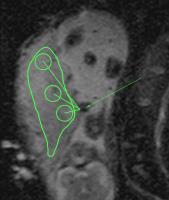
Differentiation of Pancreatic Carcinoma and Mass-forming focal
pancreatitis: assessment by dynamic contrast-enhanced MRI
combined with diffusion-weighted imaging 
Tingting Zhang1 and
Dengbin Wang1
1Department of Radiology, Xinhua Hospital,
Shanghai Jiao Tong Medical University, Shanghai, China,
Shanghai, China, People's Republic of
This study is to access the differential value in Pancreatic
Carcinoma (PC) and Mass-Forming focal Pancreatitis (FP) with
qualitative and quantitative analysis of DCE-MRI and DWI.
Pancreatic TIC types and sub-types from DCE-MRI and ADC
value and tumor-to-pancreas contrast ratio of ADC value from
DWI were compared between PC and FP. We found significant
differences in TIC and ADC value and ADC tumor-to-pancreas
contrast ratio between PC and FP. DCE-MRI and DWI were
discovered to provide reliable information for
differentiating PC from FP, while the combination of them
can achieve a higher sensitivity and specificity.
|
| |
10:42
|
0827.
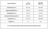 |
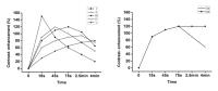
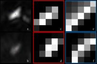
The potential of apparent diffusion coefficient in
differentiating various sub-types of breast tumors and its
association with hormonal status
Khushbu Agarwal1, Rani Gupta Sah1, Uma
Sharma1, Smriti Hari2, Sandeep Mathur3,
Vurthaluru Seenu4, Rajinder Parshad4,
and Naranamangalam R Jagannathan1
1Department of NMR and MRI Facility, All India
Institute of Medical Sciences, Delhi, India, 2Department
of Radio-diagnosis, All India Institute of Medical Sciences,
Delhi, India, 3Department
of Pathology, All India Institute of Medical Sciences,
Delhi, India, 4Department
of Surgical Disciplines, All India Institute of Medical
Sciences, Delhi, India
Potential of apparent diffusion coefficient (ADC) in
differentiating various sub-types of malignant and benign
breast tumors using diffusion weighted MRI and its
association with different hormonal status in breast cancer
patients was studied. A significantly lower ADC of malignant
compared to benign and healthy breast tissues was observed.
The ADC of fibroadenomas was lower compared to fibrocystic
with fibroadenoma and cystic lesions and higher in cystic
and fibrocystic lesions than benign ductal epithelial cells.
No association of ADC with molecular biomarkers ER, PR and
Her2neu was seen. Results showed the utility of ADC in
differentiating various types of breast tissues.
|
| |
10:54
|
0828.
 |
Intravoxel Incoherent Motion DWI and Aquaporins MR Imaging of
Transplanted Kidneys at 3.0 T
Yanjun Li1, Shumin Tao1, Dandan Zheng2,
Yong Zhang2, and Guangming Lu1
1Medical Imaging, Jingling Hospital, School of
Medicine, Nanjing University, Nanjing, China, People's
Republic of, 2GE
Healthcare, MR Research China, Beijing, China, People's
Republic of
Diffusion-weighted imaging (DWI) in human transplantation
was regarded as a promising indicator for dectecting graft
dysfunction.According to the IVIM theory, ADC integrates the
effects of both diffusion of water molecules and
microcirculation of blood in capillaries. By separating
diffusion and perfusion, we could observe each component’s
contribution to the changes of ADC values. Furthermore, the
exchanges of intracellular water molecules with
extracellular’s may have impact on ADC. Thus, AQP ADC might
reflect quantitative water channel expression.
|
| |
11:06
|
0829.
 |
The Diffusion and Perfusion Characteristics of Placenta with
Differential Fetal Growth Restriction Types Using Intravoxel
Incoherent Motion MR Imaging
TANG Min1, Xiaoqin LIU1, Xiaoling
ZHANG1, Kaining Shi2, and Kaining Shi2
1Shaanxi Provincial People`s Hospital, Xi’an,
China, People's Republic of, 2Clinical
science, Philips Healthcare China, Xi’an, China, People's
Republic of
The purpose of this study is to explore the feasibility of
assessing perfusion and diffusion information of placenta
with different FGR types using Intravoxel Incoherent Motion
MR Imaging .we find that there are different diffusion and
perfusion characteristic in different in placenta of FGR
hypotype.
|
| |
11:18
 |
0830.
 |
Improved IVIM MRI of Small Lesions in the Liver by Deformable
Image Registration with Modality Independent Neighborhood
Descriptor
Yihao Guo1, Zhentai Lu1, Yingjie Mei2,
Jing Zhang3, Yikai Xu3, Feng Huang4,
Ed. X. Wu5,6, and Yanqiu Feng1,5,6
1School of Biomedical Engineering and Guangdong
Provincial Key Laboratory of Medical Image Processing,
Southern Medical University, GuangZhou, China, People's
Republic of, 2Philips
Healthcare, GuangZhou, China, People's Republic of, 3Department
of Medical Imaging Center, Nanfang Hospital, Southern
Medical University, GuangZhou, China, People's Republic of, 4Philips
Healthcare(Suzhou), Suzhou, China, People's Republic of, 5Laboratory
of Biomedical Imaging and Signal Processing, The University
of Hong Kong, Hong Kong SAR, China, People's Republic of, 6Department
of Electrical and Electronic Engineering, The University of
Hong Kong, Hong Kong SAR, China, People's Republic of
Respiration-induced misalignment between multiple b-value
liver DW image scan severely reduce the accuracy and
stability of IVIM parameter quantification, especially in
the presence of small focal lesions. These small lesions
usually exhibit significantly differentintensity in
different b-value images, but have similar structural
information. This work introduces modality independent
neighborhood descriptor to extract the structural
information of small lesions for improved realignment
between multiple b-value images. Preliminary results show
that this structure-based registration method can well
correct respiration-induced misalignment between multiple
b-value images with small lesions, improve the IVIM model
fitting quality, and reduce variance in quantified
parameters.
|
| |
11:30
 |
0831.
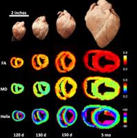 |
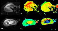
Diffusion Tensor Imaging and Histology of the Developing
Myocardium
Osama M Abdullah1, MarJanna Dahl2,
Gavin Yeip1, Julia Cortino1, Arnold
David Gomez3, Thomas Seidel1, Frank
Sachse1, Kurt Albertine2, and Edward W
Hsu1
1Bioengineering, University of Utah, Salt Lake
ity, UT, United States, 2Pediatric
Neonatology, University of Utah, Salt Lake ity, UT, United
States, 3Electrical
and Computer Engineering, Johns Hopkins, Baltimore, MD,
United States
Diffusion tensor imaging (DTI) has emerged as the method of
choice for noninvasive quantifications of myocardial
microstructure. However, the origins and behaviors of DTI
measurements as functions of myocardial remodeling during
development remain poorly understood. In this work,
conventional and bi-compartmental DTI and histological
correlation were performed on an animal model of myocardial
development to investigate the effects of tissue remodeling.
Results indicate that tissue remodeling during development
manifests in progressively increased DTI transverse
diffusivities, decreased fractional anisotropy, and
unchanged fiber orientation. The findings show that DTI can
be used to noninvasively characterize microstructural
remodeling of the myocardium during development.
|
| |
11:42
|
0832.
 |
Comparing the breath-hold, respiratory-triggered and
free-breathing techniques in the diffusion-weighted imaging for
the evaluation of focal liver lesions on a 3.0T system
Zhuo Shi1, Xinming Zhao1, Ouyang Han1,
and Lizhi Xie2
1Department Of Imaging Diagnosis,Cancer Hospital,
Chinese Academy of Medical Sciences & Peking Union Medical
College, Beijing China, Beijing, China, People's Republic
of, 2GE
Healthcare, MR Research China, Beijing, Beijing, China,
People's Republic of
DWI plays an important role in detecting and characterizing
liver lesions or tumors. There are three kinds of liver DWI
acquisitions are commonly used in hepatic DWI: breath-hold ,
respiratory-triggered,free-breathing. This work compares the
advantages and disadvantages of diagnosing focal liver
lesions, to find the best acquisition sequence for different
situations.
|
| |
11:54
|
0833.
 |
Study of Spatial Function in the Human Placenta with Diffusion
Weighted Imaging
Edward Sutherland 1,
Luís F Gonçalves1,2,3, and Yuxiang Zhou1,3
1William Beaumont Hospital School of Medicine,
Oakland University, Rochester, MI, United States, 2Department
of Obstetrics and Gynecology, Beaumont Health, Royal Oak,
MI, United States, 3Diagnostic
Radiology and Molecular Imaging, Beaumont Health, Royal Oak,
MI, United States
Magnetic resonance diffusion weighted imaging (DWI) has been
widely used to quantitatively measure the random motion of
water molecules within a voxel of tissue and represents this
information in the form of apparent diffusion coefficient
(ADC) maps. As the ADC map has been shown to be influenced
by circulatory motion and perfusion at low b-values, we
hypothesize that ADC values obtained from the placenta may
vary as a function of distance to the umbilical cord
insertion. In this retrospective study, 78 healthy placentas
were evaluated by MR-DWI. We conclude that ADC values of
placental tissues obtained at high b-values do not vary in
normal human placentas as a function of distance to
umbilical cord insertion.
|
| |
12:06
|
0834.
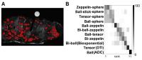 |
Comparison of multi-component restricted and anisotropic models
of diffusion in glandular breast tissue
Sisi Liang1, Narina Norddin2,
Eleftheria Panagiotaki 3,
Andre Bongers4, Peng Shi1, Laurence
Gluch5, and Roger Bourne2
1College of Engineering and Science, Victoria
University, Melbourne, Australia, 2Discipline
of Medical Radiation Sciences, Faculty of Health Sciences,
University of Sydney, Sydney, Australia, 3Center
for Medical Image Computing, University College London,
London, United Kingdom, 4Biological
Resource Imaging Laboratory, University of New South Wales,
Sydney, Australia, 5The
Strathfield Breast Centre, Strathfield, Australia
DWI signal attenuation measured in biological tissue is
widely observed to be non-monoexponential. One important
diffusion characteristic underlying that complex behavior is
restriction and hindrance to water diffusion. This study
compared multi-component restricted and unrestricted models
of diffusion in the glandular part of breast tissue. The
results show that multi-component restricted and anisotropic
models explain the data best. This finding is consistent
with the presence of distinct diffusion microenvironments in
breast tissue. Development of clinical DWI methods that
incorporate these features may improve breast cancer
assessment.
|
| |
12:18
|
0835.
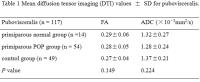 |
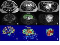
Evaluation of the levator ani muscle in primiparas six weeks
after vaginal delivery using diffusion tensor imaging and fiber
tractography
Can Cui1, Yujiao Zhao1, Yu Zhang 2,
and Wen Shen3
1Radiology, Tianjin First Center Clinical
College, Tianjin Medical University, Tianjin, China,
People's Republic of, 2Philips
Healthcare, Beijing, China, People's Republic of, 3Radiology,
Tianjin First Center Hospital, Tianjin, China, People's
Republic of
The aim of this study was to investigate the clinical
application of DTI fiber tractography on evaluating the
levator ani injury after first vaginal delivery. Thirty-five
primiparous women with 6 weeks after vaginal delivery and
twenty-five age-matched nulliparous women volunteers as
control group were included. The primiparas women were
divided into 2 subgroups by the existence of pelvic organ
prolapse. DTI with fiber tractography provided satisfactory
3D representation of pubovisceralis while the study of
iliococcygeus was more difficult. No significant differences
of FA and ADC values were found among primiparous normal
group, primiparous POP group and control group for
pubovisceralis.
|
|