| |
08:00
|
1078.
 |
Multi-compartment microscopic diffusion anisotropy imaging
brought into clinical practice 
Enrico Kaden1, Nathaniel D. Kelm2,
Robert P. Carson3, Mark D. Does2, and
Daniel C. Alexander1
1Centre for Medical Image Computing, University
College London, London, United Kingdom, 2Institute
of Imaging Science, Vanderbilt University, Nashville, TN,
United States, 3Departments
of Neurology and Pediatrics, Vanderbilt University,
Nashville, TN, United States
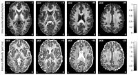
This work introduces a multi-compartment model for
microscopic diffusion anisotropy imaging using an
off-the-shelf pulse sequence achievable on standard clinical
scanners. In particular, we will provide estimates of
microscopic features specific to the intra- and extra-neurite
compartments unconfounded by the effects of fibre crossings
and orientation dispersion, which are ubiquitous in the
brain. The new imaging technique is demonstrated in a large
cohort of healthy young adults as well as for the detection
of microstructural tissue alterations in a preclinical
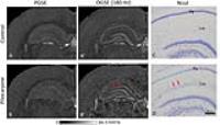
animal model of tuberous sclerosis complex.
|
| |
08:12
|
1079.
 |
The lifespan trajectory of white matter microstructure detected
by NODDI
Jiaying Zhang1, Aurobrata Ghosh1,
Daniel C Alexander1, and Gary Hui Zhang1
1Computer Science and Centre for medical image
computing, University College London, London, United Kingdom
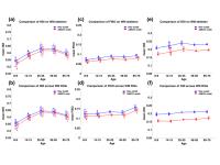
The structure and function of human brain evolve across the
lifespan. The microstructural white matter changes across
lifespan have been studied using Diffusion tensor imaging.
Whilst sensitive, DTI parameters have no direct tissue
specificity. Here, given the availability of high-quality
HCP lifespan dataset, we aim to study the lifespan
trajectory of microstructural WM changes using NODDI and
evaluate another NODDI fitting framework - Accelerated
microstructure imaging via convex optimization (AMICO). We
found U-shaped neurite density changes across lifespan and
feasibility of AMICO NODDI parameters in capturing the
similar lifespan trajectory as the standard fitting.
|
| |
08:36
|
1080.
 |
WITHDRAWN
Nicholas G Dowell1, Simon L Evans2,
Sarah L King2, Naji Tabet3, and
Jennifer M Rusted2
1Clinical Imaging Sciences Centre, Brighton and
Sussex Medical School, Brighton, United Kingdom, 2Psychology,
University of Sussex, Brighton, United Kingdom, 3Centre
for Dementia Studies, Brighton and Sussex Medical School,
Brighton, United Kingdom
The APOE-e4 gene is the best known genetic risk factor for
late-onset Alzheimer's Disease. However, carriers of this
gene have demonstrated behavioural differences compared to
non-carriers on a number of cognitive tasks at young age. In
this study, we demonstrate for the first time using the
advanced diffusion imaging technique, NODDI, that there are
detectable genotype-dependent structural differences in the
brain of young healthy volunteers. The strongest differences
are in the measure of orientation dispersion (ODI) of
neurites, where e4 carriers show higher ODI than non-e4
carriers in the white matter.
|
| |
08:48
|
1081.
 |
Microscopic Anisotropy Imaging at 7T Using Asymmetrical Gradient
Waveform Encoding
Filip Szczepankiewicz1, Carl-Fredrik Westin2,
Freddy Ståhlberg1, Jimmy Lätt3, and
Markus Nilsson4
1Dept. of Medical Radiation Physics, Lund
University, Lund, Sweden, 2Dept.
of Radiology, Brigham and Women’s Hospital, Harvard Medical
School, Boston, MA, United States, 3Center
for Medical Imaging and Physiology, Skåne University
Hospital, Lund, Sweden, 4Lund
University Bioimaging Center, Lund University, Lund, Sweden
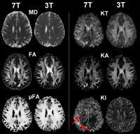
Diffusion MRI that goes beyond DTI is challenging at 7T due
to the short transverse relaxation time. We address this
inherent limitation of 7T by employing asymmetric gradient
waveforms for diffusion encoding, and demonstrate that
imaging of microscopic diffusion anisotropy is feasible at a
7T system.
|
| |
09:00
 |
1082.
 |
Chronic mild stress induces changes in neurite density in the
amygdala as revealed by diffusion MRI and validated with novel
histological analyses
Ahmad Raza Khan1, Andery Chuhutin1,
Ove Wiborg2, Christopher D Kroenke3,
Jens R Nyengaard4, Brian Hansen1, and
Sune Nørhøj Jespersen1,5
1Center of Functionally Integrative Neuroscience,
Aarhus University, Aarhus, Denmark, 2Centre
for Psychiatric Research, Aarhus University, Aarhus,
Denmark, 3Advanced
Imaging Research Center, Portland, OR, United States, 4Stereology
and Electron Microscopy Laboratory, Aarhus University,
Aarhus, Denmark, 5Department
of Physics and Astronomy, Aarhus University, Aarhus, Denmark
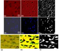
Biophysical modelling of diffusion MRI data allows detection
of specific tissue microstructures such as neurite density.
However, histological validation of MR-derived indication of
microstructural alteration is limited due extensive time
labour and invasive character, even though histological
validation is crucial because it remains the gold standard.
The present study applies Matlab based image processing and
analysis tools to compute histological neurite density to
validate diffusion MRI based neurite density changes in the
amygdala of chronic mild stress rat brains. The image
processing and analyses provides novel tools to validate
diffusion data robustly.
|
| |
09:12
|
1083.
 |
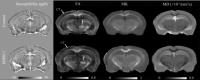
Altered hippocampal microstructure in the epileptogenic rat
brain revealed with diffusion MRI using oscillating field
gradients
Manisha Aggarwal1, Olli Gröhn2, and
Alejandra Sierra2
1Department of Radiology, Johns Hopkins
University School of Medicine, Baltimore, MD, United States, 2Department
of Neurobiology, A. I. Virtanen Institute for Molecular
Sciences, University of Eastern Finland, Kuopio, Finland
We investigate changes in the temporal diffusion spectrum
sampled using oscillating gradient spin-echo (OGSE)
acquisitions at increasing gradient frequencies in the
epileptogenic rat brain. PGSE and OGSE data at discrete
oscillation frequencies were acquired from
pilocarpine-treated and control rat brains (n=5 each) with a
spectral resolution of 60 Hz (f = 60 Hz, 120 Hz,
180 Hz). Our findings reveal significant changes in the
frequency-dependent modulation of apparent diffusion
coefficient (ADC) in specific areas of the pilocarpine
brain, which were found to correspond to region-specific
gliosis and neuronal loss respectively. Using comparison
with histological findings, our results show unique
sensitivity of OGSE diffusion MRI to probe specific
cellular-level alterations in the epileptogenic brain.
|
| |
09:24
|
1084.
 |
Detecting Disrupted-in-Schizophrenia-1 Gene Related
Microstructural and Molecular Alterations using Diffusion
Kurtosis Imaging and Quantitative Susceptibility Mapping
Nan-Jie Gong1, Russell Dibbs2, Kyle
Decker2, Mikhail V. Pletnikov3, and
Chunlei Liu1
1Brain Imaging and Analysis Center, Duke
University, Durham, NC, United States, 2Center
for In Vivo Microscopy, Duke University, Durham, NC, United
States, 3Behavioral
Neurobiology and Neuroimmunology Laboratory, Johns Hopkins
University, Baltimore, MD, United States
DKI method provided sensitive metrics for reflecting
microstructural changes in not only the anterior commissure
but also relatively isotropic gray matter regions of
hippocampus, cerebral cortex and caudate putamen. Further
relating DKI findings to molecular compositions measured by
QSM enabled clearer interpretations of myelin content and
cellular density related mechanisms. Further validations
that establish the relationship between imaging metrics and
histological measurements such as neuronal cell body
density, myelin thickness and g-ratio are needed.
|
| |
09:36
|
1085.
 |
White Matter Changes in Elderly Patients Suffer from
Post-operative Cognition Disorders : Evidence from Diffusion
Kurtosis Magnetic Resonance Imaging
Bing Yu1, Na Chang1, Xiaoxue Ge1,
Yueren Wang1, and Qiyong Guo1
1Shengjing Hospital of China Medical University,
Shenyang, China, People's Republic of
In the present study, we reconstructed the white matter
skeleton of the brain using tract-based spatial statistics
(TBSS) and compared differences in diffusion kurtosis
imaging (DKI) parameters within the skeleton between
patients sufferde from postoperative cognitive dysfunction
(POCD) and healthy controls to detect white matter
abnormalities in POCD.
|
| |
09:48
|
1086.
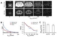 |
Rapid Estimation of Spinal Cord Injury Severity in Rats using
Double Diffusion Encoded Magnetic Resonance Spectroscopy
Nathan P Skinner1,2,3, Shekar N Kurpad3,4,
Brian D Schmit5, L Tugan Muftuler3,
and Matthew D Budde3,4
1Biophysics Graduate Program, Medical College of
Wisconsin, Milwaukee, WI, United States, 2Medical
Scientist Training Program, Medical College of Wisconsin,
Milwaukee, WI, United States, 3Department
of Neurosurgery, Medical College of Wisconsin, Milwaukee,
WI, United States, 4Clement
J. Zablocki Veteran's Affairs Medical Center, Milwaukee, WI,
United States, 5Department
of Biomedical Engineering, Marquette University, Milwaukee,
WI, United States
Diffusion tensor imaging (DTI) is frequently applied to
spinal cord injury, yet suffers from poor detection of
axonal integrity changes caused by conflicting extracellular
processes. A double diffusion encoding (DDE) sequence was
developed for the spinal cord to remove non-neuronal signal
contribution by applying a strong diffusion weighting
perpendicular to the spinal cord. A parallel diffusion
gradient then sampled diffusivity along the spinal cord.
Application in a rat model showed DDE parameters
outperformed DTI in sensitivity to injury severity with
substantially reduced acquisition and post-processing time.
Thus, this technique shows potential for rapid, sensitive
determination of spinal cord injury severity.
|
| |
09:48
|
1087.
 |
Measurement of restricted and hindered anisotropic diffusion
tissue compartments in a rat model of Wallerian degeneration
Benoit Scherrer1, Damien Jacobs2,
Maxime Taquet1,2, Anne des Rieux3,
Benoit Macq2, Sanjay P Prabhu1, and
Simon K Warfield1
1Department of Radiology, Boston Children's
Hospital, Harvard Medical School, Boston, MA, United States, 2ICTEAM,
Universite catholique de Louvain, Louvain-La-Neuve, Belgium, 3LDRI,
Université catholique de Louvain, Brussels, Belgium
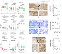
The DIAMOND model has been recently proposed to model the
heterogeneity of tissue compartments in diffusion
compartment imaging. However, it did not enable the
characterization of the intra-axonal volume fraction (IAVF),
a critical measure to more accurately characterizing axonal
loss in abnormal tissues. In this work we investigated
mathematical extensions to DIAMOND that model both the IAVF
and the heterogeneous nature of in-vivo tissue. We validated
our approach using both Monte-Carlo simulations and
histological microscopy with an animal model of Wallerian
degeneration. We show that our novel model better predicts
the signal and provides additional parameters to further
describe tissues.
|
|