| |
14:15
|
0137.
 |
Fully Automatic Left Atrium and Pulmonary Veins Segmentation for
Late Gadolinium Enhanced MRI Combining Contrast Enhanced MRA 
Qian Tao1, Esra Gucuk Ipek2, Rahil
Shahzad1, Floris F. Berendsen1, Saman
Nazarian2, and Rob J. van der Geest1
1Department of Radiology, Leiden University
Medical Center, Leiden, Netherlands, 2Department
of Cardiology, The Johns Hopkins University School of
Medicine, Baltimore, MD, United States
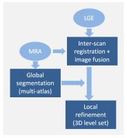
The extent and distribution of left atrial (LA) scar,
visualized by LGE MR, can provide important information for
treatment of atrial fibrillation (AF) patients. However, in
current practice, to extract such information requires
substantial manual effort and expertise. In this study, a
fully automatic method was developed to segment LA and PV’s
in LGE-MRI, combining robust multi-atlas segmentation and
flexible level-set based segmentation optimization. The
method demonstrated comparable accuracy to manual
segmentation, with improved 3D continuity. The method
enables automated generation of patient-specific LA and PV
geometry models, and potentially objective LA scar
assessment for individual AF patients.
|
| |
14:27
|
0138.
 |
Dark Blood Late Gadolinium Enhanced Imaging of Myocardial Scar
using First-Moment-Nulled Motion Sensitized Driven Equilibrium
(m2MSDE)
Gregory J Wilson1, Niranjan Balu1,
Jinnan Wang1,2, Chun Yuan1, and
Jeffrey H Maki1
1University of Washington, Seattle, WA, United
States, 2Bayer
Healthcare, Whippany, NJ, United States
A novel black-blood pre-pulse is described that darkens
intraventricular blood pool signal in late gadolinium
enhanced (LGE) imaging of myocardial scar. The pre-pulse is
m1-nulled motion-sensitized driven equilibrium
(m2MSDE) with user-specified motion-sensitizing direction.
The pre-pulse nulls blood signal while maintaining good
myocardial image quality. Preliminary results are described.
|
| |
14:39
|
0139.
 |
Visual quality assessment of 3D High Resolution Late Gadolinium
Enhancement with Compressed-Sensing in a Clinical Setting: the
impact of patient factors
Charlene Liew1,2, Tamer Basha1, Mehmet
Akcakaya1, Connie Tsao1, Francesca
Delling1, Kraig Kissinger1, Beth Goddu1,
Sophie Berg1, Warren Manning1,3, and
Reza Nezafat1
1Division of Cardiology, Department of Medicine,
Beth Israel Deaconess Medical Center, Boston, MA, United
States, 2Department
of Radiology, Changi General Hospital, Singapore, Singapore, 3Department
of Radiology, Beth Israel Deaconess Medical Center, Boston,
MA, United States
Compressed sensing can be used to reduce 3D LGE scan time by
factor of 5 with isotropic spatial resolution. However,
clinical feasibility and overall image quality of 3D LGE
with compressed sensing is still unknown. In this study, we
sought to assess the image quality of 3D LGE with isotropic
spatial resolution of 1-1.5 mm3 in
268 consecutive patients with known or suspected
cardiovascular disease and investigate the impact of patient
characteristics on overall image quality.
|
| |
14:51
|
0140.
 |
Detection of myocardial infarcts without contrast agent
injection: Comparison of spin-lock with magnetization transfer
MR imaging
Joep van Oorschot1, Martijn Froeling1,
Thijs van den Broek2, Frebus van Slochteren2,
Steven Chamuleau2, Peter Luijten1, Tim
Leiner1, and Jaco Zwanenburg1
1Radiology, University Medical Center Utrecht,
Utrecht, Netherlands, 2Cardiology,
University Medical Center Utrecht, Utrecht, Netherlands
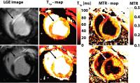
Two promising techniques for endogenous myocardial infarct
detection are Magnetization Transfer and T1ρ-MRI. Goal of
the study was to compare the ability to detect and quantify
myocardial scar tissue in a chronic infarct model using MT
and T1ρ mapping. In vivo MRI was performed on a clinical 1.5
MR scanner in 3 anesthetized pigs, 4 weeks after 90 minutes
occlusion of the LAD. The MTR was significantly lower in the
infarcted region (0.27±0.01 ms), compared to remote
myocardium (0.38±0.01 ms).The T1ρ relaxation time was
significantly higher in the infarcted region (87.0±1 ms),
compared to healthy remote myocardium (56.4± 1 ms).
|
| |
15:03
|
0141.
 |
Free-breathing 3D late gadolinium enhancement cardiovascular
magnetic resonance using outer volume suppressed projection
navigators: Development and clinical validation
Rajiv G Menon1, G Wilson Miller2, Jean
Jeudy1, Sanjay Rajagopalan3, and
Taehoon Shin1
1Diagnostic Radiology and Nuclear Medicine,
University of Maryland, Baltimore, Baltimore, MD, United
States, 2Department
of Radiology and Medical Imaging, University of Virginia,
Charlottesville, VA, United States, 3Division
of Cardiovascular Medicine, University of Maryland,
Baltimore, Baltimore, MD, United States
We developed a free-breathing, 3D late gadolinium
enhancement (FB 3D-LGE) cardiovascular magnetic resonance
technique based on outer volume suppressed 1D-projection
navigators and a stack-of-spirals acquisition. The
free-breathing 3D-LGE and conventional breath-hold 2D-LGE
scans were performed on 29 cardiac patients. 2D and 3D
techniques showed no significant differences in overall
image quality scores and image artifact scores (P > 0.1).
There was a significant correlation in the average
difference in fractional scar volume (r=0.96). The FB 3D-LGE
is a viable option for patients, particularly in acute
settings or in patients who are unable to comply with
breath-hold instructions.
|
| |
15:15
|
0142.
 |
Cardiac 31P MRS in breast cancer patients undergoing
chemotherapy
Gillian Macnaught1,2, Christopher Rodgers3,
Martin Denvir4, Olga Oikonomidou5,6,
Annette Cooper1, William Clarke3,
Heather McVicars6, Larry Hayward6,
Saeed Mirsadraee1, and Scott Semple1,4
1Clinical Research Imaging Centre, University of
Edinburgh, Edinburgh, United Kingdom, 2the
MRC Centre for inflammation Research, University of
Edinburgh, Edinburgh, United Kingdom, 3RDM
Cardiovascular Medicine, University of Oxford, Oxford,
United Kingdom, 4BHF
Centre for Cardiovascular Science, University of Edinburgh,
Edinburgh, United Kingdom, 5Edinburgh
Cancer Research Centre, University of Edinburgh, Edinburgh,
United Kingdom, 6Edinburgh
Cancer Centre, NHS Lothian, Edinburgh, United Kingdom
Anthracyclines are chemotherapy agents widely used to treat
cancer but that can also induce cardiotoxicity. Techniques
are required to provide an earlier warning of cardiotoxicity
before irreversible myocardial damage. 9 subjects were
recruited to this on-going 31P MRS study to detect changes
in cardiac energetics of breast cancer patients undergoing
chemotherapy. Between pre- and mid-chemotherapy four
subjects experienced a greater than 20% decrease in their
cardiac PCr/ATP ratio, 1 subject experienced a 13.8%
decrease in left ventricular ejection fraction (LVEF) and
all had increased troponin levels. Ultimately this study
aims to determine whether changes in PCr/ATP precede changes
in LVEF.
|
| |
15:27
|
0143.
 |
Significant improvement of survival by T2* MRI in thalassemia
major
Antonella Meloni1, Caterina Borgna-Pignatti2,
Giovanni Carlo Del Vecchio3, Maria Antonietta
Romeo4, Maria Rita Gamberini5,
Federico Bonetti6, Maria Giovanna Neri1,
Elisabetta Chiodi7, Vincenzo Positano1,
and Alessia Pepe1
1Fondazione G. Monasterio CNR-Regione Toscana,
Pisa, Italy, 2Università
di Ferrara, Ferrara, Italy, 3Uiversity
of Bari, Bari, Italy, 4University
of Catania, Catania, Italy, 5Arcispedale
"S.Anna", Ferrara, Italy,6Policlinic Foundation
San Matteo IRCCS, Pavia, Italy, 7Arcispedale
“S. Anna”, Ferrara, Italy
The introduction of T2* CMR for the reproducible and
non-invasive assessment of myocardial iron overload reduced
the likelihood of developing decompensated cardiac failure,
allowing the reduction of cardiac mortality in chronically
transfused TM patients
|
| |
15:39
|
0144.
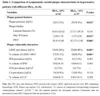 |
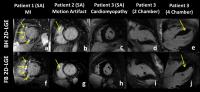
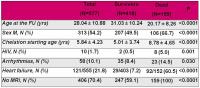
Elevated Hemoglobin A1c(HbA1c) Is Independently Associated with
Large Lipid-Rich Necrotic Cores in Hypertensive Patients with
Symptomatic Carotid Atherosclerosis: A 3.0T MRI Study
Huilin Zhao1, Beibei Sun1, Xiaosheng
Liu1, Xihai Zhao2, Yongming Dai3,
Chun Yuan4, and Jianrong Xu1
1Radiology, Renji Hospital, Shanghai Jiao Tong
University School of Medicine, Shanghai, China, People's
Republic of, 2Center
for Biomedical Imaging Research, Tsinghua University School
of Medicine, Beijing, China, People's Republic of, 3Philips
Healthcare, Shanghai, China, People's Republic of, 4Radiology,
University of Washington, Seattle, WA, United States
Further understanding of the association of hemoglobin A1c(HbA1c)
levels with symptomatic carotid plaque characteristics will
be helpful for stroke risk stratification and treatment
strategy modification. This study sought to investigate the
associations of HbA1c levels
with MR-identified carotid plaque characteristics in
hypertensive patients with acute stroke. Our key findings
are that elevated HbA1c was
associated with carotid plaque presence, higher HbA1c level
tended to exhibit an increased plaque burden and larger
lipid-rich necrotic core, independent of other
cardiovascular risk factors. Our findings indicate that
elevated HbA1c may
contribute to the development of advanced carotid plaques in
stroke patients with hypertension.
|
| |
15:51
|
0145.
|
Cardiac Magnetic Resonance detects an association between aortic
stiffness and epicardial fat volume in patients with increased
cardiovascular risk - Permission Withheld
Rami Homsi1, Alois Martin Sprinkart1,
Jürgen Gieseke1,2, Julian Luetkens1,
Michael Meier-Schroers1, Darius Dabir1,
Daniel Kuetting1, Christian Marx1,
Hans Schild1, and Daniel Thomas1
1Radiology, University Hospital Bonn, Bonn,
Germany, 2Philips
Healthcare, Hamburg, Germany
In a Cardiac Magnetic Resonance based approach the study
reveals a relationship between epicardial fat and aortic
stiffness which are both associated with cardiovascular risk
and disease.
|
| |
16:03
|
0146.
 |
Intradialytic MRI for the assessment of Cardiovascular Function
Charlotte E Buchanan1,2, Azharuddin Mohammed2,
Eleanor F Cox1, Maarten W Taal2,
Nicholas M Selby2, Susan T Francis1,
and Christopher W McIntyre3
1Sir Peter Mansfield Imaging Centre, School of
Physics and Astronomy, University of Nottingham, Nottingham,
United Kingdom, 2Division
of Medical Sciences and Graduate Entry Medicine, University
of Nottingham, Nottingham, United Kingdom, 3Schulich
School of Medicine and Dentistry, University of Western
Ontario, London, ON, Canada
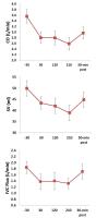
We perform the first study of intradialytic MRI to assess
cardiovascular stress during dialysis. A significant
reduction in cardiac output (CO), stroke volume (SV) and IVC
flux was seen during dialysis. Myocardial strain measures
revealed significant stunned segments in the long axis in
all individuals. No significant change in coronary artery
flow was evident, and both myocardial perfusion and T1 measures
in a single short axis slice showed no significant change.
The change in CO and SV was negatively correlated with
dialysis ultrafiltration volume. This work demonstrates MRI
can be used to assess cardiac stress during dialysis.
|
|