|
|
|
Plasma # |
|
0541.
 |
1 |
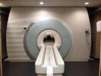
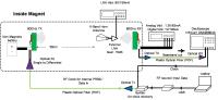
Porcine Imaging in a 10.5T Whole-Body Human MRI 
Lance DelaBarre1, Russell L. Lagore1,
Yigitcan Eryaman1, Gregor Adriany1,
and J. Thomas Vaughan1
1Center for Magnetic Resonance Research -
University of Minnesota, Minneapolis, MN, United States
Recently, our 10.5T whole-body MRI magnet achieved field
strength and its installation was completed. While waiting
for IRB and IDE approval, a human-sized porcine model serves
as a surrogate for later human studies, thus allowing
development of techniques in vivo. Using an 8-channel head
coil on a porcine head, the first in vivo images from the
10.5T whole-body MRI were acquired.
|
 |
0542.
 |
2 |
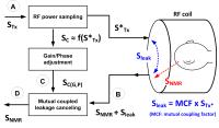
The first demonstration of simultaneous transmit and receive MRI
in vivo - Permission Withheld
Sung-Min Sohn1, J. Thomas Vaughan1,
Michael Garwood1, and Djaudat Idiyatullin1
1Center for Magnetic Resonance Research,
University of Minnesota, Minneapolis, MN, United States
This is the first demonstration of in vivo human MR imaging
with simultaneous transmit and receive using continuous mode
SWIFT at 4T. Due to a large RF power difference between Tx
and Rx working at the same frequency, the difficulties to
obtain the high and stable Tx/Rx isolation, and the
sensitivity of the Tx/Rx isolation to the loading
conditions, in vivo images using the simultaneous RF pulse
transmission and signal acquisition have not been reported.
This work proposed the simultaneous Tx/Rx system with highly
minimized effects from variation of coil loading, which
allowed us to acquire the first in vivo images with
continuous SWIFT at 4T.
|
 |
0543.
 |
3 |
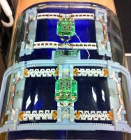
Anatomically adaptive local coils for MR Imaging - Evaluation of
stretchable antennas at 1.5T - Permission Withheld
Bernhard Gruber1 and
Stephan Zink2
1Medical Engineering - School of Applied Health
and Social Sciences, University of Applied Sciences Upper
Austria, Linz, Austria, 2R&D
HW LC, Siemens Healthcare GmbH, Erlangen, Germany
This abstract is a first investigation on antenna materials
and designs for anatomically adaptive local coils for MR
Imaging. To overcome the SNR losses by poorly loaded and
non-fitting RF coils, we proposed a stretchable antenna
design. Each loop has the ability to reversible stretch up
to 100% of its original size, to be anatomically adaptive to
different shapes and sizes in three dimensions. Through
bench measurements and MR Imaging at 1.5T we investigated
different stretchable antenna materials, that fit the
defined requirements. The results of stretchable loops
showed an in average SNR loss of under 10% in comparison to
standard loops, but we suppose that the improved filling
factor will lead to much higher SNR of the adaptive loops.
Further research may consider different improvements.
|
|
0544.
 |
4 |
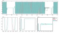
Gradient response harvesting for continuous system
characterization during MR sequences - Permission Withheld
Bertram J. Wilm1, Benjamin E. Dietrich1,
Jonas Reber1, S. Johanna Vannesjo1,
and Klaas P. Pruessmann1
1Institute for Biomedical Engineering, University
of Zurich and ETH Zurich, Zurich, Switzerland
Gradient impulse response functions were recently proposed
to characterize MR gradient systems with high accuracy.
However, changes of the impulse response, e.g. due to
thermal drifts, can limit its accuracy and hence
applicability. To overcome this problem, we present a novel
method where the gradient response is continuously
characterized during MR sequences from repeatedly performed
field probe measurements. The benefit of this method is
demonstrated by obtaining the continuous gradient output of
MR sequences and first imaging results are presented.
|
|
0545.
 |
5 |
A Wireless MRI system using mm-Wave Transmission
Kamal Aggarwal1, Kiran Raj Joshi1,
Yashar Rajavi1,2, Mazhareddin Taghivand1,2,
Ada S. Y. Poon1, John M. Pauly1, and
Greig Scott1
1Electrical Engineering, Stanford University,
Stanford, CA, United States, 2Qualcomm
Atheros, San Jose, CA, United States
High path loss and availability of wide bandwidth make
mm-waves an ideal candidate for short range, high data rate
transmission for wireless MRI applications. The proposed
system uses a custom designed integrated chip (IC) radio
that uses mm-waves (60 GHz) as the radio frequency carrier.
We report link tests up-to 500 Mb/s for distances up-to 50cm
in the MRI bore. The addition of time division multiplexing
(TDM) circuitry allows multiple wireless links to be created
simultaneously with minimal inter-channel interference. This
leads to a highly scalable, low-power solution for wireless
MRI.
|
|
0546.
 |
6 |
A Broadband Spectrometer for Simultaneous Multinuclear Magnetic
Resonance Imaging and Spectroscopy
Stephen Ogier1, John C Bosshard1, and
Steven M Wright1
1Electrical and Computer Engineering, Texas A&M
University, College Station, TX, United States
In this abstract we report progress and results towards
developing a fully broadband spectrometer for multi-coil
multi-nuclear MRI/MRS. This may be of interest for
hyperpolarized MRI and MRS studies due to the very limited
lifetime of the magnetization, as well as for quantitative
MRI. A prototype spectrometer has been developed and tested
by simultaneous 1H/2H imaging on a 1.0T magnet and
simultaneous 1H/23Na/2H spectroscopy on a 4.7T magnet. The
system is capable of acquiring data from four channel array
coils for these and other nuclei.
|
|
0547.
 |
7 |
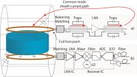
MR Probe Design with On-Coil Digital Receiver - Permission Withheld
David Otto Brunner1, Benjamin Sporrer2,
Christian Vogt3, Jonas Reber1, Josip
Marjanovic1, Luca Bettini2, Lianbo Wu2,
Thomas Burger2, Gerhard Troester3,
Qiuting Huang2, and Klaas P Pruessmann1
1Institute for Biomedical Engineering, University
and ETH Zurich, Zurich, Switzerland, 2Integrated
Systems Laboratory, ETH Zurich, Zurich, Switzerland, 3Electronics
Laboratory and Wearable Computing Group, ETH Zurich, Zurich,
Switzerland
RF receivers placed directly on coil in conjunction with
fibre-optical data transmission can provide various
advantages for the design of array coils in terms of
avoidance of dangerous sheath currents, common-mode noise
and unwanted coil to coil interactions, as well as reduction
of cable weight and routing problems. This helps to further
increase channel counts but also usability or even
wear-ability of RF receive arrays. Here we present first
results from coil designs employing fully integrated (in
130 nm CMOS technology) digital receivers with a form factor
and power requirements to be placed directly on the coil
footpoint.
|
|
0548.
 |
8 |
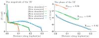
Ultra-fast MRI based transfer function determination for the
assessment of implant safety.
Janot Tokaya1, A.J.E. Raaijmakers1,
J.F. Bakker2, P.R. Luijten1, and
C.A.T. van den Berg1
1Imaging Division, UMC Utrecht, Utrecht,
Netherlands, 2Medtronic,
Eindhoven, Netherlands
Tissue heating induced by sharply peaked scattered electric
fields at the tip of elongated implants is a severe safety
hazard refraining patients with active implants from
undergoing MRI examinations. Transfer functions (TFs) are
widely used in modern safety standards to assess implant
safety. Currently, dedicated setups are required to
determine TFs in challenging and time consuming experiments.
We introduce a new experimental technique based on the
principle of reciprocity and exploiting the ability to map
induced currents with MRI. The proposed method can
accurately determine TFs with high spatial resolution in a
single, quick and relatively simple measurement. It
furthermore has the potential to be applied in heterogeneous
media allowing safety assessment in more realistic scenarios
where the conventional methods become inapplicable.
|
|
0549.
 |
9 |
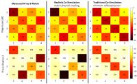
Improving Peak Local SAR Prediction in Parallel Transmit Using
In-situ S-matrix Measurements
Matthew Restivo1, Alexander Raaijmakers1,
Cornelis A.T. van den Berg1, Pedro Crespo-Valero2,
Peter Luijten1, and Hans Hoogduin1
1Center for Imaging Sciences, University Medical
Center Utrecht, Utrecht, Netherlands, 2Zurich
Med Tech, Zurich, Switzerland
We propose a technique where we measure the real S-matrix of
the array/subject setup in-situ and then closely match it in
simulation using circuit co-simulation with a modified cost
function. We show that by accurately simulating coupling,
the B1+ and thus the SAR can be better predicted using FDTD
simulations. Better pTx SAR predictions will ensure RF
safety while reducing the overly conservative pTx SAR
predictions that are used currently.
|
|
0550.
 |
10 |
Percentage of change in the calculated SAR values in human head
during 3T MRI of patients with deep brain stimulation implants:
A computational study of realistic vs. simplified lead
trajectories
Laleh Golestanirad1, Maria Ida Iacono2,
Leonardo M Angelone2, and Giorgio Bonmassar1
1Radiology, Massachusetts General Hospital,
Charlestown, MA, United States, 2Division
of Biomedical Physics, Office of Science and Engineering
Laboratories, Center for Devices and Radiological Health, US
Food and Drug Administration, Silver Spring, MD, United
States
Each year approximately 300,000 patients with medical
implants including deep brain stimulation (DBS) devices are
denied magnetic resonance imaging (MRI) examination due to
safety concerns. One of the major contraindications of MRI
for DBS patient population is due to the potential for
permanent injuries from excessive tissue heating. One open
question when evaluating RF-induced heating with DBS is the
effect of the lead path and the need for patient-specific
information. Using finite element method, we report results
of calculated SAR maps for patient-specific lead paths based
on CT images, and compare them to simplified path
trajectories.
|
|
0551.
 |
11 |
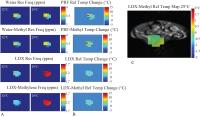
Accurate MR Thermometry by Hyperpolarized 129Xe
Le Zhang1,2, Alex Burant2,3, Andrew
McCallister2,3, Karl Koshlap4, Simone
Degan5, Michael Antonacci2,3, and Rosa
Tamara Branca2,3
1Department of Applied Physical Sciences,
University of North Carolina at Chapel Hill, Chapel Hill,
NC, United States, 2Biomedical
Research Imaging Center, University of North Carolina at
Chapel Hill, Chapel Hill, NC, United States, 3Department
of Physics and Astronomy, University of North Carolina at
Chapel Hill, Chapel Hill, NC, United States, 4Eshelman
School of Pharmacy, University of North Carolina at Chapel
Hill, Chapel Hill, NC, United States, 5Center
for Molecular and Biomolecular Imaging, Duke University,
Durham, NC, United States
A new thermometry method based on the temperature dependence
of lipid-dissolved 129Xe
was proposed, while its accuracy was assessed by direct
comparison with Proton Resonance Frequency (PRF) based MR
thermometry methods. The temperature dependences of chemical
shifts of lipid-dissolved 129Xe,
water and methylene spins were first measured in
vitro with
high accuracy on various fat-rich tissues. The results were
then used to obtain relative temperature maps in
vivo in mice
acclimated at different temperatures. Lipid-dissolved 129Xe
based MR thermometry demonstrated superior accuracy in both in
vivo and in
vitro results
when compared to PRF based MR thermometry in fatty tissues.
|
|
0552.
 |
12 |
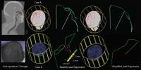
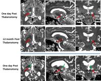
MR Guided Focused Ultrasound Thalamotomy for Essential Tremor -
Maryland Experience
Rao P Gullapalli1, Jiachen Zhuo1,
Dheeraj Gandhi1, Charlene Aldrich2,
Erma Owens1, John Hebel1, Paul Fishman3,
Howard Eisenberg2, and Elias Melhem1
1Diagnostic Radiology & Nuclear Medicine,
University of Maryland School of Medicine, Baltimore, MD,
United States, 2Neurosurgery,
University of Maryland School of Medicine, Baltimore, MD,
United States, 3Neurology,
University of Maryland School of Medicine, Baltimore, MD,
United States
In the context of the remarkable reduction in tremors and
improvement in quality of life at one year following MRgFUS
thalamotomy procedure to treat Essential Tremors in a
recently concluded multi-center trial, we examined pre- and
post-imaging data including an assessment of the accuracy of
MRgFUS targeting of the VIM nuclei and lesion evolution over
12 months. Lesions generated by this procedure were
accurately placed, and matched well with the known location
of VIM nucleus based on anatomical atlases. The lesions
appear to regress in size over a 12 month period but the
therapeutic effect is maintained.
|
|
0553.
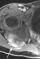 |
13 |
Magnetic Resonance-Guided Focused Ultrasound Treatment of
Extra-Abdominal Desmoid Tumors: A Retrospective Multicenter
Study
Pejman Ghanouni1, Andrew Dobrotwir2,
Alberto Bazzocchi3, Matthew Bucknor4,
Rachelle Bitton1, Jarrett Rosenberg1,
Kristen Telischak5, Maurizio Busacca3,
Stefano Ferrari6, Ugo Albisinni3,
Shannon Walters1, Kristen Ganjoo7,
Alessandro Napoli8, Kim Butts Pauly1,
and Raffi Avedian9
1Radiology, Stanford University, Stanford, CA,
United States, 2Radiology,
The Royal Women's Hospital, Parkview, Australia, 3Diagnostic
and Interventional Radiology, The Rizzoli Orthopaedic
Institute, Bologna, Italy, 4Radiology
and Biomedical Imaging, University of California, San
Francisco, San Francisco, CA, United States, 5Anesthesiology,
Perioperative and Pain Medicine, Stanford University,
Stanford, CA, United States, 6Oncology,
The Rizzoli Orthopaedic Institute, Bologna, Italy, 7Medicine,
Stanford University, Stanford, CA, United States, 8Radiology,
Sapienza University, Rome, Italy,9Orthopaedic
Surgery, Stanford University, Stanford, CA, United States
Desmoid tumors are benign, but can result in pain and
dysfunction. Surgery, radiation and chemotherapy are only
only partially effective and can cause significant
morbidity. MR guided focused ultrasound (MRgFUS) was used to
treat patients with desmoid tumors, sometimes in lieu of
surgery, radiation or chemotherapy. This retrospective
multicenter feasibility study of 15 patients demonstrates
that MRgFUS is safe and that this technique may be used to
control the growth of symptomatic desmoid tumors.
|
|
0554.
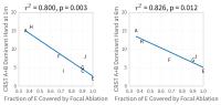 |
14 |
White-Matter-Nulled MP-RAGE Predicts Clinical Outcome of Focused
Ultrasound Thalamic Ablation for Essential Tremor
Jason Su1, Christian Federau2, Thomas
Tourdias3, Manojkumar Saranathan4,
Casey Halpern5, Jaimie Henderson5,
Veronica Santini6, Kim Butts-Pauly2,
Pejman Ghanouni2, and Brian Rutt2
1Electrical Engineering, Stanford University,
Stanford, CA, United States, 2Radiology,
Stanford University, Stanford, CA, United States, 3Neuroradiology,
Bordeaux University Hospital, Bordeaux, France,4Radiology,
University of Arizona, Tucson, AZ, United States, 5Neurosurgery,
Stanford University, Stanford, CA, United States, 6Neurology,
Stanford University, Stanford, CA, United States
This retrospective analysis of MR-guided focused ultrasound
ablation for essential tremor (ET) treatment is centered on
clinical outcome (CRST A+B) and segmentation of ablation
lesions using the white-matter-nulled MP-RAGE contrast.
There is no significant correlation between the volume of
ablation and clinical outcome at 1 month. We identify a new
potential target region based on the best-responding patient
and compute the percent coverage of that region by each
subject’s ablation via nonlinear registration. This measure
correlates with the outcome after 1 month in 8 subjects with
r2=0.8 and p=0.003, a remarkable association that
may aid future targeting strategies in ET.
|
|
0555.
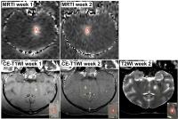 |
15 |
Evaluation of thermal ablation with a 230 kHz transcranial
MRI-guided focused ultrasound system in a large animal model
Nathan McDannold1, Jonathan Sutton1,
Natalia Vykhodtseva1, and Margaret Livingstone2
1Radiology, Brigham and Women's Hospital, Boston,
MA, United States, 2Neurobiology,
Harvard Medical School, Boston, MA, United States
This work evaluated the feasibility of thermal ablation in
the brain in nonhuman primates using a 230 kHz transcranial
MRI-guided focused ultrasound system. We aimed to determine
whether using this low frequency can expand the treatment
envelope where focused ultrasound can be used in the brain
without overheating the skull. We found that focal heating
was increased and skull heating decreased compared to prior
work in macaques that tested a higher frequency version of
this system, suggesting that it can indeed increase this
envelope. Furthermore, closed-loop feedback maintained a low
level of cavitation activity.
|
|
0556.
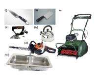 |
16 |
MR safety screening - Is it really worth the time investment?
Derek K Jones1, John Evans1, and
Richard G Wise1
1CUBRIC, Cardiff University, Cardiff, United
Kingdom
Safety screening is considered essential to any MR lab's
working practice. However, it is time-consuming and reduces
participant throughput. Here, we capitalise on the rare
opportunity to experiment with a 3T system prior to it being
decommissioned. We test the hypothesis that large
ferrous-containing objects, if released into the magnet with
a participant inside, do indeed inflict pain and injury. A
selection of house-hold objects was used and a subjective
pain rating employed to quantify the response. Our results
are highly consistent with the main hypothesis, lending
support to continued safety screening. However, we discuss
alternative options to improve workflow
|
|