|
|
|
Plasma # |
|
0091.
 |
1 |
Phaseless Encoding 
Franciszek Hennel1 and
Klaas P. Pruessmann1
1Institute for Biomedical Engineering, University
of Zurich and ETH Zurich, Zurich, Switzerland
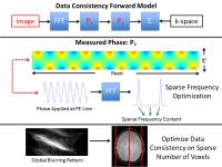
Classically encoded MRI signals are complex and therefore
sensitive to uncontrolled phase variations. We propose an
alternative spatial encoding method which leads to real
positive signals and allows phase fluctuations to be removed
by a simple magnitude calculation before the Fourier
transform. The phase immunity of the method is demonstrated
by recovering an image from a scan with unknown random
receiver phase.
|
|
0092.
 |
2 |
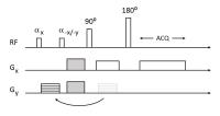
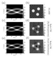
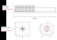
Rabi Modulated Continuous Wave Imaging
James C Korte1, Bahman Tahayori1,
Peter M Farrell1, Stephen M Moore2,3,
and Leigh A Johnston1
1Dept. Electrical and Electronic Engineering,
University of Melbourne, Melbourne, Australia, 2IBM
Research, Melbourne, Australia, 3Dept.
Mechanical Engineering, University of Melbourne, Melbourne,
Australia
The observable periodic magnetisation induced in a spin
system excited by Rabi modulated Continuous Wave excitation
is exploited in this work to construct a new imaging
paradigm. Localised frequency information is encoded in the
steady-state Rabi harmonics, reconstructed as radial
projections of proton density and back-projected to form
images. This form of imaging has the potential to image
samples with ultra-short T2 decay,
which is beneficial for the diagnosis of muscular skeletal
injury and disease.
|
|
0093.
 |
3 |
Gradient Free MRI with a rotating magnet and receiver fields
Somaie Salajeghe1, Paul Babyn2, Logi
Vidarsson3, and Gordon E. Sarty1
1Biomedical Engineering, University of
Saskatchewan, Saskatoon, SK, Canada, 2Medical
Imaging, University of Saskatchewan, Saskatoon, SK, Canada, 3LT
Imaging, Toronto, ON, Canada
Portable MRI can be possible by eliminating gradient coils
and B0 homogeneity
requirements. Relaxing the B0 homogeneity
requirements leads to non-uniform B0 field.
In-homogeneous B0 fields have the potential to
encode spatial information in one direction for use in novel
image encoding schemes. We investigated the possibility of
image reconstruction of the signal from a non-uniform
rotating magnetic field and two rotating RF receivers. Our
results indicate that this is a feasible approach.
|
|
0094.
 |
4 |
Cyclic Continuous Max-Flow: Phase Processing Using the Inherent
Topology of Phase
John Stuart Haberl Baxter1, Zahra Hosseini1,
Junmin Liu2, Maria Drangova3, and
Terry M Peters1
1Biomedical Engineering Graduate Program, Western
University, London, ON, Canada, 2Imaging
Laboratories, Robarts Research Institute, London, ON,
Canada, 3Department
of Medical Biophysics, Western University, London, ON,
Canada
Tissue susceptibility differences manifest in MR phase
images as high-frequency changes in an otherwise smooth
phase background. Two paradigms currently exist for
isolating these changes: one involves phase unwrapping
followed by filtering; the other involves filtering the
complex signal. Both rely on a linear topology, which can
result in artifacts such as phase wraps and shadowing, as
phase is inherently cyclic. This paper introduces the cyclic
continuous max-flow (CCMF) method, which uses optimization
over a cyclic topology to process phase information. More
robust field maps are generated using this approach compared
to the traditional paradigms.
|
 |
0095.
 |
5 |
a-f BLAST: A Non-Iterative Radial k-t BLAST Reconstruction in
Radon Space
Madison Kretzler1, Jesse Hamilton2,
Mark Griswold2,3, and Nicole Seiberlich2,3
1Electrical Engineering, Case Western Reserve
University, Cleveland, OH, United States, 2Biomedical
Engineering, Case Western Reserve University, Cleveland, OH,
United States, 3Radiology,
University Hospitals, Cleveland, OH, United States
This abstract presents a-f BLAST, a non-iterative approach
to non-Cartesian k-t BLAST for radial trajectories, and
demonstrates its use for accelerated cardiac imaging.
|
|
0096.
 |
6 |
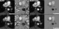
Model-based Reconstruction for Real-Time Phase-Contrast Flow MRI
- Improved Spatiotemporal Accuracy
Zhengguo Tan1, Volkert Roeloffs1, Dirk
Voit1, Arun Joseph1, Markus
Untenberger1, Klaus-Dietmar Merboldt1,
and Jens Frahm1
1Biomedizinische NMR Forschungs GmbH,
Max-Planck-Institute for Biophysical Chemistry, Goettingen,
Germany
The proposed model-based reconstruction technique jointly
computes a magnitude image, a phase-contrast map, and a set
of coil sensitivities from every pair of flow-compensated
and flow-encoded datasets obtained by highly undersampled
radial FLASH. Real-time acquisitions with 5 and 7 radial
spokes per image resulted in 25.6 and 35.7 ms measuring time
per phase-contrast map, respectively. It yields
quantitatively accurate phase-contrast maps with improved
spatial acuity, reduced phase noise, reduced partial volume
effects, and reduced streaking artifacts.
|
|
0097.
 |
7 |
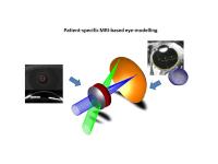
Acquisition of high resolution three-dimensional ocular images
at 7 Tesla to generate patient-specific eye-models for clinical
ray-tracing
Jan-Willem Beenakker1, Lucia Hervella2,
Juan Tabarnero2, Dennis Shamonin1,
Andrew Webb1, Gregorius Luyten1, and
Pablo Artal2
1Leiden University Medical Centre, Leiden,
Netherlands, 2University
of Murcia, Murcia, Spain
Patient-specific three-dimensional eye models obtained using
very high resolution scans on a human 7T MRI system have
been shown to form a much more accurate input for ray
tracing algorithms than the current state-of-the-art
generalized eye models used for clinical ophthalmology.
Using a cued-blink protocol, custom-built phased array coil
and segmentation software, accuracy of less than one-half
dioptre can be achieved using the MRI data. These
patient-specific models should provide much improved input
for therapeutic procedures such as intra-ocular lens
replacement for post-cataract surgery.
|
|
0098.
 |
8 |
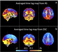
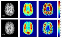
Perfusion map derived from resting state fMRI
Yunjie Tong1, Kimberly P Lindsey1, Lia
M Hocke2, Gordana Vitaliano1,
Dionyssios Mintzopoulos1, and Blaise B Frederick1
1McLean Hospital/Harvard Medical School, Belmont,
MA, United States, 2Hotchkiss
Brain Institute, University of Calgary, Calgary, AB, Canada
Previously, we have demonstrated that we can extract
systemic low frequency oscillation (sLFO) from resting state
(RS) fMRI data and map its dynamic patterns as it moves
through the brain. We have hypothesized that the dynamic
patterns represent the cerebral blood flow. In this study,
we tested this hypothesis by conducting both Dynamic
Susceptibility Contrast scan (bolus tracking) and RS fMRI
scan in health subjects. By comparing the flow patterns of
the bolus with that of sLFO, we found that the flow of sLFO
does represent the blood flow, however, mostly in the
capillaries and veins.
|
|
0099.
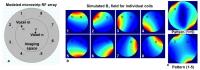 |
9 |
Nonlinear RF spatial encoding with multiple transmit coils based
on Bloch-Siegert shift
Yuqing Wan1, Maolin Qiu1, Gigi Galiana1,
and R. Todd Constable1
1Radiology and Biomedical Imaging, Yale
University, New Haven, CT, United States
We developed a nonlinear encoding method with multiple RF
coils based on the Bloch-Siegert shift. Simulated
reconstructions showed that higher B1 fields and lower
off-resonance frequency shift improves reconstruction
quality. This approach is potentially promising as a
replacement for conventional gradient encoding providing
excellent spatial encoding with essentially silent imaging.
|
|
0100.
 |
10 |
The role of brain viscoelasticity in chronically shunted
hydrocephalus using Magnetic Resonance Elastography
Kristy Tan1, Adam L. Sandler2, Avital
Meiri1, Rick Abbott2, James T.
Goodrich2, Eric Barnhill3, and Mark E.
Wagshul1
1Gruss MRRC, Albert Einstein College of Medicine,
Bronx, NY, United States, 2Department
of Neurological Surgery, Albert Einstein College of
Medicine/Children’s Hospital at Montefiore, Bronx NY, Bronx,
NY, United States, 3Clinical
Research Imaging Centre, University of Edinburgh, Edinburgh,
United Kingdom
Hydrocephalus patients with functioning shunts are often
faced with severe headache disorders. This is believed to be
due to a change in brain viscoelasticity. MRE uses external
mechanical vibrations to induce waves and estimates
viscoelasticity from the wave propagation. This study found
a significant decrease of brain viscoelasticity in
patients (N=14)
compared to controls (N=12) (G* white matter, controls:
1407.82 (SD=111.3) Pa vs patients: 1099.33 (SD=262.86) Pa, p
=0.0001). Additionally, an inverse correlation between
ventricular volume and viscoelasticity in corresponding
lobes was found indicating that brain viscoelasticity may
play a role in hydrocephalus patient’s symptoms such as
headaches.
|
|
0101.
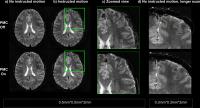 |
11 |
Prospective Motion Correction With NMR Markers Using Only Native
Sequence Elements
Alexander Aranovitch1, Maximilian Haeberlin1,
Simon Gross1, Thomas Schmid1, and
Klaas Paul Pruessmann1
1Institute for Biomedical Engineering, ETH Zurich
and University of Zurich, Zurich, Switzerland
A field-detection based method for prospective motion
correction is proposed which uses the sequence itself for
localizing NMR field probes. No additional gradients or
increase of the sequence duration are required to apply this
method to various MR sequences, such as clinically relevant
spin-warp sequences. The proposed method collects
high-frequency information present due to gradient switching
from multiple short, temporally separated snippets within
one TR. A precision on the order of 10µm and 0.01° (RMS) for
translational and rotational degrees of freedom is obtained.
The method is demonstrated in-vivo with high-resolution
T2*-weighted gradient echo scans.
|
|
0102.
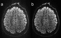 |
12 |
Whole-brain quantitative diffusion MRI at 660 µm resolution in
25 minutes using gSlider-SMS and SNR-enhancing joint
reconstruction
Justin P Haldar1, Qiuyun Fan2, and
Kawin Setsompop2
1Electrical Engineering, University of Southern
California, Los Angeles, CA, United States, 2A.
A. Martinos Center for Biomedical Imaging, Department of
Radiology, Massachusetts General Hospital, Charlestown, MA,
United States
We propose a novel approach to data acquisition and image
reconstruction that achieves high-quality in
vivo whole-brain
human diffusion imaging at (660 µm)3 resolution
in 25 minutes. The approach uses a powerful acquisition
strategy (generalized SLIce Dithered Enhanced Resolution
Simultaneous MultiSlice, or gSlider-SMS) that enables
high-resolution whole-brain imaging in 25 minutes (64
diffusion weightings + 7 b=0
images), but the resulting images suffer from low SNR
without averaging. To address the SNR problem, we utilize a
regularized reconstruction/denoising approach that leverages
the shared spatial structure of different diffusion images. In
vivo results
demonstrate the effectiveness of this approach.
|
|
0103.
 |
13 |
Joint K-space Trajectory and Parallel Imaging Optimization for
Auto-calibrated Image Reconstruction
Stephen Cauley1,2, Kawin Setsompop1,2,
Berkin Bilgic1, Himanshu Bhat3, Borjan
Gagoski2,4, Thomas Witzel1,2, and
Lawrence L. Wald1,2,5
1MGH/HST, Athinoula A. Martinos Center for
Biomedical Imaging, Charlestown, MA, United States, 2Harvard
Medical School, Boston, MA, United States, 3Siemens
Medical Solutions Inc, Malvern, PA, United States, 4Fetal-Neonatal
Neuroimaging & Developmental Science Center, Boston
Children's Hospital, Boston, MA, United States, 5Harvard-MIT
Division of Health Sciences and Technology, MIT, Cambridge,
MA, United States
Fast MRI acquisitions often rely on efficient traversal of
k-space, e.g. Spiral, EPI, and Wave-CAIPI. Limitations in
hardware and other physical effects cause these trajectories
to deviate from the theoretical path, and additional
measurements are typically used to approximate
discrepancies. We propose a joint optimization to directly
estimate trajectory discrepancies simultaneously with the
underlying image, without need for additional
characterization measurements. Model reduction schemes are
introduced to make this optimization computationally
efficient and ensure final image quality. We demonstrate our
approach for a clinically relevant Wave-CAIPI acquisition,
where we accurately optimize across >6million unknowns in
30s on standard vendor hardware.
|
|
0104.
 |
14 |
Looping star: A novel, self-refocusing zero TE imaging strategy
Ana Beatriz Solana1, Anne Menini1, and
Florian Wiesinger1
1GE Global Research, Garching bei Muenchen,
Germany
Zero TE is an extremely efficient 3D pulse sequence which
also has the advantages of low geometrical distortion,
reduced acoustic noise and the capacity of imaging short T2
structure. However, its native contrast is proton density.
Here we present a novel method that allows gradient
refocusing at echo times suitable for fMRI or susceptibility
weighted imaging. As a proof of concept, this new imaging
strategy is tested in phantom experiments.
|
|
0105.
 |
15 |
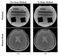
Real-time SENSE reconstruction using pre-scan and E-maps
sensitivities
Muhammad Faisal Siddiqui1, Abubakr Shafique2,
Yousif Rauf Javed2, Talha Ahmad Khan2,
Hamza Naeem Mughal2, Ahmed Wasif Reza1,
Hammad Omer2, and Jeevan Kanesan1
1Electrical Engineering, University of Malaya,
Kuala Lumpur, Malaysia, 2Electrical
Engineering, COMSATS Institute of Information Technology,
Islamabad, Pakistan
FPGA (Field Programmable Gate Array) based application
specific hardware, for real-time Sensitivity Encoding
(SENSE) reconstruction, embedded on the receiver coil system
may provide reconstruction without transferring the data to
the MRI server. This may dramatically decrease the
transmission cost of the system and the image reconstruction
time. This paper proposes an FPGA implementation of SENSE
algorithm using two different sensitivity maps estimation
methods (pre-scan and E-maps). The results show that the
proposed system consumes only 145.64 μs for SENSE
reconstruction (acceleration factor=2), while maintaining
the quality of the reconstructed images with good mean SNR
(29+ dB) and significantly less artefact power (<9×10⁻⁴)
values.
|
|
0106.
 |
16 |
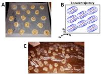
Do try this at home: the role of CAIPIRINHA and non-Cartesian
techniques for increased throughput and aesthetic enhancement in
baking (or vice versa)
Benedikt A Poser1
1Faculty of Psychology and Neuroscience,
Maastricht University, Maastricht, Netherlands
Parallel imaging with controlled aliasing has revolutionised
the way we do MRI, and this may directly translate to the
way we bake. In this work CAIPIRINHA principles are
successfully applied to the baking of cinnamon rolls.
Furthermore, the question is considered of whether
CAIPIRINHA may have been inspired by established baking
practices in the first place.
|
|