|
|
|
Plasma # |
|
0468.
 |
16 |
Dual-modal cardiovascular in vivo assessment in rats using a
highly integrated MPI-MRI hybrid system – initial result 
Jochen Franke1,2, Nicoleta Baxan3,
Ulrich Heinen1, Alexander Weber1,4,
Heinrich Lehr1, Martin Ilg1, Wolfgang
Ruhm1, Michael Heidenreich1, and
Volkmar Schulz2
1Preclinical Imaging Devision, Bruker BioSpin MRI
GmbH, Ettlingen, Germany, 2Physics
of Molecular Imaging Systems, University RWTH Aachen,
Aachen, Germany, 3Biomedical
Imaging Centre, Imperial College London, London, United
Kingdom, 4Institute
of Medical Engineering, University of Lübeck, Lübeck,
Germany
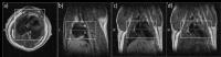
Using a highly integrated Magnetic Particle Imaging –
Magnetic Resonance Imaging hybrid system, a dual-modal
cardiovascular in vivo assessment in rodents under the usage
of a non-toxic Resovist dosage was performed successfully.
The subject was imaged sequentially in both modality modes,
whereas neither subject repositioning nor anesthesia
interruption were required. Complementary datasets were
acquired within a single seamless multi-modal study using
ParaVison6 (Bruker BioSpin, Germany) allowing direct
MRI-based MPI Field-of-View planning. After reconstructing
time-resolved (TR=21.45 ms) 3D MPI images of the
bolus-passage they were successfully fused with a
high-resolution static 3D MRI dataset and visualized as
combined 4D/3D hybrid dataset.
|
 |
0453.
 |
1 |
Hybrid Interleaved Multi-contrast Imaging (HIMI) for
Simultaneous Brain and Carotid Vessel Wall Imaging
Shuo Chen1, Zechen Zhou1, Rui Li1,
Xihai Zhao1, Huijun Chen1, Changwu
Zhou1,2, Bida Zhang3, and Chun Yuan1,4
1Center for Biomedical Imaging Research,
Department of Biomedical Engineering, School of Medicine,
Tsinghua University, Beijing, China, People's Republic of, 2Department
of Radiology, Yangzhou First People's Hospital, Yangzhou,
China, People's Republic of, 3Healthcare
Department, Philips Research China, Shanghai, China,
People's Republic of, 4Vascular
Imaging Laboratory, Department of Radiology, University of
Washington, Seattle, WA, United States
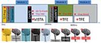
The aim of this study was to develop a Hybrid Interleaved
Multicontrast Imaging (HIMI) sequence for simultaneous brain
and carotid vessel wall imaging. The proposed HIMI sequence
takes advantage of the long delay time in conventional 3D
FLAIR sequence to acquire multi-contrast carotid vessel wall
images. Four healthy volunteers were recruited in this
study. The results indicate that HIMI can generate a
comparable FLAIR image with conventional FLAIR sequence and
three more different contrast weighted (T1w, T2w, gray
blood) carotid vessel wall images with the same scan time as
a single conventional 3D FLAIR sequence.
|
|
0454.
 |
2 |
How Accurately and Precisely Are we Measuring Coronary
Endothelial Function with Radial MRI?
Jerome Yerly1,2, Danilo Gubian3,
Jean-Francois Knebel2,4, Thomas Robin5,
Giulia Ginami1, and Matthias Stuber1,2
1CardioVascular Magnetic Resonance (CVMR)
research center, Department of Radiology, University
Hospital (CHUV) and University of Lausanne (UNIL), Lausanne,
Switzerland, 2Center
for Biomedical Imaging (CIBM), Lausanne, Switzerland, 3University
Hospital (CHUV), Lausanne, Switzerland, 4Laboratory
for Investigative Neurophysiology (The LINE), Departments of
Radiology and Clinical Neurosciences, University Hospital
(CHUV) and University of Lausanne (UNIL), Lausanne,
Switzerland, 5Transport
and Mobility Laboratory (TRANSP-OR), Swiss Federal Institute
of Technology of Lausanne (EPFL), Lausanne, Switzerland
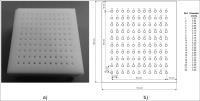
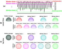
MRI with isometric handgrip exercise was recently proposed
to non-invasively assess coronary endothelial function.
However, the sensitivity of this technique has not yet been
fully investigated. To address this need, we have designed a
phantom that simulates a physiological range of coronary
cross-sectional areas. Radial cine MR images with different
spatial resolutions were acquired under moving conditions.
Cross-sectional areas were automatically measured and
compared to the known nominal values. Statistical analysis
suggests that MRI is capable of distinguishing area changes
in the order of 0.2-0.3mm2, which correspond to a
percentage coronary area change of 3-4% for a 3mm baseline
diameter.
|
|
0455.
 |
3 |
Evaluation of lower extremity arteries with severe wall
calcification in peripheral arterial disease (PAD); comparison
of Fresh blood imaging (FBI) with CT angiography with using a
commercially available calcification removable tool
Katsumi NAKAMURA1,2, Akiyoshi Yamamoto1,
Hiroki Matoba1, Yuji Shintani1, Daiji
Uchiyama1, Seigo Yoshida1, and Mitsue
Miyazaki3
1Radiology, Tobata Kyoritsu Hospital, Kitakyushu,
Japan, 2Nexus
Image Lab, Kitakyushu, Japan, 3Toshiba
Medical Research Institute USA, Inc., Vernon Hills, IL,
United States
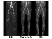
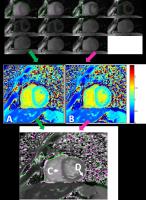
We compared the diagnostic ability of FBI with that of CTA
with using a calcification removal tool in the evaluation
of the lower-extremity arteries with wall calcifications. In
all segments, FBI provided diagnostic images regardless of
the degree of wall calcification. On the contrary, CTA-MIP
and CTA-MIP w/o Ca were strongly affected by
calcification. The diagnostic ability of FBI was
significantly superior to that of CTA-MIP and CTA-MIP w/o Ca
in the moderate to severe calcified arterial segments. In
conclusion, FBI is an accurate and noninvasive alternative
to CTA for the assessment of aortoiliac and lower extremity
arteries in patients with PAD.
|
|
0456.
 |
4 |
A Novel Concept for Motion Suppression Applied to Free-Breathing
3D Whole-Heart Coronary MRA: Respiratory Motion-Resolved
Reconstruction
Davide Piccini1,2, Li Feng3, Gabriele
Bonanno2, Simone Coppo2, Jérôme Yerly2,4,
Ruth P. Lim5, Juerg Schwitter6, Daniel
K. Sodickson3, Ricardo Otazo3, and
Matthias Stuber2,4
1Advanced Clinical Imaging Technology, Siemens
Healthcare, Lausanne, Switzerland, 2Department
of Radiology, University Hospital (CHUV) and University of
Lausanne (UNIL), Lausanne, Switzerland,3Center
for Advanced Imaging Innovation and Research, New York
University School of Medicine, New York City, NY, United
States, 4Center
for Biomedical Imaging (CIBM), Lausanne, Switzerland,5Department
of Radiology, Austin Health and The University of Melbourne,
Melbourne, Australia, 6Division
of Cardiology and Cardiac MR Center, University Hospital of
Lausanne (CHUV), Lausanne, Switzerland
We hypothesize that sparse reconstruction algorithms can be
exploited to reconstruct respiratory motion-resolved 3D MRA
images of the heart without the need for breath-holding,
navigators, or self-navigated respiratory motion correction.
Phantom, volunteer, and patient acquisitions were performed
and image quality was compared to 1D self-navigation for
vessel sharpness, length and diagnostic quality. Respiratory
motion-resolved reconstruction effectively suppresses
respiratory motion artifacts with superior results with
respect to self-navigation. Instead of discarding data or
enforcing motion models for motion correction,
motion-resolved reconstruction makes constructive use of all
respiratory phases to improve image quality, and may lead
coronary MRA closer to clinical practice.
|
|
0457.
 |
5 |
Preliminary Results: Cardiac Cine “Watermark” MRI provides both
Anatomical Function via Magnitude Cine and 2D Myocardial Strain
via Spatially Modulated Phase
Ronald J Beyers1, Davis M Vigneault2,
Dean Schwartz3, Nouha Salibi1,4, David
A Bluemke2, and Thomas Denney1
1MRI Research Center, Auburn University, Auburn
University, AL, United States, 2Radiology
and Imaging Sciences, National Institutes of Health,
Bethesda, MD, United States, 3Anatomy,
Physiology and Pharmacology, Auburn University, Auburn
University, AL, United States, 4MR
R&D, Siemens Healthcare, Malvern, PA, United States
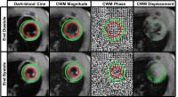
We developed a Cine Watermark (CWM) cine sequence that
produces normal cine magnitude images, plus a grid pattern
of tags added only in the phase for quantitative cine
strain, while requiring no extra operator effort. Using
spatial cosine modulation combined with k-space
sum/differencing produced separate normal magnitude cine and
unique phase-only grid-tags for strain calculation. In
vivorat and human scans demonstrated good magnitude
cine and phase-only quantified displacement. Calculated by
Farneback optical flow algorithm, the peak principle strain,
averaged around the LV for rat = -16.5±2.4 % and human =
-17.8±6.2 % (mean±StdDev).
|
|
0458.
 |
6 |
Fetal cardiac cine imaging from motion-corrected
super-resolution reconstruction of highly-accelerated real-time
MRI
Joshua FP van Amerom1, Maria Kuklisova Murgasova1,
Anthony N Price1, Shaihan J Malik1,
Paul Aljabar2, David A Lloyd1, Kuberan
Pushparajah1,3, Maelene Lohezic1,
Matthew J Fox2, Joanna M Allsop2, Mary
A Rutherford1,2, Reza Razavi1,3, and
Joseph V Hajnal1
1Division of Imaging Sciences & Biomedical
Engineering, King's College London, London, United Kingdom, 2Centre
for the Developing Brain, King's College London, London,
United Kingdom, 3Department
of Congenital Heart Disease, Evelina London Children's
Hospital, London, United Kingdom
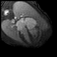
Motion is a key limiting factor in fetal cardiac MRI as the
small, rapidly beating heart is subject to various periodic
and spontaneous motions. Highly accelerated real-time
imaging with high temporal resolution was used to obtain
serial ‘snapshots’ of the fetal heart and surrounding
anatomy that could be motion-corrected and reassembled,
combining several cardiac cycles into a single heartbeat. A
super-resolution reconstruction was applied to increase the
visibility of dynamic anatomical features in the densely
sampled data. The resulting cine images provide a clear
depiction of dynamic cardiac features.
|
|
0459.
 |
7 |
A Golden-Angle Acquisition Coupled with k-t Sparse SENSE
Reconstruction for Fetal Self Retro-Gated Cine Cardiac MRI: an
In Vivo Feasibility Study
Jerome Chaptinel1, Yvan Mivelaz2,
Jerome Yerly1,3, Leonor Alamo1, Milan
Prsa2, Yvan Vial4, François Gudinchet1,
Gregoire Berchier1, Jean-Baptiste Ledoux1,
and Matthias Stuber1,3
1Department of Radiology, University Hospital
(CHUV) and University of Lausanne (UNIL), Lausanne,
Switzerland, 2Department
of Pediatrics, University Hospital (CHUV) and University of
Lausanne (UNIL), Lausanne, Switzerland, 3Center
for Biomedical Imaging (CIBM), Lausanne, Switzerland, 4Department
of Gynecology-Obstetrics, University Hospital (CHUV) and
University of Lausanne (UNIL), Lausanne, Switzerland
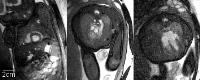
Fetal cardiac cine MRI is challenging due to the lack of an
ECG trigger signal, fetal motion, and the need for both a
high spatial and temporal resolution. To overcome these
hurdles, we have developed and tested a new
acquisition-reconstruction paradigm: data collection was
performed with a continuous radial golden-angle acquisition
and cine images were reconstructed with a k-t sparse SENSE
algorithm. A cardiac gating signal was extracted from the
images themselves and supported self retro-gated
reconstructions in which motion-corrupted data were
excluded. Fetal self retro-gated cardiac cine images with
high temporal and spatial resolution were successfully
obtained in pregnant patients.
|
|
0460.
 |
8 |
Accurate T1 mapping in patients with Pulmonary Hypertension and
age matched volunteers using synthetic image based registration
Laura Claire Saunders1, Neil J Stewart1,
Charlotte Hammerton1, David Capener1,
Valentina O Puntmann2, David G Kiely3,
Martin J Graves4, Andy Swift1, and Jim
M Wild1
1Academic Unit of Radiology, The University of
Sheffield, Sheffield, United Kingdom, 2Department
of Cardiovascular Imaging, Kings College London, London,
United Kingdom, 3The
University of Sheffield, Sheffield, United Kingdom, 4University
of Cambridge School of Clinical Medicine, University of
Cambridge, Cambridge, United Kingdom
Patients with suspected pulmonary hypertension (n=94) and
healthy volunteers (n=26) underwent T1 mapping of the right
ventricle with a Modified Look Locker inversion recovery
(MOLLI) sequence at 1.5T. MOLLI images were registered using
pairwise registration to synthetic images produced using a
simplified inversion recovery model to correct cardiac or
respiratory motion. 89% of patients and 100% of healthy
volunteers were successfully registered, with mean T1s of
1.00±0.10s and 0.97±0.06s (septal), 1.05±0.11s and
0.97±0.06s (right ventricular insertion point) and
1.02±0.11s and 1.04±0.13s (right ventricular free wall)
respectively.
|
|
0461.
 |
9 |
Towards a quantitative MRI-based measure of disease burden in
patients with atrial fibrillation
Maurce Pradella1, Sven Knecht2,
Michael Kühne2, Aline Mühl2, Tobias
Reichlin2, Gian Voellmin2, David Conen2,
Jens Bremerich1, Stefan Osswald2,
Christian Sticherling2, and Bram Stieltjes1
1Department of Radiology, University of Basel
Hospital, Basel, Switzerland, 2Department
of Cardiology, University of Basel Hospital, Basel,
Switzerland
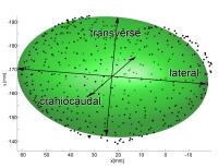
Atrial fibrillation (AF) is a common disease and associated
with myocardial infarction, stroke and dementia. We propose
a new approach based on the sphericity of fitted ellipsoids
in left atriums of patients with AF. Our results show a
strong correlation between sphericity of these ellipsoids
and burden of disease and may serve as an objective
surrogate parameter in the future.
|
|
0462.
 |
10 |
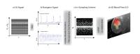
Joint Processing of Highly Accelerated Multi-Directional PC-MRI
Data Using ReVEAL
Adam Rich1, Lee C. Potter1, Ning Jin2,
Juliana Serafim da Silveira3, Orlando P.
Simonetti3, and Rizwan Ahmad3
1Electrical and Computer Engineering, The Ohio
State University, Columbus, OH, United States, 2Siemens
Medical Solutions, The Ohio State University, Columbus, OH,
United States, 3Dorthy
M. Davis Heart and Lung Research Institute, The Ohio State
University, Columbus, OH, United States
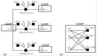
Phase-contrast magnetic resonance is a powerful tool for
study of cardiac flow, but clinical application is limited
to planar imaging of one velocity component. This abstract
demonstrates three-directional flow imaging using a single
breath-hold acquisition. Imaging is accomplished by jointly
processing all encodings and frames; Bayesian reconstruction
leverages image structure via both wavelet compression and
statistical relations among velocity encoded images. Digital
phantom results show accurate estimation of stroke volume
and peak velocity, with significant reductions in bias and
variance, as well as over 30% increase in Pearson
correlation coefficient, compared to L1-SENSE. In vivo
results demonstrate repeatable flow estimation.
|
|
0463.
 |
11 |
A new hybrid approach for quantitative multi-slice myocardial
DCE perfusion
Edward DiBella1, Devavrat Likhite1,
Ganesh Adluru1, Chris Welsh1, and
Brent Wilson1
1University of Utah, Salt Lake City, UT, United
States
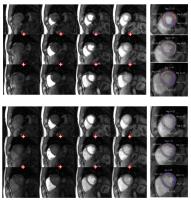
Here we propose a unique perfusion acquisition that uses one
saturation pulse per heartbeat. This combined with
simultaneous multi-slice (SMS) methods allows for acquiring
the same set of slices continuously through the cardiac
cycle. This has a number of advantages including the ability
to retrospectively reconstruct an accurate arterial input
function (AIF) and optimized systolic/diastolic frames, or
other portions of the cardiac cycle. The approach proposed
here acquires both k-space rays that reflect the influence
of the saturation pulse and other rays that reflect the
steady-state GRE contrast, and thus is termed the “hybrid”
method. Preliminary quantitative results including
comparisons to more standard methods in two subjects show
the promise of this SMS approach.
|
|
0464.
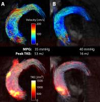 |
12 |
Added Value of Phase-Contrast MRI based Turbulent Kinetic Energy
Quantification for the Assessment of Aortic Stenosis Severity - Permission Withheld
Alexander Gotschy1,2, Christian Binter1,
Simon H Sündermann3, Michelle Frank2,
Felix C Tanner2, Robert Manka2, and
Sebastian Kozerke1
1Institute for Biomedical Engineering, University
and ETH Zurich, Zurich, Switzerland, 2Department
of Cardiology, University Hospital Zurich, Zurich,
Switzerland, 3Division
of Cardiovascular Surgery, University Hospital Zurich,
Zurich, Switzerland
Aortic stenosis (AS) is the most prevalent valvular heart
disease. Risk stratification and the decision for valve
replacement are mostly based on echocardiography and
symptomaticity. This work investigates the additional value
of quantifying Turbulent Kinetic Energy (TKE) for the
assessment of AS severity beyond echocardiographic measures.
TKE was confirmed to be significantly elevated in patients
with AS compared to controls. While TKE showed only weak
correlation with the echocardiographic Mean Pressure
Gradient, TKE allowed to discriminate the impact of bicuspid
aortic valves and aortic dilatation on energy loss in AS
patients; effects which are not assessable by standard
echocardiographic measures.
|
|
0465.
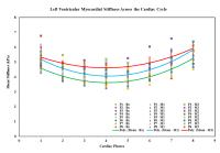 |
13 |
In-Vivo Quantification of Myocardial Stiffness in Heart Failure
with Preserved Ejection Fraction Using Magnetic Resonance
Elastography: Assessment in a Porcine Model
Ria Mazumder1,2, Samuel Schroeder2,3,
Xiaokui Mo4, Bradley D Clymer5,
Richard D White2,6, and Arunark Kolipaka2,6
1Department of Electrical and Computer
Enginerring, The Ohio State University, Columbus, OH, United
States, 2Department
of Radiology, The Ohio State University, Columbus, OH,
United States,3Department of Mechanical
Engineering, The Ohio State University, Columbus, OH, United
States, 4Department
of Biomedical Informatics, The Ohio State University,
Columbus, OH, United States,5Department of
Electrical and Computer Engineering, The Ohio State
University, Columbus, OH, United States, 6Department
of Internal Medicine-Division of Cardiovascular Medicine,
The Ohio State University, Columbus, OH, United States
Left ventricular (LV) myocardial stiffness (MS) is elevated
in heart failure with preserved ejection fraction (HFpEF)
and hence has the potential to be used as a diagnostic tool.
Current clinical techniques to estimate LV MS are invasive
in nature and provides global stiffness measurements.
Therefore, in this study, we implement cardiac magnetic
resonance to investigate temporal alteration in LV MS over a
two month period of disease progression in a porcine model
induced with HFpEF. The alteration in LV MS is compared
against change in mean LV pressure, LV thickness,
circumferential strain and MRI relaxometry parameters.
|
|
0466.
 |
14 |
Free breathing self-gated PC-MRI with Pseudo Random sampled
kt-Sparse-Sense
Volker Herold1, Patrick Winter1,
Philipp Mörchel2, Fabian Gutjahr1, and
Peter Michael Jakob1
1Department of Experimental Physics 5, University
of Wuerzburg, Wuerzburg, Germany, 2Research
Center for Magnetic Resonance Bavaria e.V., Wuerzburg,
Germany
Phase-Contrast (PC) cine MRI is an established method for
the assessment of blood flow and tissue motion patterns in
cardiovascular MRI. In this paper we presented a highly
accelerated self-gated PC-MRI-sequence based on free
breathing random sampled data acquisition. Data acquired
during respiratory motion as well as any other source of
undesirable motion can be excluded from the post-processing.
Moreover ECG-signal acquisition which is prone to
distortions especially at higher field strength can be
avoided. The high flexibility of data processing would also
allow the correction of unstable heart rate during the
measurement.
|
|
0467.
 |
15 |
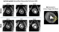
End-systolic Myocardial Perfusion MRI Using a Hybrid 2D/3D
Steady-State Acquisition Scheme: Towards Reliable Detection of
Subendocardial Ischemia in Coronary Microvascular Dysfunction
Behzad Sharif1, Rohan Dharmakumar1,
Daniel Berman2, Debiao Li1, and Noel
Bairey Merz2
1Biomedical Imaging Research Institute, Dept of
Biomedical Sciences, Cedars-Sinai Medical Center, Los
Angeles, CA, United States, 2Heart
Institute, Cedars-Sinai Medical Center, Los Angeles, CA,
United States
A significant portion of patients with ischemic heart
disease suffer from coronary microvascular dysfunction.
Despite intense interest and several recent advancements,
reliable diagnosis of coronary microvascular dysfunction on
the basis of stress first-pass perfusion (FPP) cardiac MRI
is an ongoing challenge. We hypothesized that
high-resolution systolic FPP imaging can detect diffuse
vasodilator-induced subendocardial defects and transmural
perfusion gradients consistent with microvascular
dysfunction in a swine model of diet-induced diabetes with
no obstructive disease. To this end, we developed,
optimized, and tested a new high-resolution FPP method with
hybrid 2D/3D excitation capable of imaging all myocardial
slices at the end-systolic phase.
|
|