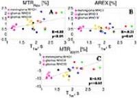
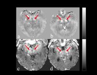
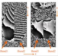
ISMRM 24th Annual Meeting & Exhibition • 07-13 May 2016 • Singapore

Traditional Poster Session: Contrast Mechanisms

1470 -1482 Perfusion & Permeability: Contrast Agent Methods
1483 -1499 Arterial Spin Labeling
1500 -1511 Endogenous CEST & MT
1512 -1526 CEST: Agents & Methods
1527 -1542 Contrast Mechanisms: Relaxation Based
1543 -1561 Magnetic Susceptibility
1562 -1573 Contrast Mechanisms: Novel Ways of Imaging


1470.   
3D CMRO2 mapping in human brain with direct 17O-MRI and proton-constrained iterative reconstructions
Dmitry Kurzhunov1, Robert Borowiak1,2, Marco Reisert1, Philipp Wagner1, Axel Krafft1,2, and Michael Bock1
1University Medical Center Freiburg, Dept. of Radiology - Medical Physics, Freiburg, Germany, 2German Cancer Research Center (DKFZ), German Cancer Consortium (DKTK), Heidelberg, Germany
This work presents a comparison analysis of different reconstruction techniques for quantification of 3D maps of the cerebral metabolic rate of oxygen consumption (CMRO2) in human brain. Several 17O-MR 3D data sets of a healthy volunteer’s brain were acquired at a clinical 3 Tesla MR system with inhalation of 70%-enriched 17O2 gas. Iterative image reconstruction procedures, e.g. where different co-registered 1H MR image data sets of high spatial resolution act as edge-preserving constraints, are compared and used to improve the image quality and the precision of CMRO2 mapping. Anisotropic Diffusion as non-Homogeneous Constraint (ADHOC) is shown to be superior.


1471.   
Hyperemic Blood-Oxygen Level Dependent MRI of the foot for identifying perfusion defects in those with peripheral arterial disease
Tomoki Fujii1, Krishna R. Singh2, Bill Bordeau3, Joao A. Lima1, and Bharath Ambale-Venkatesh1
1Johns Hopkins University, Baltimore, MD, United States, 2Prairie Vascular Institute, Springfield, IL, United States, 3Zimmer Biomet Biologics, Warsaw, IN, United States
Peripheral artery disease is a major public health concern particularly among the elderly. Although measures such as ankle-brachial index and segmental pressures have been used to characterize disease severity, MRI techniques allow us to assess subclinical vascular function and morphology and may help improve our understanding of vascular adaptations. In a small pilot study, we test whether hyperemia-induced BOLD oxygenation changes are a viable measure of tissue oxygenation in the foot and if they represent the reduced oxygenation seen in PAD.


1472.   
Contrast Enhanced MRI Reveals Perplexing T2 effect of Aggregate Forming Compounds In the Murine Placenta
Marina Lysenko1, Noam Ben-Eliezer1, Inbal E Biton2, Joel R Garbow3, and Michal Neeman1
1Biological Regulation, Weizmann Institute of Science, Rehovot, Israel, 2Veterinary Resources, Weizmann Institute of Science, Rehovot, Israel, 3Biomedical Magnetic Resonance Laboratory, Mallinckrodt Institute of Radiology, Washington University, St. Louis, MO, United States
The murine placenta is a complex organ, consisting of different cell compartments that greatly influence its blood-flow pattern. Dynamic contrast enhanced (DCE) MRI of murine placental perfusion has been reported previously using both low and high MW contrast media. In this study, we used high-MW, albumin-based macromolecular contrast agent that does not cross the placental barrier, but, instead, forms contrast-based aggregates that accumulate in the maternal vasculature simultaneously with active contrast internalization by trophoblast cells in the labyrinth .To interpret the observed data, we suggest a novel model for describing feto-maternal processing and aggregate formation of labeled albumin in placental DCE-MRI experiments.


1473.   
Tissue Partial Volume Correction of Perfusion Maps in Dynamic Susceptibility Contrast MRI
André Ahlgren1, Ronnie Wirestam1, Freddy Stĺhlberg1,2,3, and Linda Knutsson1
1Department of Medical Radiation Physics, Lund University, Lund, Sweden, 2Department of Diagnostic Radiology, Lund University, Lund, Sweden, 3Lund University Bioimaging Center, Lund University, Lund, Sweden
Partial volume effects (PVE) can significantly affect parameter estimates in perfusion MRI. In contrast to arterial spin labeling (ASL), the impact of PVEs in dynamic susceptibility contrast MRI (DSC-MRI) has not yet been well established. In this work, we assess and compare partial volume correction (PVC) of DSC-MRI and ASL data in 20 healthy subjects. PVC reduced the tissue volume dependence of perfusion estimates in DSC-MRI and ASL. White matter perfusion maps were of higher quality for DSC-MRI. However, for PVC of DSC-MRI we used several assumptions which need further evaluation.


1474.   
Improved Vascular Transport Function Characterization in DSC-MRI via Deconvolution with Dispersion-Compliant Bases
Marco Pizzolato1, Rutger Fick1, Timothé Boutelier2, and Rachid Deriche1
1Athena Project-Team, Inria Sophia Antipolis - Méditerranée, Sophia Antipolis, France, 2Olea Medical, La Ciotat, France
Bolus dispersion phenomena affect the residue function computed via deconvolution of DSC-MRI data. Indeed the obtained effective residue function can be expressed as the convolution of the true one with a Vascular Transport Function (VTF) that characterizes the dispersion. The state-of-the-art technique CPI+VTF allows to estimate the actual residue function by assuming a model for the VTF. We propose to perform deconvolution representing the effective residue function with Dispersion-Compliant Bases (DCB) without assumptions on the VTF, and then apply the CPI+VTF on DCB results. We show that DCB improve robustness to noise and allow to better characterize the VTF.


1475.   
Feasibility and Value of Quantitative Dynamic Contrast Enhancement MR imaging in Evaluation of Orbital Masses in Adults
Liyuan Song1, Lizhi Xie2, and Junfang Xian1
1Department of Radiology,Beijing Tongren Hospital,Capital Medical Universityy, Beijing, China, People's Republic of, 2GE Healthcare, MR Research China, Beijing, Beijing, China, People's Republic of
This work assessed the feasibility of quantitative parameters derived from dynamic contrast enhanced MR imaging (DCE-MRI) and evaluate the value of quantitative dynamic contrast enhanced MR imaging in the diagnosis and differential diagnosis of orbital masses in adults. From the result we can see that it is feasible that quantitative parameters of orbital masses can be derived from DCE-MRI. ROI onf the earliest and most enhanced area was optimal for distinguishing benign masses from malignant masses in orbit.


1476.   
Assessing the repeatability and reproducibility of contrast time courses from a dynamic MRI flow phantom: initial results and experiences
Jacob M. Johnson1, Leah C. Henze Bancroft2, James H. Holmes3, Edward F. Jackson1,2, Frank R. Korosec1,2, Courtney K. Morrison2, Roberta M. Strigel1, Kang Wang3, and Ryan J. Bosca1
1Radiology, University of Wisconsin, Madison, WI, United States, 2Medical Physics, University of Wisconsin, Madison, WI, United States, 3GE Healthcare, Madison, WI, United States
The recent development of a multi-modality, commercially available, dynamic flow phantom has provided a means of assessing the repeatability, reproducibility, and fidelity of contrast concentration time courses. In this work, we aimed to develop and evaluate a methodology for assessing the repeatability and reproducibility contrast concentration time courses derived from dynamic contrast-enhanced MR images of this dynamic flow phantom.


1477.   
Improved Image Quality when estimating Perfusion Parameters using Bayesian Fitting Algorithm
Irene Klćrke Mikkelsen1, Anna Tietze1,2, Lars Ribe1, Anne Obel3, Mikkel Bo Hansen1, and Kim Mouridsen1
1CFIN, Aarhus University, Aarhus, Denmark, 2Dept. of Neuroradiology, Aarhus University Hospital, Aarhus, Denmark, 3Neuroradiology, Aarhus University Hospital, Aarhus, Denmark
Dynamic Contrast Enhanced Perfusion Imaging (DCE) allows for quantification of the blood-brain barrier integrity in tumor patients. A key post-processing step is to fit a hemodynamic model to DCE data. The fitting procedure can, however, cause spurious voxels and image degradation. We compared the widely used Levenberg-Marquardt fitting algorithm to a Bayesian algorithm. Image quality was assessed in 42 tumor patients. The Bayesian approach provided the highest image quality scores. This was confirmed in simulated data with fewer outliers (spurious voxels) when using the Bayesian approach. The hemodynamic two-compartment model that separates cerebral blood flow and leakage, provides reliable Ve images, when the robust Bayesian fitting algorithm is used.


1478.   
DCE-MRI at high temporal resolution using undersampled radial FLASH: A phantom study
Jost Michael Kollmeier1, Volkert Roeloffs1, and Jens Frahm1
1Biomedizinische NMR Forschungs GmbH, Max-Planck-Institut für biophysikalische Chemie, Göttingen, Germany
We present a DCE-MRI experiment using a commercial perfusion flow phantom for quantitative analysis of image series with high spatial and high temporal resolution (107 ms). Both can be obtained by current real-time MRI methods, i.e. radial undersampled radial FLASH and image reconstruction by nonlinear inversion (NLINV). Contrast agent bolus tracking with high CNR and quantitative parameter maps are presented.


1479.   
Dynamic contrast-enhanced MRI in primary rectal cancer: correlation with histologic prognostic factors
Zhe Han1,2, Juan Chen2, Min Chen2, Chen Zhang2, and Dandan Zheng3
1Chinese Academy of Medical Science and Peking Union Medical College, Beijing, China, People's Republic of, 2Department of Radiology, Beijing Hospital, Beijing, China, People's Republic of, 3GE Healthcare, MR Research China, Beijing, China, People's Republic of
In this study we compared the association of dynamic contrast-enhanced (DCE)-derived quantitative parameters with the histologic grade, N-stage, epidermal growth factor receptor (EGFR) expression and K-RAS gene mutation of primary rectal cancer.Significant correlations were found between Ktrans values and N-stage, Ktrans values and EGFR expression, Kep values and EGFR expression. DCE-derived quantitative parameters may be a promising imaging biomarker of tumor aggressiveness and prognosis.


1480.   
Temporal resolution improvement of calibration-free dynamic contrast-enhanced MRI with compressed sensing optimized turbo spin echo: The effects of replacing turbo factor with compressed sensing accelerations
SoHyun Han1 and HyungJoon Cho1
1Biomedical Engineering, Ulsan National Institute Science and Technology, Ulsan, Korea, Republic of
In vivo estimation of Gd-concentration in dynamic contrast enhanced (DCE)-MRI are often compromised from non-negligible T2* effect and limited temporal resolution. In this study, we introduce compressed sensing assisted turbo spin echo (CS-TSE) acquisition to provide accurate Gd-concentration estimation without the need of additional signal calibration, and to achieve a sub-second temporal resolution with extended slice coverage. Phantom verification followed by in vivo arterial input function (AIF) studies validated the faithful concentration estimation of CS-TSE. Robust measurement of first-pass kidney feeding AIF with increased temporal resolution was demonstrated with sub-second temporal resolution.


1481.   
Gradient optimization using active contour for rapid breast DCE-MRI
Pavan poojar1, Bikkemane Jayadev Nutandev2, Nithin N Vajuvalli1, C.K. Dharmendra Kuman2, Ramesh Venkatesan3, and Sairam Geethanath1
1Medical Imaging Research Centre, Dayananda Sagar College of Engineering, Bangalore, India, 2Bangalore, India, 3Wipro-GE Healthcare, Bangalore, India
In dynamic scans, the significant values of k-space dependent on the shape of the organ which leads to arbitrary k-space trajectories. Gradient optimization for arbitrary k-space trajectory using active contour is a new acquisition technique that has been applied on six DCE breast data. The arbitrary k-space trajectory was obtained by active contour and gradients are optimized by employing convex optimization based on hardware constraints. Image reconstruction was performed using Fourier transform with density compensation.  $$$K^{trans}$$$ and Ve maps were generated for different acceleration factors (1x, 2x, 3x, 4x and 10x) on tumor region to demonstrate utility of the method.


1482.   
Dictionary based approach for accelerated determination of Pharmacokinetic maps using Partial Least Square regression
Nithin N Vajuvalli1, Shivaprasad Ashok Chikop1, and Sairam Geethanath1
1Medical Imaging Research Centre, Dayananda Sagar Institutions, Bangalore, India
This study is of relevance to MR researchers interested in DCE-MRI. Tofts model is a well-established two compartment model to determine the PharmacoKinetic (PK) maps, which is time consuming due to the presence of iterative curve fitting for each voxel. Current work focuses on the application of Partial Least Square (PLS) regression modelling to determine PK maps. PLS is a statistical method based on PCA and linear regression that provides the relationship between the predictor and response variables. We report 95-98% reduction in time as compared to curve fitting approaches for in silico phantoms and in vivo breast DCE data.

1483.   
Reproducibility and Variability of a Look-Locker FAIR ASL Sequence for Quantitative Measurement of Myocardial Blood Flow in Healthy Human Volunteers at 3T
Graeme A Keith1, Christopher T Rodgers1, Michael A Chappell2, and Matthew D Robson1
1Oxford Centre for Clinical Magnetic Resonance Research, University of Oxford, Oxford, United Kingdom, 2Institute of Biomedical Engineering, University of Oxford, Oxford, United Kingdom
A previously presented arterial spin labelling (ASL) method was tested for reproducibility and variability. These measures are important to consider when planning a clinical study. The results presented show that the method has the sensitivity required to detect changes in MBF in pathology and under stress. The variation in individuals is shown to be less than across the sample as a whole. This knowledge will be useful in the planning of future clinical research studies.


1497.   
Multiphase pCASL for imaging blood flow in rodent brains
James R Larkin1, Manon A Simard1, Alexandre A Khrapitchev1, Kevin J Ray1, James A Meakin2, Paul Kinchesh1, Sean Smart1, Peter Jezzard2, Michael A Chappell3, and Nicola R Sibson1
1CRUK and MRC Oxford Institute for Radiation Oncology, Department of Oncology, University of Oxford, Oxford, United Kingdom, 2FMRIB Centre, University of Oxford, Oxford, United Kingdom, 3Institute of Biomedical Engineering, Department of Engineering, University of Oxford, Oxford, United Kingdom
Arterial spin labelling perfusion imaging in the rodent brain is easily confounded by off-resonance effects at the tagging plane. These effects are a consequence of the higher field strengths used pre-clinically and the nearby air cavities in the rodent head and neck, something not as problematic in the clinic. By implementing a multiphase pCASL sequence with eight phases spaced at 45° and lying between 0 and 315°, it is possible to obtain data to allow fitting thereby accounting for any off-resonance effects. This process dramatically improves image quality without excessively affecting acquisition time. 


1484.   
T2 Relaxation of Human Blood  at 3T Revisited:  In Vivo and In Vitro Meaurement using TRUST MRI
Adam Bush1, Jon Detterich2, Thomas Coates3, Herbert Meiselman4, and John Wood1
1Biomedical Engineering/ Cardiology, University of Southern California/ Children's Hospital Los Angeles, Los Angeles, CA, United States, 2Cardiology, Children's Hospital Los Angeles, Los Angeles, CA, United States,3Hematology, Children's Hospital Los Angeles, Los Angeles, CA, United States, 4Physiology and Biophysics, University of Southern California, Keck School of Medicine, Los Angeles, CA, United States
Precise knowledge of the T2 of blood (T2b) is required for spin-echo based blood oxygenation determination methods such as TRUST (T2 Relaxation Under Spin Tagging).  In this study we measure the T2b in vivo and vitro using TRUST MRI.  After correcting for physiologic variable we found that our model of T2b is statistically significantly different from the models used by other groups.  We conclude those model lead to errors in derived parameters including oxygen saturation, oxygen extraction fraction and cerebral metabolic rate.


1485.   
Reproducibility of abdominal perfusion imaging using velocity selective arterial spin labeling
Marijn van Stralen1, Esben Thade Petersen1,2, Jeroen Hendrikse1, and Clemens Bos1
1University Medical Center Utrecht, Utrecht, Netherlands, 2Danish Research Center for MR, Hvidovre, Denmark
Abdominal perfusion imaging using contrast media injection is potentially nephrotoxic. Arterial spin labeling (ASL), employing endogenous contrast, was shown using spatially selective labeling strategies. We investigated the reproducibility of velocity selective ASL (VS-ASL), which eliminates delicate label planning and possibly improves perfusion SNR by labeling closer to the target tissue. We show that abdominal VS-ASL is feasible in healthy volunteers and overcome labeling artifacts by pacing and triggering the acquisition with good temporal SNR. However, VS-ASL is sensitive to motion during readout, deteriorating reproducibility. It could benefit from outlier rejection techniques and retrospective motion correction.  


1486.   
Whole brain volumetric perfusion imaging with high spatial resolution using simultaneous multi-slab (SMSB) 3D GRASE pCASL
Yi Wang1, Xingfeng Shao1, Steen Moeller2, and Danny JJ Wang1
1Neurology, UCLA, Los Angeles, CA, United States, 2Center of Magnetic Resonance Research, University of Minnesota, Minneapolis, MN, United States
The temporal SNR of simultaneous multi-slice (SMS) 2D EPI ASL has been shown to be inferior to that of 3D background suppressed GRASE. In this work, we present a novel simultaneous multi-slab (SMSB) 3D GRASE sequence for volumetric pCASL imaging with high spatial resolution. The image quality of SMSB-GRASE was evaluated and compared to a standard 3D GRASE pCASL sequence. Preliminary results demonstrated the feasibility for whole-brain volumetric perfusion imaging at a high spatial resolution, although the drop in RF slice profiles in the overlapped boundary slices still needs to be addressed in future work.


1487.   
A Perfusion Phantom for Arterial Spin Labeled MRI
Hyo Min Lee1,2, Marta Vidorreta3,4, Yulin Vince Chang3, and John Alan Detre3,4
1Bioengineering, University of Pennsylvania, Philadelphia, PA, United States, 2Institute for Biomedical Engineering, University and ETH Zürich, Zürich, Switzerland, 3Radiology, University of Pennsylvania, Philadelphia, PA, United States, 4Neurology, University of Pennsylvania, Philadelphia, PA, United States
ASL MRI is an appealing biomarker for clinical research and management, but ASL MRI sequences are difficult to calibrate because a reliable phantom for simulating tissue-specific perfusion has yet to be developed. In this work, we describe a prototype perfusion phantom based on 3D printed vessels and mock parenchyma that may allow reliable, ex-vivo assessments of ASL sequences.


1488.   
Improved Pseudo Continuous Arterial Spin Labeling Efficiency Robustness to Off Resonance and High Velocity
Li Zhao1 and David C Alsop1
1Radiology, Beth Israel Deaconess Medical Center, Boston, MA, United States
Pseudo continuous arterial spin labeling (pCASL) studies can be degraded by magnetic field variations at the labeling plane. We demonstrate through simulations that high velocity efficiency is particularly vulnerable to field offsets. By changing labeling parameters from published recommendations and/or introducing a new RF pulse, the off-resonance sensitivity and peak systolic velocity sensitivity of pCASL can be reduced. Preliminary experimental comparisons of parameters are reported.


1489.   
3D Arterial Spin Labeling in breast cancer: A case study.
Thorsten Honroth1, Suzan Vreemann2, Marco Vicari1, Hendrik Laue1, Ritse Mann2, and Matthias Günther1,3,4
1Fraunhofer MEVIS, Bremen, Germany, 2Radboud University Medical Center, Nijmegen, Netherlands, 3University of Bremen, Bremen, Germany, 4mediri GmbH, Heidelberg, Germany
A single-shot 3D arterial spin labeling (ASL) sequence has been developed and optimized for breast cancer imaging. In a case study, its ability to measure the perfusion of a tumor without contrast agents is demonstrated. The resulting ASL perfusion-weighted image of the tumor shows high correspondence with the subtraction image of the contrast-enhanced measurement.


1490.   
Priors-guided adaptive outlier cleaning for arterial spin labeling perfusion MRI
Ze Wang1,2
1Hangzhou Normal University, Hangzhou, China, People's Republic of, 2Psychiatry and Radiology, University of Pennsylvania, PHILADELPHIA, PA, United States
ASL CBF signal is derived from the difference between successive labeling and no-labeling images. The low signal-to-noise-ratio and the pairwise subtraction can then result in outliers, which can significantly degrade CBF quantification quality in a typical several minutes scan. A priors-guided adaptive outlier cleaning algorithm was verified in this study. Our results showed that the proposed method improved both CBF quantification quality and CBF measurement stability.


1491.   
Improving SNR in pulsed arterial spin labeling using multiple inversion modules (MM-PASL)
Jia Guo1, Richard B. Buxton1, and Eric C. Wong1,2
1Radiology, UC San Diego, La Jolla, CA, United States, 2Psychiatry, UC San Diego, La Jolla, CA, United States
The bolus duration in pulsed arterial spin labeling (PASL) is typically short, resulting in low SNR. We propose using multiple inversion pulses to increase the total bolus duration for improved SNR. In this study, a wedge-shaped inversion was combined with a regular slab inversion and a QUIPSS II pulse to lengthen the total bolus duration while keeping the ASL signal quantitative. The preliminary in vivo results showed an SNR improvement of 54% in gray matter, in good agreement with theory, compared to a regular PASL scan. The mean GM CBF values were consistent with PCASL reference scans. This new labeling method should benefit studies using PASL. 


1492.   
A simple and reliable perfusion phantom to measure precise and repeatable arterial spin labeled quantitative perfusion
Joshua S. Greer1,2, Keith Hulsey2, Robert E. Lenkinski2,3, and Ananth J. Madhuranthakam2,3
1Bioengineering, University of Texas at Dallas, Richardson, TX, United States, 2Radiology, UT Southwestern Medical Center, Dallas, TX, United States, 3Advanced Imaging Research Center, UT Southwestern Medical Center, Dallas, TX, United States
Arterial spin labeling (ASL) is a rapidly growing area of interest, primarily because of its ability to provide quantitative perfusion maps non-invasively. But, for the technique to be adopted for clinical use, these quantitative measurements need to be accurate and robust, which will require a quality controlled perfusion phantom to ensure consistency for different magnet strengths and manufacturers. In this study, we demonstrate a simple perfusion flow phantom that can be used to test the precision and repeatability of ASL perfusion measurements.


1493.   
Considerations of cardiac phase can improve ASL quality in multiple settings
Yang Li1,2, Deng Mao1,2, Zhiqiang Li3, Michael Schär1, James G. Pipe3, and Hanzhang Lu1
1Russell H. Morgan Department of Radiology and Radiological Science, Johns Hopkins University, Baltimore, MD, United States, 2Graduate School of Biomedical Sciences, University of Texas Southwestern Medical Center, Dallas, TX, United States, 3Imaging Research, Barrow Neurological Institute, Phoenix, AZ, United States
Recent studies have identified a cardiac-pulsation induced signal modulation in recommended ASL implementation (pseudocontinuous labeling 1.8s/post-labeling delay 1.8s/background-suppression/3D acquisition). In pCASL with single-shot readout, the ASL signal fluctuation could be reduced by cardiac-triggering scheme. Here is this study, we aim to extend the scope and provide possible solutions to other pCASL settings that suffer from pulsation effect, such as pCASL with 3D segmented readout, perfusion change detecting in CBF manipulations (e.g. hypercapnia), and regular pCASL when cardiac-triggering sequence is not available. We have demonstrated that considerations of cardiac phase can improve ASL data quality in multiple settings.


1494.   
Assessment of Readout Performance in Arterial Spin Labeling Using Statistical 3D Mapping.
Jalal B. Andre1, Swati Rane1, Zhiqiang Li2, James G. Pipe2, Michael N. Hoff1, Donna J. Cross1, and Satoshi Minoshima3
1Radiology, University of Washington, Seattle, WA, United States, 2Imaging Research, Barrow Neurological Institute, Phoenix, AZ, United States, 3Radiology, University of Utah, Salt Lake City, UT, United States
In this pilot project, we evaluated the effect of various readout schemes on specific ASL imaging metrics assessed by statistical 3D stereotactic surface projection, and applied to a pseudocontinuous labeling scheme that was conserved across all evaluated sequences. We conclude that descriptive statistical 3D mapping can offer insight into the performance of the five differing readout methods. 


1495.   
SAR comparison between CASL and pCASL at high magnetic field (9.4T). Evaluation of the benefit of a separate labeling coil.
Lydiane Hirschler1,2, Jérome Voiron2, Sascha Köhler2, Nora Collomb1,3, Emmanuel L. Barbier1,3, and Jan M. Warnking1,3
1Université Grenoble Alpes, Grenoble Institute of Neuroscience, Grenoble, France, 2Bruker Biospin, Ettlingen, Germany, 3Inserm, U836, Grenoble, France
Arterial Spin Labeling (ASL) is a non-invasive technique to obtain quantitative maps of perfusion. At higher magnetic fields, it benefits from both higher signal-to-noise ratio and longer T1 but could suffer from higher RF power deposition and thus temperature increase. The latter issue has however not been characterized in animals. In this study, the specific absorption rate (SAR) delivered to a rat was measured in vivo at 9.4T using continuous ASL (CASL) and pseudo-continuous ASL (pCASL) with and without a dedicated labeling coil.


1496.   
Deformation and resolution issues in partial volume correction of 2D arterial spin labeling data
Jan Petr1, Henri JMM Mutsaerts2, Enrico De Vita3,4, Jens Maus1, Jörg van den Hoff1, and Iris Asllani5
1Institute of Radiopharmaceutical Cancer Research, Helmholtz-Zentrum Dresden-Rossendorf, Dresden, Germany, 2Sunnybrook Research Institute, Toronto, ON, Canada, 3Lysholm Department of Neuroradiology, National Hospital for Neurology and Neurosurgery, London, United Kingdom, 4Academic Neuroradiological Unit, Department of Brain Repair and Rehabilitation, UCL Institute of Neurology, London, United Kingdom,5Department of Biomedical Engineering, Rochester Institute of Technology, Rochester, NY, United States
Partial volume (PV) effects are a well-recognized confounder in arterial spin labeling due to its limited spatial resolution. Several algorithms exist to correct for these errors. Nevertheless, PV-correction is rarely used, mainly because the PV maps obtained from segmented T1-weighted images are regarded as not being sufficiently reliable when transformed into ASL space. Here, we show the impact of spatial deformation and resolution in the PV-maps used for PV-correction in the calculation of mean total gray matter (GM) cerebral blood flow (CBF). We also show how the deformations affect the calculation of PV-uncorrected mean GM CBF.


1498.   
Does cardiac triggering improve pCASL signal stability? Isolation of the effect of the last labeled spins by end-of-labeling triggering and extremely long labeling durations
Jasper Verbree1 and Matthias J.P. van Osch1
1Radiology Department; Leiden Institute for Brain and Cognition; C.J. Gorter Center for High-field MRI, Leiden University Medical Center, Leiden, Netherlands
In pCASL, the blood tagged at the end-of-labeling period is expected to contribute most to the perfusion signal due to T1 recovery of the labeled spins. The influence on PCASL of cardiac triggering at the end-of-labeling was assessed with simulations and subsequently applied in volunteers. Simulations predict a 9% variation in ASL-signal over the cardiac cycle. In-vivo measurements were unable to show the predicted effect nor a difference in tSNR. Combining with earlier findings concerning cardiac triggering, neither triggering start- or end of labeling triggering improves signal stability, suggesting that cardiac triggering is not beneficial for pCASL.


1499.   
Neurophysiological effects and dose response curve of tDCS stimulation assessed by pseudo-continuous Arterial Spin Labeling
Mayank V Jog1, Kay Jann2, Lirong Yan2, and Danny JJ Wang2
1Biomedical Engineering, University of California Los Angeles, Los Angeles, CA, United States, 2Neurology, University of California Los Angeles, Los Angeles, CA, United States
Transcranial Direct Current Stimulation (tDCS) is one of such neuromodulation techniques that applies a small current (1-2mA) using scalp electrodes. Though tDCS has been shown to improve cognition as well as clinical symptoms, the mechanism of action is still unclear.

In this study, we sought to evaluate the neurophysiological effects of tDCS in a typical bilateral motor montage through concurrent Cerebral Blood Flow (CBF) measurements using arterial spin labeling (ASL). We were able to reliably detect increased blood flow under the anode as well as CBF changes in brain-wide networks.


1500.   
REGIONAL AND STRUCTURAL CHANGES OF THE SPINAL CORD TISSUE ENCOUNTERED IN AMYOTROPHIC LATERAL SCLEROSIS (ALS): A PRELIMINARY AND PROMISING CHARACTERIZATION USING DTI and ihMT.
Henitsoa Rasoanandrianina1,2,3,4, Aude-Marie Grapperon5, Manuel Taso1,2,3,4, Olivier M. Girard1,2, Guillaume Duhamel1,2, Elisabeth Soulier1,2, Lauriane Pini1,2, Audrey Rico6, Bertrand Audoin6, Maxime Guye1,2, Jean-Philippe Ranjeva1,2, and Virginie Callot1,2,3
1CRMBM UMR 7339, Aix-Marseille Université, CNRS, Marseille, France, 2CEMEREM, AP-HM, Pôle d'Imagerie Médicale, Hopital de La Timone, Marseille, France, 3iLab-Spine International Associate Laboratory, Marseille/Montréal, France, 4LBA UMR T 24, Aix-Marseille Université, IFSTTAR, Marseille, France, 5Service de Neurologie et Maladies neuro-musculaires, AP-HM,Hopital de La Timone, Marseille, France, 6Service de Neurologie et Unité NeuroVasculaire, AP-HM,Hopital de La Timone, Marseille, France
In this study, regional alteration of the spinal cord (SC) tissue encountered in amyotrophic lateral sclerosis (ALS) were investigated using dedicated SC templates and 3T-multiparametric MRI techniques, in particular diffusion tensor imaging (DTI) and the emerging myelin-specific inhomogeneous magnetization transfer (ihMT) technique. Results collected on 9 patients showed significant alteration of the DTI metrics compared to age-matched controls. They also demonstrated impairment of the MT metrics in the bilateral corticospinal tracts, as well as in the dorsal sensory tracts and the anterior gray matter horns. Combined with reduced ihMT metric variations, this suggests increase of the macromolecular pool, without pronounced demyelination. The structural changes we observed suggest a complex chrono-physiopathology that need to be further investigated. 


1501.   
Mapping the myelin g-ratio: promises and pitfalls
Jennifer SW Campbell1, Ilana R Leppert1, Mathieu Boudreau1, Sridar Narayanan1, Julien Cohen-Adad2,3, G B Pike1,4, and Nikola Stikov2,5
1Montreal Neurological Institute, Montreal, QC, Canada, 2Ecole Polytechnique, University of Montreal, Montreal, QC, Canada, 3Functional Neuroimaging Unit, University of Montreal, Montreal, QC, Canada,4Hotchkiss Brain Institute, University of Calgary, Calgary, AB, Canada, 5Montreal Heart Institute, Montreal, QC, Canada
The aggregate myelin g-ratio is a function of the myelin volume fraction (MVF) and the fiber volume fraction (FVF).  While this relationship holds in theory, obtaining precise and accurate MRI measures of the MVF and FVF remains a challenge.  Most MVF mapping techniques have been linearly correlated with histology, but the literature suggests that the slope and intercept are acquisition dependent. In this work, we focus on three magnetization transfer (MT) derived MVF metrics (MTR, MT_sat and qMT) and explore how improper calibration of the MVF estimates propagates to the aggregate g-ratio.   The result of an incorrect MVF calibration is not simply loss in sensitivity to g-ratio changes, but rather g-ratio trends that are statistically significant, incorrect, and highly dependent on the fiber volume fraction changes.


1502.   
Measurement of the Resonance Frequency of Macromolecular Protons in Brain
Xu Jiang1,2, Peter van Gelderen1, and Jeff H. Duyn1
1AMRI, LFMI, NINDS, NIH, Bethesda, MD, United States, 2Physics, University of Maryland, College Park, MD, United States
Studying the spectral asymmetry in Magnetization Transfer (MT) is essential for precise estimation of MT-related parameters from off-resonance MT experiments. Measurement of the delay-dependent water proton saturation following composite MT pulses was used to determine parameters for a 2-pool exchange model. These parameters were further used to calculate saturation levels of macromolecular protons (MPs) following off-resonance MT pulses. The off-resonance frequency for MPs is found to be -2.7ppm from water for fixed marmoset brain, and -2.56ppm for human brain. This is consistent with previous studies.


1503.   
Validation of Provotorov theory of RF saturation to describe inhomogeneous magnetization transfer (ihMT)
Scott D. Swanson1
1Department of Radiology, University of Michigan, Ann Arbor, MI, United States
This study shows that Provotorov theory of RF saturation provides an accurate description of inhomogeneous MT (ihMT) in model systems. These results help understand how long proton T1D times lead to large ihMT signals in model systems and in tissues.


1510.   
A Study on CrCEST Mapping in Human Brain at 7T MRI
Anup Singh1,2, Mohammad Haris3, Kejia Cai4, Hari Hariharan5, and Ravinder Reddy5
1Centre for Biomedical Enineering, Indian Institute of Technology Delhi, New Delhi, India, 2Biomedical Engineering, AIIMS Delhi, New Delhi, India, 3Research Branch, Sidra Medical and Research Center, Doha, Qatar,4Department of Radiology, University of Illinois at Chicago, Chicago, IL, United States, 5Department of Radiology, University of Pennsylvania, Philadelphia, PA, United States
Creatine(Cr) is a significant brain metabolite and its alterations has been reported in various disease conditions. In this study, chemical-exchange-saturation-transfer (CEST) MRI of Creatine(CrCEST) was performed in human brain at 7T MRI scanner. Numerical simulations were also carried out for evaluating contributions from other brain metabolites to CrCEST and a method to reduce this contamination was proposed based upon simulated data observations. Using, conventional CESTasy method, CrCEST has ~50% contribution from Cr. Using proposed subtraction based approach it is feasible to reduce contaminations from other metabolites/molecules and hence making CrCEST more specific to Cr.


1509.   
Monitoring therapeutic response on medullary thyroid carcinoma in chemotherapy by amide proton transfer (APT) imaging in an orthotopic mouse model
Keisuke Ishimatsu1, Karine Pozo2, Shanrong Zhang1, Koji Sagiyama1, Osamu Togao1, James Bibb3, and Masaya Takahashi1
1Advanced Imaging Research Center, University of Texas Southwestern Medical Center, Dallas, TX, United States, 2Department of Psychiatry, University of Texas Southwestern Medical Center, Dallas, TX, United States,3Department of Psychiatry / Harold C. Simmons Comprehensive Cancer Center / Department of Neurology and Neurotherapeutics, University of Texas Southwestern Medical Center, Dallas, TX, United States

The objective is to investigate whether amide proton transfer (APT) imaging is useful for evaluation of anticancer treatment responses in chemotherapy. We compared the temporal changes of APT signal with the different treatment strategies using two new drugs, administered individually or in combination, in a mouse model of medullary thyroid carcinoma.


1508.   
Amide Proton Transfer (APT) imaging of brain tumors at 7T: the role of tissue water T1-relaxation properties
Vitaliy Khlebnikov1, Daniel Polders2, Jeroen Hendrikse1, Pierre A Robe1, Eduard H Voormolen1, Peter R Luijten1, Dennis WJ Klomp1, and Hans Hoogduin1
1Radiology, University Medical Center Utrecht, Utrecht, Netherlands, 2Philips Healthcare, Best, Netherlands
The purpose of this study was to provide insight into the effect of water-T1-relaxation (T1w) on Amide Proton Transfer (APT) contrast in tumors. To this end, three different metrics of APT contrast, (mainly novel magnetization transfer ratio (MTRRex), relaxation-compensated MTRRex (AREX) and traditional asymmetry (MTRasym)) were compared in normal and tumor tissues in a variety of intracranial tumors at 7T. The strong correlation of MTRRex and MTRasym with T1w and the absence thereof in AREX suggests that much of APT contrast in tumors at 7T originates from the inherent tissue water-T1-relaxation  properties.


1511.   
Quantitative analysis of the evolution of Multiple Sclerosis (MS) lesion MT and NOE pool concentrations using CEST analysis via MR fingerprinting
Nicolas Geades1, Amal Samaraweera2, William Morley1, Matthew Cronin3, Nikos Evangelou2, Penny Gowland1, and Olivier Mougin1
1Sir Peter Mansfield Imaging Centre, University of Nottingham, Nottingham, United Kingdom, 2Division of Clinical Neuroscience, Queen’s Medical Centre, University of Nottingham, Nottingham, United Kingdom,3Brain Imaging and Analysis Centre, Duke University, Durham, NC, United States
This study presents a method of acquiring quantitative MT and NOE concentration percentages of MS lesions over a period of 30 weeks. MT and NOE mean percentages were compared for WM lesion ROIs and NAWM ROIs, showing a clear drop of both MT and NOE when a lesion appears, followed by a gradual increase in concentrations in the following weeks, indicating remyelination. The fitting results are backed by a parallel repeatability study which shows the repeatability of the method and its noise levels. The results indicate that NOE fitting is very robust against variations in B1 compared to fitting MT.


1507.   
In vivo application of lactate chemical exchange saturation transfer imaging: human exercise study
Catherine DeBrosse1, Ravi Nanga1, Puneet Bagga1, Mohammad Haris2, Hari Hariharan1, and Ravinder Reddy1
1Center for Magnetic Resonance and Optical Imaging, University of Pennsylvania, Philadelphia, PA, United States, 2Research Branch, Sidra Medical and Research Center, Doha, Qatar
Metabolic regulation is disrupted in many diseases. As a result, the levels of lactate present in the body are often affected and implicated in disease progression and clinical outcome. To better understand lactate metabolism, an imaging technique with high sensitivity and spatial resolution is required. In this study, a chemical exchange saturation transfer (CEST) magnetic resonance imaging method, based on the exchange between lactate hydroxyl proton and bulk water protons was used to image lactate. As proof-of-principle, LATEST was implemented in vivo in exercising human skeletal muscle to image the increased lactate that results from intense exercise. 


1506.   
3D Clinical APTw MRI with Improved Contrast Homogeneity
Jochen Keupp1, Jinyuan Zhou2, and Osamu Togao3
1Philips Research, Hamburg, Germany, 2Department of Radiology, Johns Hopkins University, Baltimore, MD, United States, 3Clinical Radiology, Kyushu University Hospital, Fukuoka, Japan
APTw MRI is an emerging technique for sensitive tissue characterization, in particular in oncology  (e.g. tumor grading). Fast-spin-echo(FSE)-Dixon acquisition techniques allow efficient and simultaneous acquisition of APT weighted (APTw) and ΔB0 information. An improved FSE-Dixon APTw acquisition protocol with intrinsic ΔB0 correction was implemented on a clinical MRI scanner, using multiple averages with saturation at the amide chemical shift (Δω=+3.5ppm). Contrast homogeneity was evaluated in a volunteer study and is presented together with initial clinical results on brain tumor patients.


1505.   
Estimation of the bound proton pool involved in MT using spin and stimulated echoes
Lukas Pirpamer1 and Stefan Ropele1
1Neurology, Medical University of Graz, Graz, Austria
We here present a proof of concept for a new quantitative MT mapping sequence using spin and stimulated echoes. The approach is based on the fact, that the labeled magnetization of the STEAM signal follows an double-exponential decay due to MT. With the help of a T1 map, a single acquisition with just one mixing time and an integrated spin echo allows to map the fraction of the bound proton pool.


1504.   
Ultrashort Echo Time Magnetization Transfer (UTE-MT) Imaging: Two-Pool vs Three-Pool Modeling
Yajun Ma1, Graeme Bydder1, and Jiang Du1
1Department of Radiology, UCSD, San Diego, CA, United States
Conventional MT modeling can only be applied to long T2 tissues since short T2 tissues such as cortical bone show little or no signal with clinical sequences. Ultrashort echo time magnetization transfer (UTE-MT) imaging is likely to help with this difficulty. In this study we aimed to develop and utilize UTE-MT imaging and compare two-pool with three-pool modeling of bovine cortical bone samples using a clinical 3T scanner.

1512.   
Early cancer signs detected by glucoCEST
Francisco Torrealdea1, Marilena Rega1, Sebastian Brandner1, David Thomas1, and Xavier Golay1
1Brain Repair & Rehabilitation, UCL Institute of Neurology, London, United Kingdom
In this work, the feasibility of using glucoCEST as a tool for early detection of primary brain tumours is explored. Mice bearing xenograft glioblastoma tumours were scanned longitudinally using a glucoCEST protocol. The results suggest the intriguing possibility that glucoCEST contrast may be able to detect the presence of cancer at very early stage.


1513.   
Injectable alginate hydrogel for supporting neural stem cells and imaging of survival using chemical exchange saturation transfer (CEST)
Antje Arnold1,2, Yuguo Li1,3, Guanshu Liu1,3, Peter C.M. van Zijl1,3, Jeff W.M. Bulte1,2, Piotr Walczak1,2, and Kannie WY Chan1,3
1Radiology, Johns Hopkins University School of Medicine, Baltimore, MD, United States, 2Cellular Imaging Section and Vascular Biology Program, Institute for Cell Engineering, Baltimore, MD, United States, 3FM Kirby Research Center, Kennedy Krieger Institute, Baltimore, MD, United States
Cell therapy is showing promise in treating neurological disorders, but cell survival after transplantation is usually low, which is a major limiting factor for achieving therapeutic efficacy. One of the major hurdles in translating cell therapies to patients is the lack of non-invasive approaches to monitor the cells and their microenvironment after transplantation. We developed an injectable alginate hydrogel that supports cell survival and allows monitoring of cell status using liposomes as the nanosensors after transplantation into the brain. Hydrogel embedded cells survived better as compared to the cells without the hydrogel, and cells transplanted using the nanosensor-labeled hydrogel could be imaged using CEST-MRI.


1514.   
Chemical exchange-sensitive spin-lock MRI of glucose and deoxyglucose in brain tumors
Tao Jin1, Bistra Iordanova1, Ping Wang1, and Seong-Gi Kim2,3
1University of Pittsburgh, Pittsburgh, PA, United States, 22Center for Neuroscience Imaging Research, Institute for Basic Science, Suwon, Korea, Republic of, 3Department of Biomedical Engineering, Sungkyunkwan University, Kuwon, Korea, Republic of
Glucose uptake and metabolism are important biomarkers for tumor diagnosis and prognosis. Recent studies showed that the glucose uptake and metabolism can be measured by a chemical exchange sensitive spin-lock (CESL) MRI approach with administration of non-labelled glucose or analogs (glucoCESL), providing unique advantage over the widely used position emission tomography technique. In this preliminary study, we evaluated the efficacy of glucoCESL for the study of brain tumor. The sensitivity and spatiotemporal characteristics of CESL signal with administration of D-Glucose, 2-deoxy-D-glucose and L-glucose were compared.


1515.   
On Resonance VDMP Technique for Improved glucoCEST Detection in Brain Tumors
Xiang Xu1,2, Kannie WY Chan1,2, Huanling Liu1,3, Yuguo Li1,2, Guanshu Liu1,2, Peter C.M. van Zijl1,2, and Jiadi Xu1,2
1Department of Radiology and Radiological Science, Johns Hopkins University, Baltimore, MD, United States, 2F.M. Kirby Research Center, Kennedy Krieger Institute, Baltimore, MD, United States, 3Department of Ultrasound, Guangzhou Panyu Central Hospital, Guangzhou, China, People's Republic of
An on-resonance variable delay multi-pulse (onVDMP) CEST technique was developed for the detection of fast-exchanging protons. The new method was applied to the detection of glucoCEST signal changes upon venous glucose injection in a mouse tumor model and compared with conventional cw-CEST method. Both methods highlight the tumor and the blood vessels upon glucose injection in mice brain implanted with brain tumors. However compared with cw-CEST, the onVDMP technique increased the tumor contrast to noise ratio by about 50% due to its sensitivity to total fast exchanging protons. 


1516.   
Model-based Extraction of z-spectrum Asymmetry using SYmmetric basis (EASY)
Hoonjae Lee1,2 and Jaeseok Park3
1Center for Neuroscience Imaging Research (CNIR), Institute for Basic Science (IBS), Suwon, Korea, Republic of, 2Department of Brain and Cognitive Engineering, Korea University, Seoul, Korea, Republic of,3Department of Biomedical Engineering, Sungkyunkwan University, Suwon, Korea, Republic of
CEST MRI is an indirect molecular imaging technique, in which a small molecular signal is amplified by chemical exchange phenomenon. Multiple acquisition of imaging data with varying saturation frequencies, called z-spectrum acquisition, is typically performed, and then subtraction-based MTR asymmetry analysis is employed to investigate the effect of CEST on MRI. However, since the z-spectrum is additionally convoluted by inherent asymmetric MT, NOE, etc, conventional asymmetry analysis is prone to substantial errors. To tackle these problems, in this work we introduce a new, model-based extraction method of the z-spectrum asymmetry using symmetric basis (EASY) to directly characterize the signal sources of the asymmetric z-spectrum.


1517.   
Rapid 3D spiral CEST
Bing Wu1, Rui Li2, Chien-yuan Lin3, Lin Ma2, and Zhenyu Zhou1
1GE healthcare MR Research China, Beijing, China, People's Republic of, 2PLA 301 Hospital, Beijing, China, People's Republic of, 3GE healthcare MR Research China, Taipei, Taiwan
There is a growing need for larger spatial coverage and better resolution for CEST (Chemical exchange saturation transfer). In this work, CEST acquisition based on 3D spiral was implemented and tested. Whole brain coverage could be achieved at 8s per spectral point that allows practical application. APT study showed consistent results as previous studies.  


1525.   
Model-based direct Extraction of z-spectrum Asymmetry from undersampled k-space using SYmmetric Basis (k-EASY)
Hoonjae Lee1,2 and Jaeseok Park3
1Center for Neuroscience Imaging Research (CNIR), Institute for Basic Science (IBS), Suwon, Korea, Republic of, 2Department of Brain and Cognitive Engineering, Korea University, Seoul, Korea, Republic of,3Department of Biomedical Engineering, Sungkyunkwan University, Suwon, Korea, Republic of
In chemical exchange saturation transfer (CEST) MRI, multiple acquisition of imaging data with varying saturation frequencies is typically performed, which prohibitively prolongs imaging time. Furthermore, conventional, subtraction-based MTR asymmetry analysis is prone to substantial errors, because the z-spectrum is convoluted by CEST as well as inherent asymmetric MT, nuclear Overhauser enhancement (NOE), etc. To tackle these problems, in this work we propose a new, model-based direct Extraction of the z-spectrum Asymmetry from undersampled k-space using SYmmetric basis (k-EASY) that incorporates main field inhomogeneity correction and z-spectrum asymmetry analysis into a framework of compressed sensing.


1526.   
A CEST signal quantification method for non-steady state
Yi Wang1, Bing Wu2, Yang Fan2, and Jia-Hong Gao1
1School of Physics, Peking University, Beijing, China, People's Republic of, 2MR Research group, GE Healthcare China, Beijing, China, People's Republic of
For quantitative analysis of CEST signal, it is crucial to decrease or eliminate the influence of parameters unrelated to chemical exchange thus emphasizing chemical exchange weight. Recently inverse Z-spectrum method realized analytical calibration but only in situation of steady state. We propose a novel analytical calibration method suitible to non-steady state situation, calculating new indexes which reflect chemical exchange weight better than those commonly used, and verifying its performance in phantom and in vivo experiment.This calibration method will be greatly helpful in quantitative CEST data analysis. 


1518.   
Enhanced sensitivity of renal pH measurement with MR-CEST ratiometric imaging of Iopamidol in a normal rodent model at 4.7 T
Yin Wu1,2, Iris Yuwen Zhou1, Takahiro Igarashi1, Yingkun Guo1, Lin Li1, and Phillip Zhe Sun1
1Athinoula A. Martinos Center for Biomedical Imaging, Department of Radiology, Massachusetts General Hospital and Harvard Medical School, Charlestown, MA, United States, 2Paul C. Lauterbur Research Centre for Biomedical Imaging, Shenzhen Key Laboratory for MRI, Shenzhen Institutes of Advanced Technology, Chinese Academy of Sciences, Shenzhen, China, People's Republic of
Renal pH was recently quantified with MR-CEST ratiometric imaging of Iopamidol at 7 T. However, the two exchangeable proton groups of Iopamidol would substantially overlap at lower magnetic field, leading to inaccurate pH quantification. Here, we investigated a Lorentzian-based decoupling algorithm to resolve the two saturation transfer effects for improved ratiometric pH measurement in rodents at 4.7 T. Results exhibits substantially enhanced range and sensitivity of pH measurements. The obtained renal pH maps are consistent with the published results. Therefore, the proposed method provides a novel way for reliable renal pH mapping, which benefits pH quantification at clinical field strengths.


1519.   
Insight into the Quantitative Metrics of Chemical Exchange Saturation Transfer (CEST) Imaging
Hye-Young Heo1,2, Dong-Hoon Lee1, Yi Zhang1, Xuna Zhao1, Shanshan Jiang1, and Jinyuan Zhou1,2
1The Russell H. Morgan Department of Radiology and Radiological Science, Johns Hopkins University School of Medicine, Baltimore, MD, United States, 2F.M. Kirby Research Center for Functional Brain Imaging, Kennedy Krieger Institute, Baltimore, MD, United States
Amide proton transfer (APT) imaging is a novel chemical exchange saturation transfer (CEST)-based MRI modality that can detect various endogenous mobile proteins and peptides in tissue, such as those in the cytoplasm. The APT quantification results depend on the CEST metrics, which is undesirable. In this study, four CEST metrics: (i) CEST ratio (CESTR), (ii) CESTR normalized with the reference value (CESTRnr), (iii) inverse Z-spectrum-based (MTRRex), and (iv) apparent exchange-related relaxation (AREX), were compared using five-pool Bloch equation-based simulations with varied RF saturation powers and magnetic field strength, and in an in vivo rat tumor study at 4.7 T.


1520.   
Fast Whole-Brain Spiral-CEST Encoding with Spectral and Spatial B0 Correction
Sugil Kim1,2 and Jaeseok Park3
1Center for Neuroscience Imaging Research (CNIR), Institute for Basic Science (IBS), Suwon, Korea, Republic of, 2Department of Brain and Cognitive Engineering, Korea University, Seoul, Korea, Republic of,3Department of Biomedical Engineering, Sungkyunkwan University, Suwon, Korea, Republic of
To develop fast whole-brain spiral-CEST encoding with spectral and spatial correction of magnetic field inhomogeneities 


1524.   
Highly-accelerated CEST Measurements in Three Dimensions with Linear Algebraic Modeling
Yi Zhang1, Hye-Young Heo1, Dong-Hoon Lee1, Shanshan Jiang1, Xuna Zhao1, Paul Bottomley1, and Jinyuan Zhou1,2
1Division of MR Research, Department of Radiology, Johns Hopkins University, Baltimore, MD, United States, 2F. M. Kirby Research Center for Functional Brain Imaging, Kennedy Krieger Institute, Baltimore, MD, United States
CEST MRI can provide valuable molecular level information in vivo, but its translation to routine clinics is hindered by long imaging times. Regional average CEST measurements often suffice for quantitative evaluation, diagnosis, and treatment assessment, while allowing much shorter scan times. Recently, the spectroscopy with linear algebraic modeling (SLAM) method was adapted for CEST MRI in two dimensions (2D), directly obtaining compartmental-average measurements manifold faster than conventional CEST. Here, the SLAM CEST method is extended from 2D to 3D, and applied to patients with brain tumors with acceleration factors of up to 98-fold.


1523.   
Selective Amide- and NOE-CEST- MRI in Prostate at 7T using a Multi-transmit system
Catalina S. Arteaga de Castro1, Hans J.M. Hoogduin1, Vitaliy Khlebnikov1, Peter R. Luijten1, Dennis W.J. Klomp1, and Moritz Zaiss2
1Imaging Division, University Medical Center Utrecht, Utrecht, Netherlands, 2Medical Physics in Radiology, German Cancer Research Center, Heidelberg, Germany
The feasibility of selective NOE- and amide-CEST detection in the prostate at 7T was investigated with a multi-transmit system. Both effects can be acquired simultaneously due to the increased sensitivity and spectral resolution available at 7T. Fitted NOE- and amide-CEST were reproducible within experiments. NOE-CEST was found to be more pronounced than amide-CEST in small and whole prostate ROIs and the peripheral zone showed the lowest amide- and NOE-CEST effects.


1522.   
Fast Chemical Exchange Saturation Transfer (CEST) Imaging with Variably-accelerated Sensitivity Encoding (vSENSE)
Yi Zhang1, Hye-Young Heo1, Dong-Hoon Lee1, Paul Bottomley1, and Jinyuan Zhou1,2
1Division of MR Research, Department of Radiology, Johns Hopkins University, Baltimore, MD, United States, 2F. M. Kirby Research Center for Functional Brain Imaging, Kennedy Krieger Institute, Baltimore, MD, United States
CEST imaging has numerous applications, but its widespread clinical use is hampered by relatively long acquisition times. Here, a novel variably-accelerated sensitivity encoding (vSENSE) method is proposed that provides faster CEST acquisitions than conventional SENSE. The vSENSE approach undersamples k-space variably for images acquired at different saturation frequencies to maximize acquisition speed. vSENSE was validated in a phantom and in 8 patients with brain tumors studied at 3T. The vSENSE method provided a 4-fold acceleration, compared to conventional SENSE which permitted only a 2-fold acceleration, with both compared to a full k-space reconstruction.


1521.   
Influence of tissue integrity and external field strength on the exchange-relayed NOE-CEST effect of mobile proteins
Johannes Windschuh1, Moritz Zaiss1, Jan-Eric Meissner1, Steffen Goerke1, and Peter Bachert1
1Division of Medical Physics in Radiology, German Cancer Research Center (DKFZ), Heidelberg, Germany
We investigated the dependencies of the exchange-relayed Nuclear Overhouser Effect (rNOE) observable in Chemical Exchange Saturation Transfer (CEST) experiments on tissue integrity and static magnetic field strength B0. By comparison of a homogenized and native sample of white matter tissue of animal brain we could show that the CEST signal of the aliphatic rNOE is independent of  tissue structure. The observed increase of all CEST effects on decrease of B0 probably results from relatively broader saturation bandwidth at lower field strengths. No indication for a rNOE dependency on B0 differing from that of chemical exchange effects could be found.

1527.   
T2 of cerebrospinal fluid depends on glucose concentration
Alexia Daoust1, Stephen Dodd1, Govind Nair1, Steven Jacobson1, Daniel Reich1, and Alan Koretsky1
1NINDS, NIH, Bethesda, MD, United States
There continues to be interest in using changes in CSF properties to image neurodegenerative diseases. To optimize MRI sequences that enable segmentation of CSF from tissue, we characterized the CSF relaxometric properties at various field strengths in vivo and in vitro. Our in vitro results suggest that in vivo T2 value at high field is incorrect due to residual gradients and that low field is more optimal to quantify CSF relaxivity in vivo. We have shown an important difference of in vitro CSF T2 vs saline T2 that is mostly explained by the relaxivity of glucose.


1528.   
Temperature dependence of R1, R2* and magnetic susceptibility of ferritin at 7T
Mobeen Ali1, Penny Gowland1, and Richard Bowtell1
1SPMIC, School of Physics and Astronomy, University of Nottingham, Nottingham, United Kingdom
Comparison of post mortem and in vivo MR images requires an understanding of the temperature dependence of the NMR parameters that generate relevant image contrast. Here, we therefore evaluated the temperature dependence of the susceptibility and relaxivity of ferritin-doped agar. A phantom containing cylinders doped with different ferritin concentrations was scanned at 7T at temperatures ranging from 5–35°C. R1, R2* and field maps were generated and the variation of each parameter with ferritin concentration was evaluated. The variations of susceptibility, R2* and R1 with ferritin concentration all decreased with increasing temperature with R2* showing the strongest temperature dependence.  


1529.   
Transverse Relaxometry with B1+ Constrained Stimulated Echo Correction
Reza Basiri1, Marc Lebel2, and Paolo Federico3
1Biomedical Enginnering, University of Calgary, Calgary, AB, Canada, 2Alberta Children's Hospital Research Institute, Calgary, AB, Canada, 3Foothills Hospital, Calgary, AB, Canada
Quantitative T2 mapping provides diagnostic capabilities complementing standard qualitative imaging. However, conventional fitting algorithms to estimate T2 are prone to bias. In this work, we propose a fitting method that remains applicable to existing datasets while addressing many of the imperfections and shortcomings of current methods. Our proposed method is an extension of stimulated echo correction that highly constrains the estimated transmit field. It was evaluated using simulated and experimental data. We found that variance in the T2 estimate could be reduced by ~25% in certainly realistic conditions while maintaining full accuracy relative to the current stimulated echo corrected fit. Transverse relaxometry, a quantitative T2 mapping has shown superior diagnostic capabilities compare with qualitative maps for neurological diseases. However, the conventional fitting Quantitative T2 mapping provides diagnostic capabilities complementing standard qualitative imaging. However, conventional fitting algorithms to estimate T2 are prone to bias. In this work, we propose a fitting method that remains applicable to existing datasets while addressing many of the imperfections and shortcomings of current methods. Our proposed method is an extension of stimulated echo correction that highly constrains the estimated transmit field. It was evaluated using simulated and experimental data. We found that variance in the T2 estimate could be reduced by ~25% in certainly realistic conditions while maintaining full accuracy relative to the current stimulated echo corrected fit.


1530.   
Simultaneous Multi-Angular Relaxometry of Tissue with Magnetic Resonance Imaging (SMART MRI)
Alexander L Sukstanskii1, Jie Wen1, Anne H Cross2, and Dmitriy A Yablonskiy1
1Mallinckrodt Institute of Radiology, Washington University, Saint Louis, MO, United States, 2Neurology, Washington University, Saint Louis, MO, United States
The cross-relaxation effects between “free” and “bound” water affect the gradient recalled echo (GRE) MRI signal and can bias quantitative measurements of tissue relaxation parameters. Herein we have generalized the classical Ernst equation for the GRE signal dependence on sequence parameters (echo and repetition times, flip angle) by accounting for cross-relaxation effects. The derived equation creates a basis for a new technique - Simultaneous Multi-Angular Relaxometry of Tissue with Magnetic Resonance Imaging (SMART MRI). The technique allows simultaneous quantitative measurements of longitudinal and transverse relaxation rates constants and some essential cross-relaxation parameters without utilizing off-resonance magnetization transfer pulses.


1538.   
Repeatability and sample size estimations for myelin water imaging
Thibo Billiet1, Stefan Sunaert1, Bea Van den Bergh1, Ronald Peeters1, Mathieu Vandenbulcke1, and Louise Emsell1
1KU Leuven, Leuven, Belgium
Currently, only single slice repeatability results for myelin water imaging (MWI) metrics are available. We assessed the within- and between-subject variation of the myelin water fraction (MWF); intra- and extracellular water fraction (IEWF), and intra- and extracellular water geometric mean T2 time (IEW-gmT2) in a whole cerebrum MWI sequence1 We demonstrated good within- and between subject variability, comparable to previous single-slice results. Future studies may benefit from sample size estimations documented in this work.


1539.   
Mapping Higher Order Components of the GRE Signal Decay at 7T with Short TE Data through Adaptive Smoothing
Martina F Callaghan1, Kerrin J Pine1, Karsten Tabelow2, Joerg Polzehl2, Nikolaus Weiskopf1,3, and Siawoosh Mohammadi1,4
1Wellcome Trust Centre for Neuroimaging, UCL Institute of Neurology, London, United Kingdom, 2Weierstrass Institute, Berlin, Germany, 3Department of Neurophysics, Max Planck Institute for Human Cognitive and Brain Sciences, Leipzig, Germany, 4Department of Systems Neuroscience, University Medical Center Hamburg-Eppendorf, Hamburg, Germany
In vivo histology aims to extract biologically relevant metrics from MRI data. In neuroimaging this includes characterising white matter fibres in terms of orientation, distribution and g-ratio, or determining the cortical myelo- and cyto-architecture. It has been shown, both theoretically and experimentally, that the signal decay in gradient recalled echoes (GRE) exhibits higher order temporal behaviour that is dependent on a variety of intra-voxel microstructural metrics. Here we use adaptive smoothing to generate maps of both the first and second order components of the temporal decay of the GRE signal from short TE data using a time-efficient multi-parameter mapping protocol.


1531.   
Estimation of the Macro-Molecular Proton R1 in Human Brain at 3 and 7 T
Peter van Gelderen1 and Jeff H Duyn1
1Advanced MRI, LFMI NINDS, National Institutes of Health, Bethesda, MD, United States
The longitudinal relaxation rate (R1) of MRI-invisible macro-molecular protons is an important parameter in the generation of MT and T1 contrast. Despite this, considerable uncertainty exists about its actual value. To address this MT and inversion recovery experiments were jointly analyzed with a 2-pool model of exchange, and estimates were derived for human brain at 3T and 7T.


1536.   
Fast and Accurate T2 Mapping from Multi Spin Echo Data Using Bloch-Simulation-Based Reconstruction: Investigation of intra-subject and inter-scan stability and reproducibility
Veronica Cosi1, Akio Ernesto Yoshimoto2, Timothy Shepherd2,3, KAI Tobias Block2,3, Daniel K Sodickson2,3, and Noam Ben-Eliezer2,3
1Department of Specialised, Experimental, and Diagnostic Medicine, University of Bologna, Bologna, Italy, 2Bernard and Irene Schwartz Center for Biomedical Imaging, Department of Radiology, New York University School of Medicine, New York, NY, United States, 3Center for Advanced Imaging Innovation and Research (CAI2R), New York University School of Medicine, New York, NY, United States
Accurate quantification of T2 values in vivo poses a long-standing challenge, hampered by the inherent bias of fast multi-SE protocols by stimulated and indirect echoes, non-rectangular slice profile and transmit-field inhomogeneities. This bias, moreover, is dependent on the sequence implementation and parameter-set employed, and thus varies between scanners and vendors.  We present full stability and reproducibility tests of a recently developed T2 mapping technique – the echo-modulation curve (EMC) algorithm – which uses precise Bloch simulations of the pulse-sequence scheme to deliver the true T2 value of the tissue in a manner that is independent of the parameter-set and scanner being used.


1535.   
Fast Field-Cycling NMR Relaxometry Extended in the Ultra-Low Field Region: Calibration Method and Acquisition of T1-Dispersion Curves that reach 2.3 µT
Vasileios Zampetoulas1, Lionel M. Broche1, and David J. Lurie1
1Aberdeen Biomedical Imaging Centre, School of Medicine & Dentistry, University of Aberdeen, Foresterhill, AB25 2ZD, Aberdeen, United Kingdom, Aberdeen, United Kingdom
A graph of T1 versus magnetic field obtained via Fast Field-Cycling (FFC) NMR relaxometry techniques can be developed into a new diagnostic tool thanks to the information about molecular dynamics that it provides. In this work, a novel method that compensates for the environmental fields acting on an FFC relaxometer is analysed, and applied to acquire measurements in the µT region for the study of much slower molecular motions, that was not previously possible. The results acquired from human cartilage indicate motions occurring in a slow time scale (0.1 to 10 ms), which show promise for clinical studies.  


1532.   
Temporal Changes in Calculated Values of Longitudinal and Transverse Magnetisation Time Constant Values, T1 and T2*, for Fetal and Adult Simulated Subdural Haematoma
Peter Wright1, Hannah Webley2, Andrew Fry1, and Elspeth Whitby2
1MIMP, Sheffield Teaching Hospitals NHS Foundation Trust, Sheffield, United Kingdom, 2Academic Unit of Reproductive and Developmental medicine, University of Sheffield, Sheffield, United Kingdom
Subdural haematoma (SDH) resulting from traumatic brain injury relating to non accidental head injury is unfortunately relatively common in the UK at 36 per 100000 incidence in children < 6 months old. However, adult models are used when aging SDH. This study aimed to compare calculated relaxation time constants, T2* and T1 of fetal and adult blood samples in a simulated SDH for data acquired daily over 28 days. Significant differences between fetal and adult were found in T2* and T1 values for week 1 and weeks 1, 3 and 4 respectively.


1533.   
3T longitudinal relaxation of human blood with hemoglobin S
Meher Juttukonda1, Manus Donahue1, Melissa Gindville2, and Lori Jordan2
1Radiology and Radiological Sciences, Vanderbilt University Medical Center, Nashville, TN, United States, 2Pediatrics, Division of Pediatric Neurology, Vanderbilt University Medical Center, Nashville, TN, United States
Quantitative CBF maps derived from pseudo-continuous ASL (pCASL) may be useful in assessing stroke risk in sickle cell anemia (SCA) patients, but T1 relaxation of SCA blood must first be characterized. Venous blood samples were collected from SCA patients as well as normal subjects, and an inversion recovery approach was used to quantify the T1 relaxation times ex vivo. For similar hematocrit, oxygenation, and temperature, T1 relaxation times of SCA blood appear similar to those of normal blood. Therefore, computation of CBF in SCA patients may not be affected by the assumption of normal blood T1 relaxation.


1534.   
Fast reconstruction of T2 maps with indirect echo compensation using highly undersampled radial Fast Spin Echo data
Mahesh Bharath Keerthivasan1, Lindsie Jeffries2, Diego Blew3, Jean-Philippe Galons3, Puneet Sharma3, Ali Bilgin1,2,3, Diego R Martin3, and Maria I Altbach3
1Electrical and Computer Engineering, University of Arizona, Tucson, AZ, United States, 2Biomedical Engineering, University of Arizona, Tucson, AZ, United States, 3Medical Imaging, University of Arizona, Tucson, AZ, United States
There has been increased interest in the quantitative characterization of tissue based on T2. Techniques based on spin-echo (SE) or fast spin-echo (FSE) sequences are time consuming because they require multiple acquisitions for obtaining an adequate number of TE images for accurate T2 mapping. Radial FSE based methods have been introduced for efficient T2 mapping by using TE data from a single k-space data set. In this work, we explore combining the Echo Sharing algorithm for the fast reconstruction of the TE images with SEPG model fitting to compensate for indirect echoes. 


1537.   
Highly Accelerated T2 Mapping with a Simple Dictionary
Li Zhao1, Yang Yang2, Chuan Huang3, and Craig Meyer2
1Radiology, Beth Israel Deaconess Medical Center, Boston, MA, United States, 2Biomedical Engineering, University of Virginia, Charlottesville, VA, United States, 3Departments of Radiology, Psychiatry, Stony Brook Medicine, Stony Brook, NY, United States
Parameter mapping can be acquired rapidly by MR fingerprinting. It requires a pseudo random pulse sequence to build an unique dictionary between the evolution of signal and parameters. The problem can be simplified when the dimension of the dictionary is relatively low. Here, we propose a dictionary that accelerates T2 mapping with dictionary and conventional sequence.


1541.   
SAFT: Split-Algorithm for Fast T2 Mapping
Tom Hilbert1,2,3, Jean-Philippe Thiran2,3, Reto Meuli2, Gunnar Krueger2,3,4, and Tobias Kober1,2,3
1Advanced Clinical Imaging Technology (HC CMEA SUI DI BM PI), Siemens Healthcare AG, Lausanne, Switzerland, 2Department of Radiology, University Hospital (CHUV), Lausanne, Switzerland, 3LTS5, École Polytechnique Fédérale de Lausanne, Lausanne, Switzerland, 4Siemens Medical Solutions USA, Inc., Boston, MA, United States
Numerous iterative reconstruction techniques have been published in the past, facilitating the calculation of quantitative parameter maps based on undersampled k-space data. Model-based approaches, for example, iteratively minimize a cost function that comprises a formulation of the signal behavior. Minimizing this non-linear problem yields the quantitative parameter maps, but is numerically challenging and thus accompanied with reduced robustness and long reconstruction times compared to a direct Fourier transform. Here we suggest a method to split the optimization problem of a model-based T2 mapping into sub-problems which are solved alternately. The splitting results in a more robust reconstruction with less computational cost.


1542.   
Noise Propagation of Variable Flip Angle T1 mapping with Emphasis on the Precision of RF Transmit Field Mapping
Yoojin Lee1,2, Martina F. Callaghan3, and Zoltan Nagy1
1Laboratory for Social and Neural Systems Research, University of Zürich, Zürich, Switzerland, 2Institute for Biomedical Engineering, ETH Zürich, Zürich, Switzerland, 3Wellcome Trust Centre for Neuroimaging, UCL Institute of Neurology, London, United Kingdom
Rational approximation of the SPGR signal provides a simple algebraic expression of T1 within the VFA framework. For this method, we derive an analytical solution of how the precision in T1 maps depends on the noise in the B1+ map as well as the component SPGR images. We show that the derived equation provides a good prediction of the noise in T1 measured in-vivo. Further, we show that B1+ maps can introduce as much noise into the T1 maps as the SPGR images for equal input variance.


1540.   
T2* quantitation with chemical shift and multi-echo spiral imaging
Atsushi M Takahashi1
1McGovern Institute for Brain Research, Massachusetts Institute of Technology, Cambridge, MA, United States
Quantitation of  T2* with spiral imaging sequences can be made in two distinct ways: Collecting data at various echo-times results in a measurement of the chemical shift after Fourier transformation along the echo-time dimension. Off resonance is intrinsically corrected by this processing. Alternately, multiple-echo spiral imaging can be used to quantitatively measure T2* as long as dephasing from B0 distortion is small over the duration of the spiral readout. Multiple spiral interleaves are used to reduce the readout time of the spiral. Both methods are demonstrated.

1543.   
The Role of Finite Difference Schemes in Morphology Enabled Dipole Inversion (MEDI) for Quantitative Susceptibility Mapping (QSM)
Youngwook Kee1, Kofi Mawuli Deh1, Pascal Spincemaille1, and Yi Wang1
1Weill Cornell Medical College, New York, NY, United States
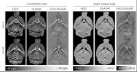
Since QSM has been recently undergoing clinical trials and the MEDI toolbox plays an important role for this purpose, numerical implementation should be consistent in the sense of continuum limit. In this abstract, we point out a numerically inconsistent finite difference scheme that has been used in the MEDI toolbox and show that by replacing it with a consistent one it drastically improves image quality.


1544.   
QUASAR: In vivo quantification of magnetic susceptibility in rodents
Ferdinand Schweser1,2, Paul Polak1, Nicola Bertolino1, and Robert Zivadinov1,2
1Buffalo Neuroimaging Analysis Center, Department of Neurology, Jacobs School of Medicine and Biomedical Sciences, The State University of New York at Buffalo, Buffalo, NY, United States, 2MRI Molecular and Translational Research Center, Jacobs School of Medicine and Biomedical Sciences, The State University of New York at Buffalo, Buffalo, NY, United States
Despite increasing exploration of quantitative susceptibility mapping (QSM) in humans and the method's potential to study tissue iron pre-clinically, only few studies have yet applied QSM in alive rodents at ultra-high magnetic field strength. In the present work we hypothesized that the low quality of pre-clinical QSM compared to human QSM is due to the combination of a similar level of non-susceptibility phase contributions with much lower susceptibility variations. Here, we propose a new type of QSM algorithm that accounts for non-susceptibility phase effects and, hence, enables pre-clinical QSM: QUAntitative Susceptibility And Residual mapping (QUASAR).


1545.   
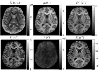
Effects of fiber orientation and myelin concentration on R2* (=1/T2*): a fiber orientation and/or myelin concentration corrected R2* map
Jingu Lee1, Woojin Jung1, Yoonho Nam2, and Jongho Lee1
1Laboratory for Imaging Science and Technology, Department of Electrical and Computer Engineering, Seoul National University, Seoul, Korea, Republic of, 2Department of Radiology, Seoul St. Mary’s Hospital, College of Medicine, The Catholic University of Korea, Seoul, Korea, Republic of
In this work, we measured the effect size of both myelin concentration and fiber orientation in R2*. Additionally, we generated the myelin concentration and/or fiber orientation bias free R2* maps which may have important applications.


1546.   
Echo time based influences on quantitative susceptibility mapping
Surabhi Sood1, Javier Urriola1, David Reutens1, Steffen Bollmann1, Kieran O'Brien2, Markus Barth1, and Viktor Vegh1
1Centre for Advanced Imaging, The University of Queensland, Brisbane, Australia, 2Siemens Ltd., Brisbane, Australia
Quantitative susceptibility mapping is an important magnetic resonance imaging tool which can help define brain structure and composition. Our work aims to explore information contained in the temporal trend by analysing the mapped magnetic susceptibility as a function of echo time from gradient recalled data acquired at 7T. Temporal susceptibility plots were studied in ten brain regions. Parameterisation of image voxel susceptibility compartments has the potential to delineate structural and chemical changes in tissue and formulate biologically meaningful measures. This in turn provides a framework for new imaging biomarker developments in neurodegenerative diseases and disorders affecting the central nervous system.  


1547.   
Application of Laplacian-based Methods to Multi-echo Phase Data for Accurate Susceptibility Mapping
Emma Biondetti1, David L. Thomas2, and Karin Shmueli1
1Department of Medical Physics and Biomedical Engineering, University College London, London, United Kingdom, 2Leonard Wolfson Experimental Neurology Centre, UCL Institute of Neurology, University College London, London, United Kingdom
In Susceptibility Mapping (SM) using multi-echo gradient-echo phase data, unwrapping and/or background-field removal is often performed using Laplacian-based methods. However, SM pipelines in the literature have applied these methods at different stages. Here, using simulated and acquired images, we compared the performance of three pipelines that apply Laplacian-based methods at different stages. We showed that Laplacian-based methods alter the linearity of the phase over time. We demonstrated that only a processing pipeline that takes this into account, i.e. by fitting the multi-echo data over time to correctly estimate a field map before applying Laplacian-based methods, gives accurate susceptibility values.


1548.   
Quantitative Susceptibility Mapping of the Substantia Nigra in Parkinson’s Disease
Xinxin Zhao1, Hedi An2, Tian Liu3, Nan Shen2, Binshi Bo4, Zhuwei Zhang4, Pengfei Weng4, Meining Chen4, Mengchao Pei4, Yi Wang3,4, Dongya Huang2, and Jianqi Li4
1Shanghai Key Laboratory of Magnetic Resonance and Department of Physics, East China Normal University, Shanghai, China, People's Republic of, 2Dongfang Hospital Neural Medical Affiliated Tongji University, Shanghai, China, People's Republic of, 3Department of Radiology, Weill Medical College of Cornell University, New York, NY, United States, 4Department of physics, Shanghai Key Laboratory of Magnetic Resonance and Department of Physics, East China Normal University, Shanghai, China, People's Republic of
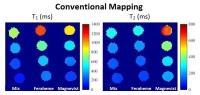
Quantitative susceptibility mapping (QSM) provides excellent contrast of iron-rich deep nuclei to quantify iron in the brains. Clinicians are interested in using QSM to diagnose PD patients. QSM and R2* values were measured in the whole substantia nigra in patients with PD and healthy controls. The significant difference between PD patients and healthy controls in the substantia nigra was found on QSM but not on R2* mapping.


1549.   
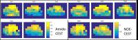
Are susceptibility-weighted imaging and quantitative susceptibility mapping suitable to gain additional information on melanoma metastasis of the brain?
Sina Straub1, Till Schneider2,3, Christian H. Ziener3, Heinz-Peter Schlemmer3, Mark E. Ladd1, Frederik B. Laun1, and Martin T. Freitag3
1Department of Medical Physics in Radiology, German Cancer Research Center (DKFZ), Heidelberg, Germany, 2Department of Neuroradiology, University of Heidelberg, Heidelberg, Germany, 3Department of Radiology, German Cancer Research Center (DKFZ), Heidelberg, Germany
The benefit of susceptibility weighted imaging (SWI) and quantitative susceptibility mapping (QSM) for the detection and quantification of bleeding of brain metastases of malignant melanoma is assessed. QSM shows paramagnetic values for hemorrhagic metastases (0.355±0.097 ppm) and less paramagnetic values (0.239±0.123 ppm) for hemorrhagic metastases that have T1w-native hyperintense signal. Moreover, our findings suggest that T1w-native hyperintense melanoma metastases have relatively diamagnetic susceptibility compared to other structures of the brain.


1550.   
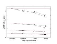
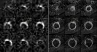
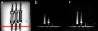
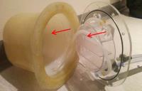
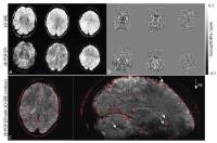
Susceptibility underestimation in a high susceptibility phantom: dependence on imaging resolution, magnitude contrast and sample orientation
Dong Zhou1, JingWei Zhang2, Pascal Spincemaille1, and Yi Wang1,2
1Radiology Department, Weill Cornell Medical College, New York, NY, United States, 2Biomedical Engineering, Cornell University, Ithaca, NY, United States
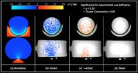
The error in digitizing the dipole convolution1 may become substantial when there is abrupt susceptibility change within a voxel. To evaluate this error, we assessed the accuracy of quantitative susceptibility mapping in a gadolinium balloon phantom with a range of large susceptibility values (0.4 – 3.2 ppm) and imaging resolutions (0.7 – 1.8 mm) at both 1.5T and 3T. Systematic underestimation of the susceptibility values was observed with decreasing imaging resolution. Numerical simulations were performed to match the experimental findings. These show that the underestimation originates directly from the changes in the voxel sensitivity function and that the amount of underestimation is affected not only by imaging resolution, but also magnitude contrast, the use of k-space filters in the image reconstruction, and details of the susceptibility inclusions such as the susceptibility value and geometry.


1551.   
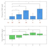
The use of quantitative susceptibility imaging for the evaluation of acute MS lesion formation
Vanessa Wiggermann1,2, Enedino Hernandez-Torres2,3, Inga C Ibs4, Stephanie M Schoerner5, Galina Vorobeychik6, Luanne Metz7, David KB Li8,9, Anthony Traboulsee9,10, and Alexander Rauscher2,9,11
1Physics and Astronomy, University of British Columbia, Vancouver, BC, Canada, 2Pediatrics, University of British Columbia, Vancouver, BC, Canada, 3UBC MRI Research Centre, University of British Columbia, Vancouver, BC, Canada, 4University of Osnabrueck, Osnabrueck, Germany, 5Technical University of Dortmund, Dortmund, Germany, 6Fraser Health MS Clinic, Burnaby, BC, Canada, 7Clinical Neurosciences, University of Calgary, Calgary, AB, Canada, 8Radiology, University of British Columbia, Vancouver, BC, Canada, 9Center for Brain Health, University of British Columbia, Vancouver, BC, Canada, 10Medicine (Neurology), University of British Columbia, Vancouver, BC, Canada, 11Child and Family Research Institute, University of British Columbia, Vancouver, BC, Canada
Using magnetic-susceptibility based MR techniques for the assessment of damage due to multiple sclerosis (MS) has been controversial, in particular in MS lesions where the underlying pathological changes are not yet fully understood. Here, we investigated the changes of the MR frequency and quantitative susceptibility signal during acute MS lesion formation. We observed that both metrics behave similarly, indicating that non-local effects have little contribution to the QSM signal increase and hence dipole inversion might not be required to assess damage during MS lesion formation accurately.


1552.   
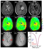
Rapid Quantitative Susceptibility Mapping with Simultaneous Multi-Band Imaging
Nan-Jie Gong1, Hing-Chiu Chang2, Hongjiang Wei1, Mark Sundman1, Nan-kuei Chen1, and Chunlei Liu1
1Brain Imaging and Analysis Center, Duke University, Durham, NC, United States, 2Diagnostic Radiology, The University of Hong Kong, Hong Kong, China, People's Republic of
We demonstrated the feasibility of using the proposed phase correction method for increasing the accuracy of QSM reconstruction from multi-band acquisitions. With multi-band acquisition, we were able to greatly shorten data acquisition time. It is expected that facilitate this method would benefit further clinical application of QSM and QSM based cerebral functional and physiological studies.


1553.   
Visibility improvement of  cerebral blood vessels by High Resolution Quantitative Susceptibility Mapping
Yuya Umemoto1, Tomohiro Ueno1, Shin-ichi Urayama2, Toshihiko Aso2, Hidenao Fukuyama2, and Naozo Sugimoto1
1Human Health Sciences, Kyoto University, Kyoto, Japan, 2Human Brain Research, Kyoto University, Kyoto, Japan
In Quantitative Susceptibility Mapping, susceptibility distribution can be obtained by deconvolution of perturbed fields with dipole fields. In our proposed method, High Resolution QSM, we employed densely sampled dipole fields to improve the quality of QSM. To verify the High Resolution QSM, we performed a human study, and acquired QSM input phase data of a healthy human subject. We compared MIP of the High Resolution QSM to that of the tricubically interpolated conventional QSM. In the High Resolution QSM, visibility of several cerebral blood vessels is improved. This means that a susceptibility map with higher spatial resolution is obtained.


1554.   
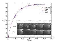
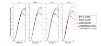
Probing the myelin water compartment with saturation recovery, multi-echo GE imaging at 7T
Elena Kleban1, Benjamin Tendler1, Penny Gowland1, and Richard Bowtell1
1The Sir Peter Mansfield Imaging Center, School of Physics and Astronomy, Nottingham, United Kingdom
The purpose of this work was to investigate the microstructural properties of white matter in the human brain using  saturation recovery multi-echo GE imaging at 7T.

Multi gradient-echo data acquired at three different flip-angles from 8 healthy subjects was fitted for corpus callosum to a three-pool model describing the axonal, myelin and external compartments and variation of the relative amplitude of the myelin water signal with flip-angle was used to assess the T1 values of the different compartments. Results show an increased frequency variation with TE and faster magnitude signal decay at higher flip-angles, consistent with reduced T­1 in the myelin water compartment.


1555.   
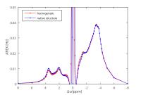
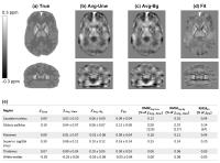
The Effect of Large Slice Thickness and Spacing and Low Coverage on the Accuracy of Susceptibility Mapping
Anita Karsa1, Emma Biondetti1, Shonit Punwani2, and Karin Shmueli1
1Medical Physics and Biomedical Engineering, University College London, London, United Kingdom, 2Centre for Medical Imaging, University College London, London, United Kingdom
Susceptibility Mapping has emerging clinical applications. To reduce scan time, clinical images are often acquired with large slice spacing/thickness and reduced coverage. The effect of these factors on susceptibility maps has not been investigated. Here, we develop a simple framework to explore the effect of low-resolution and low-coverage in the slice dimension on the accuracy of susceptibility maps. Our experiments with digital phantoms and volunteer images have shown that the error in the estimated susceptibility increases substantially with increasing slice spacing/thickness and decreasing coverage. These results underscore the need for high-resolution, full-coverage acquisitions for accurate susceptibility mapping.


1556.   
Accelerated Quantitative Susceptibility Mapping at 7T Using 3D Planes-on-a-Paddlewheel (POP) EPI
Daniel Stäb1,2, Steffen Bollmann1, Christian Langkammer3, Kristian Bredies4, and Markus Barth1
1The Centre for Advanced Imaging, The University of Queensland, Brisbane, Australia, 2Department of Diagnostic and Interventional Radiology, University of Würzburg, Würzburg, Germany, 3Department of Neurology, Medical University of Graz, Graz, Austria, 4Institute for Mathematics and Scientific Computing, University of Graz, Graz, Austria
Ultra-high field whole brain susceptibility mapping at an isotropic resolution of 1 mm was performed within 16 seconds using a 3D planes-on-a-paddlewheel (POP) EPI sequence. The non-Cartesian readout scheme is created by rotating a standard EPI readout train around its own phase encoding axis and provides higher flexibility for echo time minimization than conventional 3D EPI. Morphologic images and susceptibility maps obtained were comparable to those acquired with a conventional 4 minute 3D GRE scan.


1557.   
Quantitative Susceptibility Mapping Using Adaptive Edge-Preserving Filtering: Comparison with COSMOS in Human Brain
Toru Shirai1, Ryota Sato1, Yo Taniguchi1, Takenori Murase2, Atsushi Kuratani2, Taisei Ueda2, Takashi Tsuneki2, Yoshitaka Bito2, and Hisaaki Ochi1
1Research and Development Group, Hitachi, Ltd., Tokyo, Japan, 2Healthcare Campany, Hitachi, Ltd., Chiba, Japan
    We have proposed that a QSM reconstruction method combining an iterative least square minimization and adaptive edge-preserving filtering could generate high-quality susceptibility maps. In this study, maps calculated by the proposed method were compared qualitatively and quantitatively with those calculated by COSMOS (a calculation of susceptibility through multiple-orientation sampling) in healthy volunteers. The results from human brain experiments showed good agreement with COSMOS. The proposed QSM reconstruction of single orientation sampling is useful for generating a high-quality susceptibility map of the human brain.


1558.   
QSM at 3T: Comparison of Acquisition Methodologies
M Louis Lauzon1,2,3, Cheryl Rae McCreary1,2,3, D Adam McLean3,4, Marina Salluzzi3,4, and Richard Frayne1,2,3
1Radiology and Clinical Neurosciences, University of Calgary, Calgary, AB, Canada, 2Hotchkiss Brain Institute, Calgary, AB, Canada, 3Seaman Family MR Research Centre, Calgary, AB, Canada, 4Calgary 
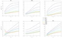
Image Processing and Analysis Centre, Calgary, AB, Canada
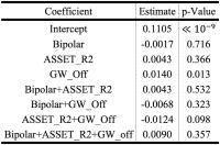
We scanned 4 volunteers 3 times each using 8 different QSM variants (unipolar/bipolar readout gradient, accelerated or not, with/without gradient warp-correction), and compared the susceptibility (average and standard deviation) in five deep gray matter tissues using linear mixed effects modeling. Gradient-warp correction was found to decrease the susceptibility estimates by 3-5%, whereas there was no statistical difference in the estimates due to readout polarity or acceleration factor.


1559.   
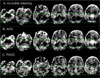
Adaptive background phase removal using knowledge-based region detection for quantitative susceptibility mapping
Taichiro Shiodera1, Takamasa Sugiura1, Yuko Hara1, Yasunori Taguchi1, Tomoyuki Takeguchi1, Masao Yui2, Naotaka Sakashita2, Yasutaka Fushimi3, Takuya Hinoda3, Tomohisa Okada3, Aki Kido3, and Kaori Togashi3
1Toshiba Corporation, Kawasaki, Japan, 2Toshiba Medical Systems Corporation, Otawara, Japan, 3Kyoto University Graduate School of Medicine, Kyoto, Japan
We propose a background phase removal method for quantitative susceptibility mapping using adaptive kernels depending on brain region. Conventional methods use distance adaptive kernel spherical mean values (SMV) to estimate background phase. However, artifacts occur where kernel sizes are not optimal for certain brain regions. Here, we adapt SMV kernel sizes depending on brain regions which are automatically detected by machine learning methods. The proposed method eliminates tissue phase artifacts near air-tissue interfaces in more central areas such as the sinus. The proposed method also eliminates streak artifacts in susceptibility images.


1560.   
Effects of concomitant gradients on Quantitative Susceptibility Mapping
Timothy J Colgan1,2, Diego Hernando1, Samir Sharma1, Debra E Horng1,2, and Scott B Reeder1,2,3,4,5
1Radiology, University of Wisconsin, Madison, WI, United States, 2Medical Physics, University of Wisconsin, Madison, WI, United States, 3Biomedical Engineering, University of Wisconsin, Madison, WI, United States,4Medicine, University of Wisconsin, Madison, WI, United States, 5Emergency Medicine, University of Wisconsin, Madison, WI, United States
MR-based Quantitative Susceptibility Mapping (QSM) techniques have multiple potential applications in brain and body imaging. QSM techniques generally rely on the removal of background field effects to obtain a local B0 map, followed by dipole inversion to estimate the underlying susceptibility distribution. However, concomitant gradients introduce significant unanticipated phase shifts in the acquired data that manifest as errors in the measured B0 field map. Our results demonstrate that CG phase corrections and/or the use of a background field removal algorithm that removes this background field component are necessary for accurate QSM.


1561.   
QSM: fast selection of optimal regularization weights
Job Gijsbertus Bouwman1 and Peter R Seevinck1
1Image Sciences Institute, University Medical Center Utrecht, Utrecht, Netherlands
Quantitative Susceptibility Mapping reconstructions may benefit from L1-regularization and magnitude weighing, however these iterative reconstruction methods are time-consuming. Recently, progression has been made in reducing the reconstruction times with Split Bregman iterations, allowing subject-specific regularization weights. Here a further reduction of the reconstruction time is reported, mostly based on accelerating the automatic selection of the optimal regularization parameter. The overall procedure reduces computational load more than threefold, without accuracy loss. Reduction of reconstruction times, may contribute to realize QSM algorithms which are either clinically feasible, or that may pave the way to include more sophisticated regularization mechanisms.

1568.   
QUANTITATIVE 1H MR TISSUE OXIMETRY (QMRO)
Scott C Beeman1, Joseph JH Ackerman1, and Joel R Garbow1
1Washington University in St. Louis, St. Louis, MO, United States
A direct and non-invasive measure of tissue O2 would be a major advance. O2 is paramagnetic and can thus, in principle, be quantified with NMR/MRI. However, such measurements are challenged/masked by two competing effects: (i) magnetization transfer between 1H spins of tissue water and the solid-like macromolecular matrix (e.g., proteins, cell membranes) and (ii) blood flow, which can bring equilibrium-polarized 1H spins into the interrogated tissue volume. We describe a strategy for mitigating these confounds and quantify the direct relationship between pO2 and the MR-measured longitudinal relaxation rate constant, R1.


1569.   
Fisher Information Matrix for Optimizing the Acquisition Parameters in Multi-Parametric Mapping Based on Fast Steady-State Sequences
romain valabregue1 and Ludovic De Rochefort2
1CENIR, ICM, Inserm U 1127, CNRS UMR 7225, Sorbonne Universités, UPMC Univ Paris 06 UMR S 1127F, Paris, France, 2IR4M (Imagerie par Résonance Magnétique Médicale et Multi-modalités), Univ. Paris-Sud, CNRS, UMR8081, Université Paris-Saclay, Orsay, France
A criterion of A-optimality was used to optimize SSFP sequence acquisition parameters in order to perform multi-parametric mapping of the physical parameters proton density, relaxation rates and apparent diffusion coefficient. A fast calculation of the steady-state was used to estimate the Fisher information matrix from which fitted parameter error was determined from its inverse. Considering a range of possible T1 and T2 values, and relevant ADC, the acquisition parameters were optimized over the four dimensions of TR, prescribed flip angle, RF phase increment and spoiling gradient to achieve the minimum error on the effectively fitted physical parameters. It is demonstrated that choosing targeted T1 and T2 values over a wide range of expected values enables defining acquisition protocols that minimize the error over this range.


1571.   
Post-mortem reperfusion of the vascular system and examination in MRI: Temperature-dependent characterisation of perfusates and contrast simulations
Bridgette Webb1, Thomas Widek1, Bernhard Neumayer1, Rudolf Stollberger2, and Thorsten Schwark1,3
1Ludwig Boltzmann Institute for Clinical Forensic Imaging, Graz, Austria, 2Institute of Medical Engineering, Graz University of Technology, Graz, Austria, 3Institute of Legal Medicine, Medical University of Graz, Graz, Austria
MRI evaluation of a post-mortem reperfused cardiovascular system requires a complete filling of vessels, acceptable contrast/image quality and consideration of temperature influences.  Assessment of the temperature dependence of viscosity, T1 and T2 of candidate perfusates (n=10) found 3 to be suitable for application in post-mortem MR angiography. Bloch equation simulations were applied to investigate contrast between these liquids and post-mortem myocardium at 1, 8.5, 16 and 23°C. For a FLASH sequence, optimal flip angles were affected by temperature variation and a decrease in contrast (max. 6-12%) was observed when flip angles optimised for one of the other temperatures were applied.


1572.   
Rapid Simultaneous Detection of Multiple Contrast Agents Using Magnetic Resonance Fingerprinting
Miko H. de Haas1,2, Huihui Ye2,3, Howard H. Chen2,4, Eric M. Gale2,4, Eszter Boros2,4, Peter Caravan2,4, Kawin Setsompop2,4, and David E. Sosnovik2,4
1Department of Biomedical Engineering, Eindhoven University of Technology, Eindhoven, Netherlands, 2Athinoula A. Martinos Center for Biomedical Imaging, Massachusetts General Hospital, Charlestown, MA, United States, 3Collaborative Innovation Center for Brain Science and the Key Laboratory for Biomedical Engineering of Education Ministry of China, Zhejiang University, Hangzhou, China, People's Republic of,4Department of Radiology, Harvard Medical School - Massachusetts General Hospital, Boston, MA, United States
MR contrast agents are typically imaged using time-consuming sequences, which allows only one parameter of relaxation to be assessed. In this research we used Magnetic Resonance Fingerprinting (MRF) to rapidly assess both T1 and T2­, and these values were then used to calculate contrast agent concentrations. The primary goal was to quantify two contrast agents residing in a mixed sample. The method  showed an accuracy  greater than 90% in most cases, indicating its feasibility. In addition,  the method was also able to quantify the bound and unbound state of a targeted contrast agent in near real-time.


1565.   
Validation of MR mapping of direct current in a phantom model
Mayank V Jog1, Robert X. Smith2, Kay Jann2, Walter Dunn3, Allan Wu2, and Danny JJ Wang2
1Biomedical Engineering, University of California Los Angeles, Los Angeles, CA, United States, 2Neurology, University of California Los Angeles, Los Angeles, CA, United States, 3Psychiatry, University of California Los Angeles, Los Angeles, CA, United States
Transcranial Direct Current Stimulation(tDCS) is a neuromodulation technique. Reported to improve clinical conditions as well as cognition, tDCS has potential as a treatment modality since it involves only simple scalp electrodes to drive mA currents. To date, only mathematical modeling has been used to visualize tDCS-applied currents.


In previous work, we used MRI field mapping in a novel paradigm to visualize in-vivo, a component of the magnetic field generated by these currents. The present work completes the picture by validating our current visualization technique via comparison between the measured and simulated current-induced fields in a specially constructed phantom.


1570.   
dcQSM: Quantitative Susceptibility Mapping by Directly Fitting Complex Images
Zhe Liu1, Pascal Spincemaille2, and Yi Wang1,2
1Biomedical Engineering, Cornell University, Ithaca, NY, United States, 2Radiology, Weill Cornell Medical College, New York, NY, United States
The quality of Quantitative Susceptibility Mapping (QSM) depends critically on a correct estimation of total magnetic field, which may sometimes be degraded by phase unwrapping failure. We propose to bypass the traditional field estimation and phase unwrapping steps and estimate both background field and local susceptibility distribution directly from complex GRE images, which is referred to as dcQSM. Since no field is explicitly existent in our method, dcQSM eliminates phase unwrapping errors in tradition methods.


1562.   
MR Imaging of Electromagnetic Field Distribution for Treatment Planning in Electrical Stimulation
Woo Chul Jeong1, Saurav ZK Sajib1, Nitish Katoch1, Bup Kyung Choi1, Hyung Joong Kim1, Oh In Kwon2, and Eung Je Woo1
1Kyung Hee University, Seoul, Korea, Republic of, 2Konkuk University, Seoul, Korea, Republic of
Electrical stimulations are widely used as therapeutic techniques that are closely related to the electromagnetic fields inside the human body. The electromagnetic field is affected by the injected currents and electrical conductivities of biological tissue, the map of voltage, current density, and magnetic flux density can provide meaningful information for determining the tissue type and current pathways. The signal intensity of current density is proportional to magnetic flux density which can be measured by MREIT. Since the biological tissues show anisotropic characteristic, we introduced a recent DT-MREIT method to better apply it to real situation.


1563.   
Simultaneous water content, electrical conductivity and susceptibility mapping in meningiomas on a 3T MR-PET scanner
YP Liao1, A.-M. Oros-Peusquens1, J. Lindemeyer1, N. Lechea1, C. Weiss2, G. Stoffels1, C. Filss1, K.J. Langen1, and N.J. Shah1,3
1Institute of Neuroscience and Medicine-4, Forschungszentrum Juelich, Juelich, Germany, 2Department of Neurosurgery, University of Cologne, Cologne, Germany, 3Department of Neurology, JARA, Faculty of Medicine, RWTH Aachen University, Aachen, Germany
The availability of combined MR-PET scanners opens new opportunities for the characterisation of the tumour environments. In this study, MR-based simultaneous water content, electrical conductivity and susceptibility mapping in meningioma patients was implemented based on a multi-echo gradient echo sequence. The information was complemented by characterisation of the tumour with simultaneous FET-PET. This is a powerful combination of parameters which reflect important aspects of tissue physiology and also characterise to a large extent, tumour electromagnetic (EM) properties. This multi-parametric information helps to understand pathological tissue and can be applied to planning nonionizing EM hyperthermia therapy.


1564.   
Coil Compression for Improved Phase Image Signal-to-Noise Ratio in Electrical Property Tomography
Kathleen M Ropella1 and Douglas C Noll1
1Biomedical Engineering, University of Michigan, Ann Arbor, MI, United States
The use of multi-channel receivers is essential for acquiring B1+ with sufficient SNR to calculate electrical properties. Combining the individual channel images prior to these calculations typically involves a SENSE-like method or the use of some reference image. In this work we present a modified version of coil compression to provide an automatic and simplified multi-channel array data combination for high SNR phase-based conductivity mapping. 


1566.   
Observation of the correlation between Electrical Conductivity and Apparent Diffusion Coefficient values
Sung-Min Gho1, Jaewook Shin1, Min-Oh Kim1, Min Jung Kim2, Sooyeon Kim2, Jun-Hyeong Kim1, and Dong-Hyun Kim1
1School of Electrical and Electronic Engineering, Yonsei University, Seoul, Korea, Republic of, 2Department of Radiology, Yonsei University College of Medicine, Seoul, Korea, Republic of
Electric conductivity and apparent diffusion coefficient (ADC) give meaningful information to the clinicians and researchers, however, studies related to the relationship of these two phenomena were not substantially proceeded.

In this abstract, we observe the correlation between electrical conductivity and ADC under various situations (i.e. phantom, in vivo brain, and breast tumor case).


1573.   
Black-Blood T2* Mapping with Delay Alternating with Nutation for Tailored Excitation
Shi Su1, Yanan Ren1, Caiyun Shi1, Xiaoyong Zhang1,2, Hairong Zheng1, Xin Liu1, and Guoxi Xie1
1Lauterbur Research Center for Biomedical Imaging, Shenzhen Institutes of Advanced Technology, Chinese Academy of Sciences, Shenzhen, China, People's Republic of, 2Centers for Biomedical Engineering, College of Information Science and Technology, University of Science and Technology of China, Hefei, China, People's Republic of
T2* mapping provides a means to quantitatively estimate the iron load of tissue, which is closely related to numerous diseases, such as thalassemia, hereditary hemochromatosis and sickle cell disease. However, blood signal would induce artifacts which lead to T2* estimation inaccurate. To address this issue, a novel black-blood T2* mapping technique utilizing Delay Alternating with Nutation for Tailored Excitation (DANTE) preparation module followed by multi-echo gradient echo (GRE) readout (DANTE-GRE) was developed to obtain blood suppressed T2* maps. The proposed method is shown to acquire more accurate T2* maps due to its high SNR and effective blood signal suppression.


1567.   
MR-based Current Density Imaging during Transcranial Direct Current Stimulation (tDCS)
Saurav ZK Sajib1, Woo Chul Jeong1, Nitish Katoch1, Bup Kyung Choi1, Hyung Joong Kim1, Oh In Kwon2, and Eung Je Woo1
1Kyung Hee University, Seoul, Korea, Republic of, 2Konkuk University, Seoul, Korea, Republic of
Quantitative visualization of induced current density by the electrical stimulation current inside the anisotropic brain region may play an important role to understand the neuro-modulatory effect during transcranial direct current stimulation (tDCS). For ensuring the clinical applications, precise approaches are required to understand the exact responses inside the human body subject to an injected currents, In this study, we reconstruct current density distribution inside the in vivo canine brain region by combing the directional information obtained from a DTI-MRI scan and the z-component of the magnetic flux density data using MREIT technique.
 

The International Society for Magnetic Resonance in Medicine is accredited by the Accreditation Council for
Continuing Medical Education to provide continuing medical education for physicians.