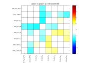
ISMRM 24th Annual Meeting & Exhibition • 07-13 May 2016 • Singapore

Traditional Poster Session: Functional MRI (Neuro)

1651 -1668 fMRI Connectivity: The Methods
1669 -1712 fMRI Connectivity: The Applications
1713 -1725 fMRI Physiology
1726 -1742 fMRI: Applications
1743 -1757 fMRI: Methods


1651.   
Directional connectivity in mouse fMRI networks
Md Taufiq Nasseef1,2, Adam Liska1,2, Stefano Panzeri1, and Alessandro Gozzi1
1Italian Institute of Technology,Center for Neuroscience and Cognitive Systems @UniTn, Rovereto, Italy, 2Center for Mind/Brain Sciences, University of Trento, Rovereto, Italy
Mouse resting-state fMRI (rsfMRI) has revealed the presence of distributed functional connectivity networks including two sets of regions exhibiting neuro-anatomical features reminiscent of the human salience (SN) and default-mode-network (DMN). Here, we applied Granger Causality to investigate the direction of information flow within mouse rsfMRI networks characterized by mono-directional and reciprocal underlying axonal connectivity. We show that multiple intrinsic rsfMRI networks of the mouse brain exhibit robust patterns of directional connectivity towards prefrontal regions, replicating topological features of human rsfMRI networks, and in agreement with higher integrative role subserved by these areas.  


1652.   
A Robust Anesthesia Regime for fMRI in Rodents
Hanbing Lu1, Julie Brynildsen1, Li-Ming Hsu1, Thomas Ross1, Elliot A Stein1, and Yihong Yang1
1Neuroimaging Research Branch, National Institute on Drug Abuse, NIH, Baltimore, MD, United States
In fMRI using animal models, it is of critical importance to develop a robust anesthetic regime that maintains neurovascular coupling, permits longitudinal experiments with minimal invasiveness, and is easy to implement. Recently, using an anesthetic method that combines low doses of dexmedetomidine and low dose of isoflurane, we have successfully identified the default mode network in rat brain, suggesting that this preparation causes minimal suppression of brain network functions. The goal of this study is to systematically characterize and to optimize physiological conditions for fMRI experiments under this anesthetic regime.


1653.   
Frontal-parietal network functional connectivity characterization in patients with end-stage renal disease by using independent component analysis
Lin Wang1, Chun-Qiang Lu1, and Shenghong Ju1
1Department of Radiology, Zhongda hospital, Medical school of Southeast university, Nanjing, China, People's Republic of
It has been reported that end-stage renal disease (ESRD) patients have cognitive decline in all aspects, especially in attention and executive function, and this is lack of objective noninvasive modalities to monitor the cognition impairment by now. Resting-stage functional MR which can detect the change of brain function as a newly-developing imaging method has been widely used to evaluate the cognitive status in many diseases. Our findings show the aberrant functional connectivity of frontal-parietal network (FPN) in ESRD patients, and supported by the results of neuropsychological tests. FPN functional connectivity may serve as a biomarker to monitor the attention and executive function impairment in patients with ESRD in the future.


1654.   
Slice-based motion metrics show stronger relationship than volume-based metrics to visual rating of motion artifact
Katherine A Koenig1, Erik Beall1, Sally Durgerian2, Christine Reece3, Stephen M Rao3, and Mark J Lowe1
1Imaging Sciences, The Cleveland Clinic, Cleveland, OH, United States, 2BrainDataDriven LLC, Milwaukee, WI, United States, 3Schey Center for Cognitive Neuroimaging, The Cleveland Clinic, Cleveland, OH, United States
A motion metric that more closely represents the amount of artifact in the signal could decrease the amount of discarded data and reduce noise in resting state studies. This work compares a sample of 455 resting state scans visually rated for motion corruption to slice-based and volume-based motion metrics. We show that the slice-based metric shows a stronger relationship to visual assessment of motion corruption.


1655.   
Brain cortical parcellation based on the anisotropy of local spatio-temporal correlation of rs-fMRI at 7T
Afonso Dias1, Marta Bianciardi2, Sandro Daniel Nunes1, Luís M. Silveira3, Lawrence L. Wald2, and Patrícia Figueiredo1
1ISR-Lisboa/LARSyS and Department of Bioengineering, Instituto Superior Técnico – Universidade de Lisboa, Lisbon, Portugal, 2Department of Radiology, A.A. Martinos Center for Biomedical Imaging, MGH and Harvard Medical School, Boston, MA, United States, 3INESC-ID, Instituto Superior Técnico – Universidade de Lisboa, Lisbon, Portugal
We propose a new metric of local functional connectivity for the parcellation of the cerebral cortex from resting-state fMRI data. It is based on the hypothesis that the anisotropy of the local spatio-temporal correlation tensor of the BOLD signal is increased in the boundaries between regions of functional segregation within gray matter. We show that the anisotropy of rs-fMRI at 7T can be used to generate cortical parcellations that are partially consistent with the results obtained using the well-established stability map. Further work is needed to investigate the validity and properties of the parcellations based on the proposed metric.


1656.   
Exploring Resting-State Functional Connectivity Invariants across the Life Span using a Novel Graph Model
Ottavia Dipasquale1,2, Paolo Finotelli3, Isa Costantini1, Giuseppe Baselli1, Francesca Baglio2, Paolo Dulio3, and Mara Cercignani4
1Department of Electronics, Information and Bioengineering, Politecnico di Milano, Milan, Italy, 2IRCCS, Don Gnocchi Foundation, Milan, Italy, 3Department of Mathematics "F. Brioschi", Politecnico di Milano, Milan, Italy, 4Clinical Imaging Sciences Centre, Brighton and Sussex Medical School, Brighton, United Kingdom
In this work we investigated resting-state functional connectivity (FC) changes and invariant properties in 133 healthy people across the life-span (6-79y) using a novel graph model that emphasizes centrality of nodes. This model estimates a weight for each node’s pair (94 cortical regions) accounting for the node degrees, anatomical distance and FC between them and penalizing the formation of long connections. Preliminary findings in two groups of 25 and 62 year-old subjects highlighted a number of interesting properties and confirmed the important role of the Precuneus and the Cingulate Gyrus, which are characterized by high functional strength and degree.


1657.   
Assessing the Reliability of Estimated Correlation During the Evaluation of Dynamic Functional Connectivity
Tuo Shi1, D Rangaprakash1, and Gopikrishna Deshpande1,2,3
1AU MRI Research Center, Department of Electrical and Computer Engineering, Auburn University, Auburn, AL, United States, 2Department of Psychology, Auburn University, Auburn, AL, United States, 3Alabama Advanced Imaging Consortium, Auburn University and University of Alabama Birmingham, Auburn, AL, United States
In this work, we propose a novel strategy for selecting the minimum window length required to capture maximum dynamics as well as reliably estimate correlation during dynamic functional connectivity analysis. Using the error in estimated correlation compared to simulated ground-truth correlation as the metric, we compared our method with (i) the fixed window length approach, and (ii) the DCC method. We show that our method can provide minimum window lengths which give more reliable correlation estimates than those obtained from DCC and fixed-window methods. Further, we show that our method can accurately track fast variations in connectivity.


1658.   
Accuracy and inter-subject reproducibility of default mode networks identified from ASL data
Felipe Barreto1,2, Xiufeng Li1, Amir Moheet3, Anjali Kumar3, Lynn Eberly4, Elizabeth Seaquist3, Fabrizio Esposito5, and Silvia Mangia1
1CMRR, Department of Radiology, University of Minnesota, Minneapolis, MN, United States, 2Department of Physics, University of Sao Paulo, Ribeirao Preto, Brazil, 3Department of Medicine, University of Minnesota, Minneapolis, MN, United States, 4Division of Biostatistics, University of Minnesota, Minneapolis, MN, United States, 5Department of Medicine and Surgery, University of Salerno, Baronissi, Italy
The present study aimed at characterizing the robustness of the default mode network (DMN) extracted at single subject level from ASL datasets with independent component analysis. Three different analyses modes were considered, including the series of perfusion weighted images, the full time series, and the pair-wise average of control/tag images (pseudo-BOLD). Results show that the three analysis modes produce DMNs with similar accuracy at a group level, but the pseudo-BOLD mode resulted in smaller inter-subject variability of the spatial distribution of the single-subject DMNs.


1659.   
Condition effects on resting-state CBF reproducibility and reliability
Marta Vidorreta1,2, Natalie N Katchmar3, Daniel H Wolf3, and John A Detre1,2
1Neurology, University of Pennsylvania, Philadelphia, PA, United States, 2Radiology, University of Pennsylvania, Philadelphia, PA, United States, 3Psychiatry, University of Pennsylvania, Philadelphia, PA, United States
Cerebral blood flow (CBF) data were collected with arterial spin labeled (ASL) perfusion MRI in a group of young, healthy subjects over two sessions, scheduled a week apart. CBF and functional connectivity metrics were derived from the CBF time series across four different resting conditions: ‘eyes open’, ‘eyes closed’, ‘fixation’ (eyes fixated on a cross), and ‘PVT’ (low-frequency psychomotor vigilance task).  Absolute CBF was highly reproducible both within and across sessions. Results suggest that ‘fixation’ is inferior to the other conditions tested for resting-state ASL reproducibility.


1660.   
Title: Distinctive relationships between functional and structural connectivity in autism spectrum disorder across different networks— a combined resting-state functional MRI and diffusion spectrum imaging study
Hsiang-Yun Sherry Chien1, Susan Shur-Fen Gau2, and Wen-Yih Isaac Tseng1,3
1Institute of Medical Device and Imaging, National Taiwan University College of Medicine, Taipei, Taiwan, Taipei, Taiwan, 2Department of Psychiatry, National Taiwan University Hospital and College of Medicine, Taipei, Taiwan, Taipei, Taiwan, 3Molecular Imaging Center, National Taiwan University, Taipei, Taiwan, Taipei, Taiwan
We conducted a data-driven approach to investigate the functional and structural connectivity (FC and SC) within three critical networks in autism spectrum disorder (ASD) and typically developing controls (TD). We found significant weaker SC within the default-mode network, e.g. the cingulum bundles, in ASD compared to TD. Furthermore, we found significant positive correlations between the FC of the right salience network and the SC within the DMN and central executive network (CEN). Given the role of salience network in modulating the switch between the DMN and CEN, our results might imply a distinctive FC-SC relationship across different networks in ASD.


1661.   
An algorithm for generating uniform random parcellations
Hu Cheng1, Andrea Koenigsberger1, Sharlene Newman1, and Olaf Sporns1
1Psychological and Brain Sciences, Indiana University, Bloomington, IN, United States
Random parcellations have some advantages over template-based parcellations in network analysis of the brain. An important criterion for assessing the “goodness” of a random parcellation is the parcel size variability. A new algorithm is proposed to create more homogeneous random parcellations than previously reported. The new algorithm takes the actual distance between voxels and local voxel density into account in placing the random seeds. With many random parcellations using our approach, global network properties exhibit normal distribution and the variability across different repetitions of the random parcellation is comparable with inter-subject variability. 


1662.   
Simultaneous PET/MR/EEG to study brain connectivity on different physiological and temporal scales in epilepsy patients
Andre Thielcke1, Adham Elshahabi2, Ilja Bezrukov1, Suril Gohel3, Mario Amend1, Holger Schmidt4, Matthias Reimold5, Holger Lerche2, Bharat Biswal3, Bernd J. Pichler1, Niels Focke2, Christian la Fougère5, and Hans F. Wehrl1
1Department of Preclinical Imaging and Radiopharmacy, Werner Siemens Imaging Center, Eberhard Karls University of Tuebingen, Tuebingen, Germany, 2Neurology & Epileptology and Werner Reichardt Centre for Integrative Neuroscience, Eberhard Karls University of Tuebingen, Tuebingen, Germany, 3Department of Biomedical Engineering, New Jersey Institute of Technology, Newark, NJ, United States, 4Interventional and Diagnostic Radiology, Eberhard Karls University of Tuebingen, Tuebingen, Germany, 5Nuclear Medicine and Werner Reichardt Centre for Integrative Neuroscience, Eberhard Karls University of Tuebingen, Tuebingen, Germany
Simultaneous PET/MR/EEG was used in humans to study brain networks in the resting state on slow, medium and fast time scales. We found that the representation of the default mode network (e.g. in terms of correlation between regions) varies between modality and time scale applied. However, for the DMN as well as other networks similarities but also differences between modalities were seen. This work opens the domain for studying brain activity on different physiological (metabolic, hemodynamic and electric) but also on different time scales.


1663.   
Estimating whole brain connectivity dynamics using spectral clustering
Ivor Cribben1
1Finance and Statistical Analysis, Alberta School of Business, Edmonton, AB, Canada
A great challenge in neuroscience is the understanding of the dynamic manner in which brain regions interact with one another in both task-based and resting-state brain imaging studies. In this work, we introduce a novel statistical method, called Network Change Point Detection (NCPD), which dynamically clusters brain regions by their functional connectivity. NCPD promises to offer deeper insight into the large-scale characterizations and mechanisms of the brain as it can be used for the dynamic modelling of a very large number of voxels or brain regions. We apply this new method to a resting-state fMRI study.


1664.   
A voxel by voxel comparison of spatio-temporal correlation tensor derived from the resting-state fMRI and diffusion tensor derived from diffusion weighted images on the human brain using the multiband EPI sequences
Jiancheng Zhuang1
1University of Southern California, Los Angeles, CA, United States
We test the validity of spatio-temporal correlation tensor method on a set of rfMRI and DTI data which are acquired by multiband EPI sequences and have the same slice and geometry parameters, so we can compare the spatio-temporal correlation tensor and diffusion tensor at a voxel by voxel level in the human brain. We find the spatio-temporal correlation tensor derived from resting-state fMRI does not match with the diffusion tensor calculated from diffusion weighted images in the white matter, which is contradictory to a previous report of spatio-temporal correlation tensor method.   


1665.   
Investigating the neural substrates of verbal working memory in children with dyslexia: An effective connectivity study
Fu Yu Kwok1, Beth Ann O'Brien2, Kiat Hong Stacey Tay3, and SH Annabel Chen1,4
1Division of Psychology, Nanyang Technological University, Singapore, Singapore, 2National Institute of Education, Nanyang Technological University, Singapore, Singapore, 3Paediatric Neurology and Developmental Paediatrics, National University Hospital, National University of Singapore, Singapore, Singapore, 4Centre for Research And Development Learning, Nanyang Technological University, Singapore, Singapore
Dynamic causal modeling was utilized to examine the effective connectivity during verbal working memory in children with dyslexia and typically developing children. Seven regions of interest—FG, IFG, IOG, IPL, thalamus, inferior cerebellum and superior cerebellum were included into the analyses. Results indicated that the effect of dyslexia led to shift in effective network connectivity. The present study furthered our understanding of the cerebro-cerebellar effective network connectivity in both children with dyslexia and typically developing children. In addition, it provided new insights about the effects of dyslexia on this network. 


1666.   
Data-driven functional sub-division of the sensory-motor network using hierarchical clustering for resting-state fMRI data.
Yanlu Wang1 and Tie-Qiang Li1,2
1Clinical Sciences, Intervention and Technology, Karolinska Institute, Stockholm, Sweden, 2Medical Physics, Karolinska University Hospital, Stockholm, Sweden
A data-driven analysis method based on hierarchical clustering was used to analyze the sensory-motor resting-state network from resting-state fMRI data. It was used to analyze the network’s functional sub-division, and intra-network functional organization, in different levels of detail. Sub-network for the sensory-motor network as obtained by hierarchical clustering is anatomically and functionally sensible. Further sub-division of the paracentral lobule network hub successfully revealed its functional sub-division in great detail. The intra-network organization of intrinsic functional connectivity derived from spontaneous activity of the brain at rest reflects consistently, the functional and neural anatomic connectivity topography of the resting-state network.


1667.   
Nonlinear registered seed selection in resting state fMRI
Wanyong Shin1 and Mark J Lowe1
1Radiology, Cleveland Clinic Founcatoin, Cleveland, OH, United States
We compared different motor cortex (M1) seed selection methods in a large sample for group resting state (rs-) fMRI analysis. We found that seed selection with non-linear registration improves the statistical power in group analysis 


1668.   
Reproducibility of seed-based rs-fMRI measures at 7 tesla
Katherine A Koenig1, Sehong Oh1, Wanyong Shin1, and Mark J Lowe1
1Imaging Sciences, The Cleveland Clinic, Cleveland, OH, United States
This work assesses the reproducibility of seed-based rs-fMRI measures at 7 tesla in a sample of five controls and three patients with multiple sclerosis. We show high reproducibility but with large variation in some subjects.

1669.   
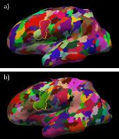
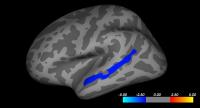
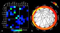
Investigation of functional baseline neuronal specificity and small-scale network in human primary motor cortex at 7T
Chan Hong Moon1, Jung-Hwan Kim1,2, and Kyongtae Ty Bae1,2
1Radiology, University of Pittsburgh, Pittsburgh, PA, United States, 2Bioengineering, University of Pittsburgh, Pittsburgh, PA, United States
Compound signal, BOLD (e.g., de-oxygenation, CBF and CBV) has different neuronal specificity depending on the major source. At high-field such as 7T, stimulus-evoked BOLD (fMRI) is known to be more localized to cortex region mainly due to suppression of short T2* signals in large draining vessels. It is question whether spontaneous-evoked BOLD during resting status (rsfMRI) can be localized to neural response and the correlation with fMRI activation. In this study, we investigated BOLD source during resting status in primary motor cortex using high-resolution 7T, and additionally the advantage of 7T rsfMRI in small-scale brain connectivity. 


1670.   
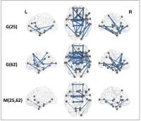
Dynamic reorganization of intrinsic functional networks in the mouse brain
Joanes Grandjean1, Maria G. Preti 2,3, Thomas AW Bolton2, Dimitri Van De Ville2, and Markus Rudin4
1ETH and University Zurich, Zurich, Switzerland, 2EPFL, Lausanne, Switzerland, 3University of Geneva, Geneva, Switzerland, 4University and ETH Zurich, Zurich, Switzerland
Dynamic functional connectivity was assessed in the mouse brain. High quality resting-state fMRI data were acquired and analysed with sliding window correlations. Re-occurring dynamic functional networks were estimated using dictionary learning from the sliding window correlation matrix.  The dynamic functional connectivity analysis reveals rich patterns of interactions, which were absent in the standard static functional connectivity analysis, and may be used to describe specific alterations in mouse models of brain disorders. In particular, the dynamic functional networks present salient features such as between and within module interactions, which complement the static functional connectivity analysis.    


1671.   
Visual Stimulation Altered Human Visual Cortical Functional Connectivity
Jie Huang1 and David C Zhu2
1Department of Radiology, Michigan State University, East Lansing, MI, United States, 2Departments of Radiology and Psychology, Michigan State University, East Lansing, MI, United States
Areas across the visual cortex are functionally connected. Certain patterns can induce perceptual illusions/distortions and visual discomfort in most people, headaches in patients with migraine, and seizures in patients with photosensitive epilepsy. This preliminary study investigated visual stimulation effect on human visual cortical functional connectivity (FC). The study found that a 25-min visual stimulation with a stressful pattern significantly enhanced the FC within the visual cortex and altered the FC to V1 in other regions too, with a lasting effect even after the cessation of the stimulation.


1672.   
Similarity in structural and functional network connectivity evolution over duration of TLE
Victoria L. Morgan1, Ahmet Cakir2, Benjamin N. Conrad1, Bassel Abou-Khalil3, Adam W. Anderson1,4, Zhaohua Ding1, and Bennett A. Landman1,2
1Institute of Imaging Science, Vanderbilt University, Nashville, TN, United States, 2Electrical Engineering and Computer Science, Vanderbilt University, Nashville, TN, United States, 3Neurology, Vanderbilt University, Nashville, TN, United States, 4Biomedical Engineering, Vanderbilt University, Nashville, TN, United States
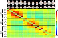
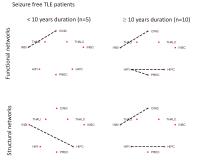
Temporal lobe epilepsy (TLE) is a common and relatively homogeneous form of epilepsy in which seizures originate in the mesial temporal regions including the hippocampus and propagate across the brain. This work represents the first step in characterizing the functional (FC) structural (SC) network connectivity evolution in TLE using MRI. We found consistent decreases in ipsilateral hippocampus and insula FC and SC primarily after 10 years of duration of disease in patients with seizure freedom after surgery. In those with seizure recurrence, there were more severe bilateral hippocampal SC decreases when compared to those with seizure freedom.


1673.   
Gender related peculiarities of amygdala deactivation during movements
Oleksii Omelchenko1, Zinayida Rozhkova2, and Mykola Makarchuk1
1Human and Animal Physiology, Taras Shevchenko National University of Kyiv, Kyiv, Ukraine, 2Medical Clinic BORIS, Kyiv, Ukraine
Men and women might display distinct characteristics of functional organization of neurocognitive brain networks. Considering gender-specific brain functioning under language, emotional and memory tasks execution, we propose fMRI visualization of the brain activated by a movement task for estimation of gender specific motor brain network peculiarities. New evidence for gender related differences in amygdala function was found. Results also give us background for further subdivision of the fMRI normative basis from which we investigate functional brain changes in patients’ population.


1674.   
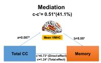
Longitudinal Study of Motor Recovery After Pontine Infarction with Resting-state fMRI:A Homotopic Connectivity Study
Yi Shan1, Chaogan Yan2, Miao Zhang1, Dongdong Rong1, Zhilian Zhao1, Qingfeng Ma3, Xinian Zuo2, Jie Lu4, and Kuncheng Li1
1Department of Radiology, Xuanwu hospital, Capital Medical University, Beijing, China, People's Republic of, 2Key Laboratory of Behavioral Science and Magnetic Resonance Imaging Research Center, Institute of Psychology, Chinese Academy of Sciences, Beijing, China, People's Republic of, 3Department of Neurology, Xuanwu hospital, Capital Medical University, Beijing, China, People's Republic of, 4Department of Nuclear Medicine, Xuanwu hospital, Capital Medical University, Beijing, China, People's Republic of
Impairment of motor function is one of the most severe deficit in ischemic stroke patients. Therefore, evaluations of brain function reorganization during spontaneous motor recovery are extremely valuable. In the present study, we used a voxel-mirrored homotopic connectivity (VMHC) method to investigate the longitudinal functional homotopic changes in patients with pontine infarction during a 180-day-period follow-up. The result shows resting-state fMRI could demonstrate dynamic whole-brain homotopic FC changes in stroke patients which might be helpful to further discuss brain reorganization after stroke. Also, VMHC between cognitive brain areas in acute stage had significant correlation with clinical behavioral performance in chronic period which might be meaningful in predicting motor outcome. 


1675.   
Exploring visual network connectivity in the mouse brain using DCM fMRI
Arun Niranjan1, Peter Zeidman2, Jack A Wells1, and Mark F Lythgoe1
1Centre for Advanced Biomedical Imaging, University College London, London, United Kingdom, 2Institute of Neurology, University College London, London, United Kingdom
Understanding effective (i.e. causal) connectivity in the brain using fMRI with dynamic causal modelling (DCM) has attracted a large amount of interest in recent years. Applications of fMRI to map brain function in the mouse are on the rise, targeting transgenic mouse models of pathology. However, DCM has not yet been applied to mouse brain fMRI, in part due to the difficulties of acquiring high quality data. In this work we demonstrate the use of DCM fMRI to understand effective connectivity in the healthy mouse visual system, showing results consistent with the underlying biology.


1676.   
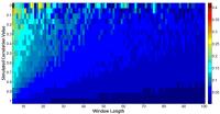
Task-related dynamic functional connectivity in fast fMRI
Ashish Kaul Sahib1, Michael Erb1, Klaus Scheffler2, Thomas Ethofer1, and Niels Focke3
1Biomedical magnetic resonance, University of tuebingen, Tuebingen, Germany, 2Max-Planck-Institute for Biological Cybernetics, Tuebingen, Germany, 3Department of Neurology/Epileptology, University of tuebingen, Tuebingen, Germany
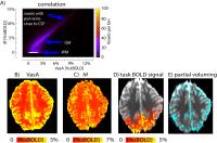
 Recent advances in simultaneous multi-slice imaging have improved the temporal resolution of fMRI. Using a sliding window approach we aimed to capture the dynamic network changes that occur during visual stimulation. We estimated the functional connectivity degree (FCD) at various stimulation lengths and window sizes. We demonstrate that the analysis of dynamic functional connectivity using a sliding window approach is an effective technique to capture whole brain temporal dynamics during a simple block-designed visual experiment (checkerboards). In summary, for the current setup, a window size of 13.s provided an optimum trade-off between temporal smoothness and FCD estimation.


1677.   
CEEMD-based Multi-Spectrum Brain Networks for Identification of MCI
Li Zheng1, Long Qian1, Dandan Zheng2, and Jiahong Gao3,4
1Department of Biomedical Engineering, College of Engineering, Peking University, Beijing, China, People's Republic of, 2GE Healthcare, MR Research China, Beijing, Beijing, China, People's Republic of, 3Beijing City Key Lab for Medical Physics and Engineering, Institute of Heavy Ion Physics, School of Physics, Peking University, Beijing, China, People's Republic of, 4Center for MRI Research, Academy for Advanced Interdisciplinary Studies, Peking University, Beijing, China, People's Republic of
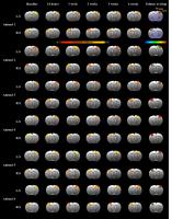
The early detection of MCI is of paramount importance for possible delay of the transition from MCI to AD. Recently, several resting-state fMRI based neural imaging studies have been applied for MCI diagnosis by the aid of pattern classification recently. In current study, CEEMD-based high-dimensional pattern classification framework was proposed to identify MCI individuals from subjects who experience normal aging with an accuracy of 93.3 percent, compared to conventional method for brain oscillation separation. In addition, the most discriminant regions selected by our method also reflected the association with MCI, to some degree.


1678.   
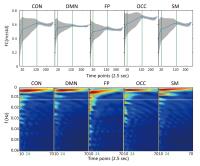
Age related fluctuation energy and variation of dynamic functional connectivity
Yuanyuan Chen1, Weiwei Wang1, Xin Zhao1, Miao Sha1, Yanan Liu1, Peng Zhou1, Hongyan Ni2, and Dong Ming1
1Tianjin University, Tianjin, China, People's Republic of, 2Tianjin First Central Hospital, Tianjin, China, People's Republic of
To reveal the age related changes of dynamic function connectivity during rest, five networks were extracted from resting stated fMRI data of 36 young people and 32 old people. The sliding window was carefully selected and the FC variation and the fluctuation energy in detailed frequency band were statistically compared.  Decreased FCV and slowing fluctuation in inter-networks were mainly found in old group. OCC and CON, OCC and FP were the most consistent inter-networks between this two age related changes. We concluded that FCV and fluctuation energy had provided a new perspective of aging research. 


1679.   
Association between structural and functional inter-subject variability of the motor and visual networks
Maxime Chamberland1,2, Gabriel Girard2, Michaël Bernier1, Michael Paquette2, David Fortin3, Maxime Descoteaux2, and Kevin Whittingstall1,4
1Nuclear Medicine and Radiobiology, Université de Sherbrooke, Sherbrooke, QC, Canada, 2Computer science, Université de Sherbrooke, Sherbrooke, QC, Canada, 3Division of Neurosurgery and Neuro-Oncology, Université de Sherbrooke, Sherbrooke, QC, Canada, 4Department of Diagnostic Radiology, Université de Sherbrooke, Sherbrooke, QC, Canada
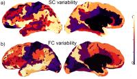
"Your brain is unique" is an unequivocal sentence that has spanned many research topics in the recent years. For example, functional connectivity (FC) based on resting-state fMRI is highly variable from one subject to the next, yet the source of this variability is unclear.  Understanding the source of FC variability is important as it is often used in clinical studies. Here, we explore how this might be explained by variability of white-matter structural connectivity (SC) derived from diffusion MRI tractography connectivity matrices. Our results show that, across multiple brain areas, motor and visual networks show the lowest inter-subject variability. This suggests that, at least in these areas, SC might explain a portion of FC variability.


1680.   
Interhemispheric Functional Connectivity Modulated by Menstrual Cycle
Xinyuan Miao1, Lin Shi1, Yan Zhuo2, and Yihong Yang3
1Department of Medicine and Therapeutics, Chinese University of Hong Kong, Hong Kong, Hong Kong, 2Institute of Biophysics, Chinese Academy of Sciences, Beijing, China, People's Republic of, 3National Institute on Drug Abuse, NIH, Baltimore, MD, United States
The functional lateralization of the brain was modulated by the menstrual cycle of women, while the mechanism of which still need to investigate.In this study, we used interhemispheric functional connectivity of the resting-state functional MRI to investigate changes in the symmetrical interhemispheric correlations in women’s different menstrual phases. Our results showed that the brainstem and cerebellum had significantly higher interhemispheric correlations in the early follicular phase than in the mid-luteal phase.


1681.   
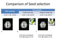
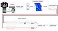
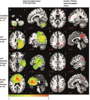
Combining Resting-State fMRI and Perfusion maps for potential Pre-Surgical Planning
Lalit Gupta1, Prativa Sahoo1, Pradeep K Gupta2, Indrajit Saha3, Rana Patir4, Sandeep Vaishya4, and Rakesh K Gupta2
1Philips India Ltd., Bangalore, India, 2Department of Radiology, Fortis Memorial Research Institute, Gurgaon, India, 3Philips India Ltd., Gurgaon, India, 4Department of Neurosurgery, Fortis Memorial Research Institute, Gurgaon, India
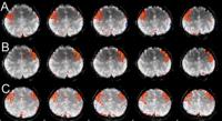
Mapping of functionally active regions for patients with mass lesions is critical for pre-surgical planning. We have developed an atlas based approach that automatically select seed points from six functional regions (motor and language regions) and computes corresponding functionally connected regions using resting state fMRI data. Functional connectivity maps were super-imposed on MR perfusion maps and structural images. Results were obtained from 22 brain tumor patients. Regions near the tumor with high correlation are seen as active regions that contribute to motor/language activities, combined with perfusion maps may help clinicians for better surgical planning.


1682.   
Memory and Learning: Visually-evoked Olfactory fMRI Activation Patterns and its Dynamics
Prasanna Karunanayaka1, Xin Zhang2, Michael Tobia1, Jianli Wang1, Bin Zhang2, Bin Zhu 2, and Qing Yang1
1Radiology, Penn State University, Hershey, PA, United States, 2The affiliated Drum Tower hospital of Nanjing university medical school, Nanjing, China, People's Republic of
Behavioral studies show that human odor perception is highly dynamic, incorporates both spatial and temporal codes, and is easily influenced by information from other sensory systems such as vision. However, the neural representation of odor perception and its dynamic processing by the brain is poorly understood. In this research, using olfactory task fMRI, we attempt to unravel how olfactory-related neural networks interact in both space and time in order to explore how the olfactory and the visual systems integrate information at the central or perceptual levels in the human brain.


1683.   
Quasi-periodic pattern of fMRI contributes to functional connectivity and explores difference between Major Depressive Disorder and control
Kai Wang1, Waqas Majeed2, Garth Thompson3, Kui Ying4, Yan Zhu5, and Shella Keilholz6
1Department of Biomedical Engineering, Tsinghua University, Beijing, China, People's Republic of, 2Department of Electrical Engineering, LUMS School of Science and Engineering, Lahore, Pakistan, 3Department of Radiology and Biomedical Imaging, Yale University, New Haven, CT, United States, 4Department of Engineering Physics, Tsinghua University, Beijing, China, People's Republic of, 5Psychiatry Department, Yu Quan Hospital, Tsinghua University, Beijing, China, People's Republic of, 6Department of Biomedical Engineering, Emory University/Georgia Institute of Technology, Atlanta, GA, United States
Quasiperiodic pattersn (QPPs) of BOLD fluctuations, first reported in [1,2] are likely contributors to functional connectivity (FC) due to their spatial and temporal structure. FC has been widely used to explore the altered brain organization in patients suffering from psychological disorders like Major Depressive Disorder (MDD). In this project, we examined the contribution of QPPs to FC in both normal subjects and MDD patients. Results showed that QPPs are a major contributor to FC, and that QPP abnormality can be a contributor to or marker of psychiatric or neurological disorders.


1684.   
Investigation of functional connectivity changes in Alzheimer's disease and amnestic mild cognitive impairment using Degree Centrality
Yong Zhang1, Naying He2, Hua-Wei Lin2, Ajit Shankaranarayanan3, Zhenyu Zhou1, and Fu-Hua Yan2
1MR Research China, GE Healthcare, Beijing, China, People's Republic of, 2Radiology, Ruijin Hospital, Shanghai Jiaotong University School of Medicine, Shanghai, China, People's Republic of, 3GE Healthcare, Menlo Park, CA, United States
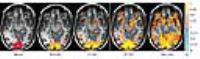
This preliminary study investigated functional connectivity changes in Alzheimer’s Disease (AD) and amnestic mild cognitive impairment (MCI) using degree centrality (DC), a novel resting-state fMRI parameter to provide voxel-wise whole brain functional connectivity measurement. Twelve AD patients, twelve MCI patients and fifteen healthy controls were recruited for comparison. As compared to normal controls, AD patients showed the deceased DC in the posterior cingulate cortex while MCI patients showed decreased DC in bilateral cuneus (visual processing) but increased DC in bilateral hippocampus (memory) and right angular gyrus (language).  The different patterns of FC changes might provide insight into disease evolvement.


1685.   
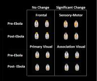
Ebola Alters Some, But Not All, Resting-State Intrinsic Functional Connectivity Networks In The Macaque Brain
Eswar Damaraju1, Margaret Lentz2, Jeffrey David Lewine1,3, David Thomasson2, Nadia Biassou4, Anna Honko2, Vince Calhoun1, and Peter Jahrling2
1Mind Research Network, Albuquerque, NM, United States, 2Integrated Research Facility/NIAID, Frederick, MD, United States, 3Lovelace Family of Companies, Albuquerque, NM, United States, 4NIH Clinical Center, Bethesda, MD, United States
Ebola has the potential to cause both acute and chronic compromise of neurological status.  To better understand the relevant neurobiology, a pilot MRI study of infected macaques was performed.  Data indicate that Ebola exposure leads to acute disruption of some, but not all, intrinsic connectivity networks, even in the absence of intraparenchymal lesions.  These studies represent the first non-invasive functional imaging studies of living, Ebola infected non-human primates. 


1686.   
Aberrant salience network and its functional coupling with default and executive networks in minimal hepatic encephalopathy: a resting-state fMRI study
Hua-Jun Chen1
1The First Affiliated Hospital of Nanjing Medical University, Nanjing, China, People's Republic of
Aberrant functional coupling of triple network in MHE


1687.   
Effect of Brain Tumours on the Default Mode Network
Sukhmanjit Ghumman1, David Fortin1, Stephen Cunnane1, and Kevin Whittingstall1
1Centre Hospitalier Universitaire de Sherbrooke (CHUS), Sherbrooke, QC, Canada
The effect of various pathologies on the default mode network (DMN) have been investigated in recent years with some encouraging results. These studies have found that some diseases of the nervous system, such as brain tumours, can have an effect on DMN connectivity. The goal of this novel research was to investigate whether tumours of certain areas of the brain or of certain histological type had disproportionately large effects on the DMN.  We believe that DMN connectivity could be developed into a prognostic score in the future which might help clinicians in making key treatment decisions for brain cancer patients.


1688.   
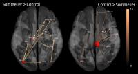
Training Induced Olfactory Network Changes in Master Sommeliers: Connectivity Analysis Using Granger Causality and Graph-theoretical Approach.
Karthik R Sreenivasan1, Xiaowei Zhuang1, Virendra Mishra1, Zhengshi Yang1, Gopikrishna Deshpande2, Sarah Banks1, and Dietmar Cordes1
1Cleveland Clinic Lou Ruvo Center for Brain Health, Las Vegas, NV, United States, 2AU MRI Research Center, Department of Electrical and Computer Engineering, Auburn University, Auburn, AL, United States
Current study used fMRI to investigate differences in effective connectivity and network topology between a group of trained master sommeliers and untrained control participants during olfactory tasks. Master sommeliers showed stronger connectivity originating from regions involved in higher-level cognitive processes than the controls. There was also increased small-world topology in the sommeliers. These findings provide unique insights into the neuroplasticity in adulthood in the olfactory network which may have added clinical importance in diseases like Alzheimer’s and Parkinson’s where early neurodegeneration is isolated to regions important in smell.


1689.   
Structural and Functional Brain Alterations in Uremic restless legs syndrome patients: A Voxel-Based Morphometry and Functional Connectivity Study
DUN DING1, PENG LI2, Ji Xin Liu2, Xue Ying Ma2, and Ming Zhang2
1XI'AN JIAO TONG UNIVERSITY, XI'AN, China, People's Republic of, 2XI'AN, China, People's Republic of
To investigate the structure and function changes in the brain in uremic RLS patients using a resting-state function magnetic resonance imaging (fMRI) paradigm, we used A voxel-based morphometry(VBM) method and a seed-based method to find the abnormiy in end-stage kidney disease patients. Our results suggest that the characteristics of the connectivity changes may reflect the pathways involved in producing uremic RLS symptoms.


1690.   
Zinc Nanoparticles Enhance Brain Connectivity in the Canine Olfactory Network: Evidence from an fMRI Study in Fully Unrestrained Conscious Dogs
Bhavitha Ramaiahgari1, Oleg M Pustovyy2, Paul Waggoner3, Ronald J Beyers1, John Schumacher4, Chester Wildey5, Edward Morrison2, Nouha Salibi1,6, Thomas S Denney1,7,8, Vitaly J Vodyanoy2, and Gopikrishna Deshpande1,7,8
1Dept of Electrical & Computer Engr, AU MRI research center, Auburn University, Auburn, AL, United States, 2Dept. of Anatomy, Physiology & Pharmacology, Auburn University, Auburn, AL, United States, 3Canine Detection Research Institute, Auburn University, Auburn, AL, United States, 4Dept. of Clinical Sciences, Auburn University, Auburn, AL, United States, 5MRRA Inc., Euless, TX, United States, 6MR R&D, Siemens healthcare, Malvern, PA, United States, 7Dept. of Psychology, Auburn University, Auburn, AL, United States, 8Alabama Advanced Imaging Consortium, Auburn University and University of Alabama Birmingham, Birmingham, AL, United States
There is intense interest in strategies for enhancing olfaction capabilities of dogs for various applications such as bomb detection. Prior fMRI studies showed increased neural activation when zinc nanoparticles were added to the odorants. In this study, we obtained fMRI data from awake and unrestrained dogs when they were exposed to odorants with and without zinc nanoparticles and zinc nanoparticles alone. We observed that zinc nanoparticles up-regulated directional brain connectivity in parts of the canine olfactory network. This provides a mechanistic explanation for previously reported enhancement in the odor detection capability of the dogs in the presence of zinc nanoparticles.


1691.   
Altered amplitude of low-frequency fluctuations and connectivities in depressed SAPHO syndrome
Jie Lu1, Yan-ping Duan2, Wen-rui Xu1, Xue-wei Zhang3, Chen Li4, and Wei-hong Zhang1
1Department of Radiology, Peking Uinon Medical College Hospital, Beijing, China, People's Republic of, 2Department of Psychology, Peking Uinon Medical College Hospital, Beijing, China, People's Republic of, 3Department of interventional radiology, China Meitan General Hospital, Beijing, China, People's Republic of, 4Traditional Chinese Medicine Department, Peking Uinon Medical College Hospital, Beijing, China, People's Republic of
To investigate depressed symptoms in SAPHO(Synovitis, acne, pustulosis, hyperostosis, osteitis ) syndrome and confirm depression in SAPHO using resting-state functional magnetic resonance imaging (rs-fMRI). We recruited twenty-four SAPHO patients and fifteen age- and gender-matched normal controls (NC). Twelve of the SAPHO patients were diagnosed with depression. Moreover, depressed SAPHO patients (D-SAPHO) were proved to have abnormal amplitude of low frequency fluctuations (ALFF) and functional connectivities (FC) involved in the regional brain changes which showed correlated with the severity of depression. These findings provide crucial information to understand the neural mechanisms of depressed SAPHO and are helpful to diagnose depression in SAPHO.


1692.   
Cocaine and the synthetic cathinone MDPV reduce small world brain network topology: a rat functional connectivity study
Luis Manuel Colon-Perez1 and Marcelo Febo1
1Psychiatry, University of Florida, Gainesville, FL, United States
Drug abuse has detrimental effects on the brain function, which lead to drug use disorders. In vivo non-invasive biomarkers are needed to determine the neurobiological outcomes of addictive drugs on the brain. Functional MRI and graph theory offer an analytical approach to address brain network changes associated with psychiatric disorders. In the present study we determined the effects of two addictive psychostimulant drugs. Comparison between saline and drug administered shows a reduction in the connectivity at 1 hr but not at 24 hrs. Acute administration of the two psychostimulants studied produce only transient effects lasting at least 1 hr.


1693.   
Resting-state functional activity and brain network abnormalities in betel nut chewers
Yu-Syuan Chou1, Ming-Chou Ho2, and Jun-Cheng Weng1,3
1Department of Medical Imaging and Radiological Sciences, Chung Shan Medical University, Taichung, Taiwan, 2Department of Psychology, Chung Shan Medical University, Taichung, Taiwan, 3Department of Medical Imaging, Chung Shan Medical University Hospital, Taichung, Taiwan
Betel nut, also known as areca, is the fourth most commonly used drug worldwide after tobacco, alcohol, and caffeine and also a stimulant and addictive substance. Previously, CM Chen et al. probed into the influence of religious affiliation on heavy betel nut chewing, and studied on the relationship between health risk perception and betel nut chewing. Feng Chen et al. analyzed gray matter abnormalities between betel nut chewers and healthy subjects with voxel-based morphometry (VBM). However, there were few studies mentioned about the functional activity and brain network changes in betel nut chewers using functional magnetic resonance imaging (fMRI). Therefore, our aim was to use resting-state fMRI (rs-fMRI) to investigate the functional differences between betel nut chewers and healthy participants with amplitude of low frequency fluctuations (ALFF) and regional homogeneity (ReHo). The graph theoretical and network-based statistic (NBS) analyses were also used to find the network difference between two groups. Our results revealed different topological organization and poor global integration of the brain network in the betel nut chewers. 


1694.   
Can Cerebral Functional Deficits Be Detected in Patients with Ankylosing Spondylitis?- A Cross-sectional Study
Jun Zhao1, Chuan-Ming Li1, Xin Wei1, and Jian Wang1
1Radiology, Southwest Hospital, Third Military Medical University, Chongqing, China, People's Republic of
This study aimed to investigate any cerebral function deficits in AS(Ankylosing spondylitis) using functional MRI technology and its possible relationship to clinical and laboratory results. Compared with normal controls, AS patients showed widespread brain activity and connectivity alterations. Functional connectivity strength of the left precuneus and the left middle temporal gyrus were closely correlated with the the BASDAI scores, ESR and hsCRP in AS patients. AS is associated with a altered cortical activity of rs-fMRI signals. Measurement of functional connectivity strength of the left precuneus and the left middle temporal gyrus may aid in the clinical detection and evaluation of AS


1695.   
Functional dysconnection between anterior cingulate cortex and thalamus in patients with Kleine-Levin syndrome
Ting-Chih Wang1, Yao-Chia Shih2,3, Hong-Huei Liu4, and Wen-Yih Issac Tseng3,5
1Department of Electrical Engineering, National Taiwan University, Taipei, Taiwan, 2Institute of Biomedical Engineering, National Taiwan University, Taipei, Taiwan, 3Institute of Medical Device and Imaging, National Taiwan University College of Medicine, Taipei, Taiwan, 4Department of Neurology, National Taiwan University Hospital, College of Medicine, National Taiwan University, Taipei, Taiwan, 5Molecular Imaging Center, National Taiwan University, Taipei, Taiwan
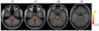
Kleine-Levin Syndrome is a rare neurological disorder characterized by recurrent episodes of excessive sleepiness and other symptoms listed in the ICSD Diagnostic Criteria for KLS. Its etiology is still unknown nowadays. The most consistent finding in KLS is abnormal thalamic function. Here, we used seed-based analysis to analyze resting state fMRI obtained from 2 patients with KLS. In bilateral thalamic seeding, both patients showed decreased connection between the thalamus and the anterior cingulate cortex. This result could be attributed to alteration of the dorsal pathway in ascending arousal system, and might also explain their attention deficits.


1696.   
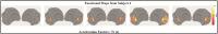
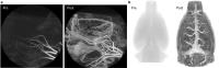
Pinpointing the rat cerebellar and medullary noxious networks with fMRI based fcMRI
Rupeng Li1, Xiping Liu2, Jason Sidabras1, Christopher Pawela3, Andrzej Jesmanowicz1, and James Hyde1
1Biophysics, Medical College of Wisconsin, Milwaukee, WI, United States, 2Dermatology, Medical College of Wisconsin, Milwaukee, WI, United States, 3Anesthesiology, Medical College of Wisconsin, Milwaukee, WI, United States
Pinpoint acquisition of high resolution, true whole brain scale fcMRI sensorimotor network using seed based analysis. We are able to greatly reduce susceptibility induced artifact in deep brain structures while keeping great SNR and depth sensitivity. fcMRI networks in cerebellum and modular areas are demonstrated with intermediate reticular nucleus (IRt) observed.


1697.   
Functional connectivity changes in attention-related networks of childhood leukemia survivors
Charlotte Sleurs1, Iris Elens2, Jurgen Lemiere1, Thibo Billiet3, Dorothée Vercruysse4, Patricia Bijttebier5, Marina Danckaerts2, Rudi D'Hooghe6, Ron Peeters3, Stefan Sunaert3, Anne Uyttebroeck1, Stefaan Van Gool7, and Sabine Deprez3
1Pediatric Hemato-Oncology, UZ Leuven, Leuven, Belgium, 2Child and Adolescent Psychiatry, UZ Leuven, Leuven, Belgium, 3Radiology, UZ Leuven, Leuven, Belgium, 4Gynaecological Oncology, UZ Leuven, Leuven, Belgium, 5School Psychology and Child and Adolescent Development, KU Leuven, Leuven, Belgium, 6Biological Psychology, KU Leuven, Leuven, Belgium, 7Pediatric Hemato-Oncology, University Hospital, Aachen, Germany
Neurocognitive sequelae in childhood leukemia survivors are often related to attentional disfunctioning. We investigated whether altered functional brain connectivity might explain neurocognitive sequelae in childhood leukemia survivors. Resting state fMRI was investigated, by using ROI-based connectivity comparisons as well as dual regression analysis at whole-brain level. We demonstrated that the Default Mode Network (DMN) and Inferior Temporal Gyrus, was less functionally connected in childhood leukemia survivors compared to controls. This suggests an altered coherence between activity of the DMN and Fronto-Parietal Network (FPN). Finally, based on this specific connectivity we could predict clearly reduced cognitive flexibility of the patients.


1698.   
Effects of long-duration isoflurane administration on default mode network of macaque brains
Chun-Xia Li1 and Xiaodong Zhang1,2
1Yerkes Imaging Center, Yerkes National Primate Research Center, Emory University, Atlanta, GA, United States, 2Division of Neuropharmacology and Neurologic Diseases, Yerkes National Primate, Atlanta, GA, United States
Long-duration anesthesia administration could cause neurocognitive decline in animals and humans. However, the potential mechanism still remains unclear. In the present study, the functional connectivity of adult rhesus monkeys under maintenance dosage of isoflurane (~1 %) for four hours was examined. The results demonstrate that long-duration isoflurane exposure resulted in decreased functional connectivity in posterior cingulate cortex (PCC) dominant default-mode network (DMN). The MRI findings suggest that the detrimental effects of isoflurane on brain connectivity may be associated with the neurocognitive decline observed in subjects after long-duration administration of isoflurane.


1699.   
Neurofeedback impact onto the brain networks interaction: fMRI study
Oleksii Omelchenko1 and Volodymyr Rogozhyn2
1Human and Animal Physiology, Taras Shevchenko National University of Kyiv, Kyiv, Ukraine, 2Radiology, Medical Clinic BORIS, Kyiv, Ukraine
Concerning the use of audio-visual stimulation (AVS) as a component of neurofeedback therapy for neuropsychiatric disorders we propose to evaluate its effect onto the brain networks interaction. We performed fMRI before and after the AVS. fMRI exams showed considerable increase of the volumes of activation after the AVS and almost complete extinction of the DMN deactivation. RS fMRI showed functional connectivity changes after the AVS (connectivity disruption in visual network, DMN frequency shift). Volume of activation increase and functional connectivity changes could be the marker for prolonged effect of AVS brain stimulation. 


1700.   
Spatial and temporal modulation of brain dynamics in response to task execution
Silvia Tommasin1,2, Daniele Mascali1,3, Tommaso Gili1,2, and Federico Giove1,2
1Enrico Fermi Centre, Rome, Italy, 2Fondazione Santa Lucia, Roma, Italy, 3Physics, Università La Sapienza, Roma, Italy
Task-related activity influences brain connectivity through a two-level pattern modulation both in attentive networks and in the default mode network. While strengthening the local homogeneity, task execution reduces regional synchronization. It produces correlation patterns with opposite large and small scale properties. Task-related activity influences also the amplitude of the low frequency fluctuations in the same networks.  The transition from resting state to steady state task execution, and the way back, causes a persisting slow drift in this quantity.


1701.   
Multi-node directed cortical network for speech processing revealed by multivariate Granger causality analysis
Yayan Yin1, Jiahong Gao1, Bing Wu2, Yang Fan2, Bingjiang lyu1, and Jianqiao Ge1
1Peking University, Beijing, China, People's Republic of, 2GE Healthcare, Beijing, China, People's Republic of
For decades, how the information flows among multiple brain regions remains unclear for speech processing, due to the challenge of mapping multi-node directed cortical pathways from brain images. In this work, multivariate Granger causality analysis is employed on functional MR images to reveal the effective connectivity of Chinese language-speech network for the first time. The results showed that left insula and posterior middle temporal gyrus were the strong driver nodes, the left middle frontal gyrus and superior temporal gyrus were the most received nodes in the network. We also found greater interhemispheric connectivity in females compared to males.


1702.   
Modular Reorganization of Resting-State Brain Network in Patients with Obstructive Sleep Apnea
Bumhee Park1, Sudhakar Tummala1, Ruchi Vig1, Daniel W Kang2, Mary A Woo3, and Rajesh Kumar1,4,5,6
1Anesthesiology, University of California at Los Angeles, Los Angeles, CA, United States, 2Medicine, University of California at Los Angeles, Los Angeles, CA, United States, 3UCLA School of Nursing, Los Angeles, CA, United States, 4Radiological Sciences, University of California at Los Angeles, Los Angeles, CA, United States, 5Bioengineering, University of California at Los Angeles, Los Angeles, CA, United States, 6Brain Research Institute, University of California at Los Angeles, Los Angeles, CA, United States
Obstructive sleep apnea (OSA) condition is accompanied by brain tissue injury and functional deficits in regions serving autonomic, neuropsychologic, and cognitive functions. Brain networks are organized into modular systems and assigning vulnerable role for each region in terms of intra- and inter-modular communication provides better understanding for functional deficits in the condition. We examined the modular reorganization of OSA functional networks, and found abnormal intra- and/or inter-modular communication roles in brain regions involved in autonomic, neuropsychologic, and cognitive regulation. The findings suggest that dysfunctions associated with OSA may be related to abnormal information flow, and can be examined with modular reorganization assessment.


1703.   
Bilateral amygdaloid functional connectivity in chronic alcoholics
Ylin Zhao1, Jun Chen2, and Hui Lin3
1Radiology, Renmin Hospital of Wuhan University, Wuhan, China, People's Republic of, 2Renmin Hospital of Wuhan University, Wuhan, China, People's Republic of, 3Healthcare,MR Research China, Beijing, China, People's Republic of
FC-MRI is a useful tool for examining functional relationships between the bilateral amygdaloid  and whole brain regions. The functional coordination of bilateral amygdala and cerebral cortex was enhanced,and the functional coordination of bilateral amygdala and cerebellum was weakened.Amygdala may be involved in regulating the function of fronto-cerebellar loops.Thus, this method shows promise as a tool for in vivo investigations of the functioning of human  fronto-cerebellar circuitry. It is our hope that in future studies this technique may provide the opportunity to examine the integrity of networks involving the brain cerebellum inpatient groups with chronic alcoholics, a major goal of our research.


1704.   
Putamen-related regional and network functional deficits in first-episode schizophrenia with auditory verbal hallucinations
Long-Biao Cui1, Yi-Bin Xi1, and Hong Yin1
1Xijing Hospital, Fourth Mililtary Medical University, Xi'an, China, People's Republic of
Our results suggest an association of abnormal regional function in the putamen and prefrontal cortex and hyperconnectivity between them with AVHs in SZ. The functional interaction of the putamen with DLPFC and Broca’s area seems to be crucial for AVHs in SZ. Additionally, the putamen-related regional and network functional deficits may also serve as a potential diagnostic biomarker of AVHs in SZ based on the direct evidence in vivo we found. In SZ patients, there is an extensive hypoconnectivity within cortical-striatal-cerebellar networks, which further supports the current thinking about disconnection hypothesis of SZ.


1705.   
Light isoflurane sedation: an excellent trade-off between anesthesia and awake condition in functional connectivity studies with rats
Jaakko Paasonen1, Raimo A Salo1, Artem Shatillo2, and Olli Gröhn1
1Department of Neurobiology, University of Eastern Finland, Kuopio, Finland, 2Charles River Discovery Services, Kuopio, Finland
Prevention of motion is a prerequisite for preclinical functional connectivity (FC) studies. However, anesthesia alters brain function, and awake protocols may induce stress. Therefore, we investigated the feasibility of using light sedation in FC studies. FC was estimated under 0.1/0.5% isoflurane (subanesthetic doses) with acclimatized rats, and under 1.3% isoflurane (anesthetic dose). Results demonstrate different FC between anesthetic and subanesthetic doses. The physiologic measures suggest, that the 0.5% rats adapted well to imaging, while the 0.1% rats did so insufficiently. Therefore, light isoflurane sedation may provide an excellent combination for FC investigations: minimal stress and motion with normal brain function.


1706.   
Visual Networks Impairments in Minimal Hepatic Encephalopathy Using Resting-State fMRI
Yun Jiao1, Xun-Heng Wang2, and Tian-Yun Tang1
1Jiangsu Key Laboratory of Molecular and Functional Imaging, Department of Radiology, Zhongda Hospital, Medical school of Southeast University, Nanjing, China, People's Republic of, 2College of Life Information Science and Instrument Engineering, Hangzhou Dianzi University, Hangzhou, China, People's Republic of
We applied dual regression to investigated functional connectivity impairments within visual networks for minimal hepatic encephalopathy (MHE) patients’ brain. Functional deficits within occipital and lateral visual networks (visual area V2 and V3) were endogenous, and significantly association with neurocognitive impairments. This maybe the reason for the compensatory enhancements within medial visual network (visual area V1) which indicated that patients with MHE had the potential to additionally recruit more neurological resource to process the spatial information from visual areas V2 and V3. Our results demonstrated the possible mechanisms for deficits in visual perception, visuo-spatial orientation, and visuo-constructive abilities in MHE patients


1707.   
Acupuncture Stimulation changes DMN functional conectivity.
Tomokazu Murase1, Masahiro Umeda1, Masaki Fukunaga2, Katsuya Maruyama3, Yuko Kawai1, Yasuharu Watanabe1, and Toshihiro Higuchi4
1Medical Informatics, Meiji University of Integrative Medicine, Nantan-shi, Japan, 2Cerebral Integration, National Institute for Physiological Sciences, Okazaki-shi, Japan, 3Research&Collaboration, Siemens Japan, Shinagawa-ku, Japan, 4Neurosurgery, Meiji University of Integrative Medicine, Nantan-shi, Japan
Clinical studies have shown that acupuncture relieves different kinds of pain. However, the effectiveness of these methods is unclear. We used rs-fMRI and FC analysis to examine the RSN activity before and after acupuncture stimulation. In the result, DMN was changed with or without manual acupuncture. The acupuncture stimulation controls a pain by modifying mechanism of pain transmission.


1708.   
Intrinsic functional connectivity in patients with presbycusis
Fei Gao1, Guangbin Wang1, Bin Zhao1, Muwei Li2, Fuxin Ren1, and Weibo Chen3
1Shandong Medical Imaging Research Institute, Shandong University, JINAN, China, People's Republic of, 2College of Electronics and Information Engineering, Sichuan University, Chengdu, China, People's Republic of, 3Philips Healthcare, Shanghai, China, People's Republic of
Presbycusis is the most common sensory deficit in the ageing population. However, little is known about whether the topological properties of brain functional networks is disrupted in patients with presbycusis. Our study demonstrates decreased clustering coefficient, local efficiency and strength in the primary auditory cortex in patients with presbycusis, as compared to age- and gender-matched healthy controls. Our study provides evidence of presbycusis-related disruptions in brain functional networks in patients with presbycusis. It is believed that our findings could be important for exploring functional changes in the central presbycusis.


1709.   
Altered small world brain function network in patients of lower back pain
Jing Liu1, Xiufen Liu2, zhizheng Zhuo3, Juan Wei4, Queenie Chan5, and Xiaoying Wang1
1Radiology, Peking University First Hospital, Beijing, China, People's Republic of, 2Anesthesiology, Peking University First Hospital, Beijing, China, People's Republic of, 3Philips Healthcare Beijing China, Beijing, China, People's Republic of, 4Philips Research China, Shanghai, China, Shanghai, China, People's Republic of, 5Philips Healthcare, China, Hongkong, China, People's Republic of
An altered functional network was found in the brain of LP here represents a less optimal network organization in the LP. It has been suggested that the small-world structure reflects an optimal balance between local processing and global integration. And the small-world structure of LP brain networks maybe destroyed due to the chronic LP.


1710.   
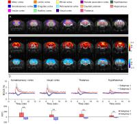
Subregion-specific Resting-State Amygdala Connectivity in Chronic Knee Osteoarthritis Pain: Towards a brain network signature of OA pain
William J Cottam1,2,3, Marianne Drabek1,2,3, Diane Reckziegel1,2,3, and Dorothee P Auer1,2,3
1Division of Clinical Neuroscience, Radiological Sciences, University of Nottingham, Nottingham, United Kingdom, 2ARUK Pain Centre, University of Nottingham, Nottingham, United Kingdom, 3Sir Peter Mansfield Imaging Centre, University of Nottingham, Nottingham, United Kingdom
Brain network connectivity analysis arguably offers the most sensitive marker to detect dysfunctional brain plasticity underlying the maladaptive nature of chronic pain. Early functional connectivity (fc) studies reveal altered functional connectivity in chronic pain states, but to the best of our knowledge no studies have focussed upon the amygdala. We aimed to investigate whether patients with painful chronic knee OA show altered amygdala connectivity compared to pain-free controls.This study identified increased functional connectivity of specific amygdala subnuclei in chronic OA pain patients compared to healthy subjects. 


1711.   
Time-shift functional connectivity MRI based on specific regional-of-interest for mapping acute ischemic Stroke
xiaokun fang1, qiang xu2, yong zhang3, zhiqiang zhang1, and guangming lu1
1Medical Imaging, Jingling Hospital, School of Medicine, Nanjing University, Nanjing, Jiangsu, China, nanjing, China, People's Republic of, 2Medical Imaging, Jingling Hospital, School of Medicine, Nanjing, nanjing, China, People's Republic of, 3MR Research China, GE Healthcare, beijing, China, People's Republic of
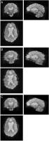
To investigate if Time-shift functional connectivity based resting-state fMRI can be used to create maps similar to time-to-maximum of  (Tmax) in acute stroke and to determine whether Maps obtained with the SSS seed (superior saggital sinus) or whole brain as the seed in Time-shift functional connectivity based resting-state fMRI be better in mapping the acute stroke.


1712.   
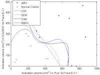
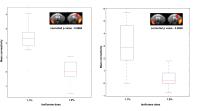
Demonstration of brain tumor-induced abnormalities on regional homogeneity (ReHo) resting state fMRI metrics KCC-ReHo & Cohe-ReHo
Shruti Agarwal1, Noushin Yahyavi-Firouz-Abadi1, Haris I. Sair1, Raag Airan1, and Jay J. Pillai1
1Division of Neuroradiology, Russell H. Morgan Department of Radiology and Radiological Science, Johns Hopkins University School of Medicine, Baltimore, MD, United States
Disruption of the normal coupling between neural activity and the consequent microvascular blood flow response (neurovascular uncoupling or NVU) may severely compromise the validity of BOLD fMRI in presurgical planning. The effects of brain tumor-related NVU on resting state BOLD fMRI (rsfMRI) using functional connectivity analysis have been previously published. In this study we evaluated regional homogeneity (ReHo) of rsfMRI data based on  Kendall's coefficient of concordance (KCC-ReHo) & Coherence (Cohe-ReHo) and compared the results with the amplitude of low-frequency fluctuation (ALFF) & standard motor tbfMRI activation to investigate regional abnormalities due to brain tumor-induced NVU in sensorimotor network. 

1713.   
Breaking ß: Understanding the ß-Value in Calibrated Functional MRI
Avery J.L. Berman1,2 and Bruce Pike2
1Montreal Neurological Institute, McGill University, Montreal, QC, Canada, 2Department of Radiology and Hotchkiss Brain Institute, University of Calgary, Calgary, AB, Canada
In calibrated functional MRI (fMRI), the parameter β is used to describe the non-linear dependence of the change in the transverse relaxation rate on the susceptibility offset (Δχ) of blood relative to tissue. Estimates of β at high field strengths have generally been assumed or obtained in post-hoc analyses. Using simulations from vessel networks, we present here a detailed description of β’s dependence on vessel radius. We show that the estimate of β is highly dependent on the range of Δχ used in the fit. This could have important implications for methods that propose to measure β-values in vivo using a contrast agent to alter Δχ.


1714.   
Flow-diffusion constrained estimation of oxygen extraction fraction and tissue oxygen tension by dual calibrated fMRI
Michael Germuska1, Alberto Merola1, and Richard G Wise1
1CUBRIC, Cardiff University, Cardiff, United Kingdom
An emerging method for quantitative mapping of OEF is by dual calibration of the BOLD signal. 1,2 However, this method is highly sensitive to measurement noise, resulting in unstable estimates of OEF. An alternative approach is to use flow-diffusion equations to calculate the biophysically supported OEF. 3 However, this approach is limited by the need to assume a tissue oxygen tension (PtO2). We propose a method for combining these two approaches, producing calibrated BOLD estimates of OEF that are constrained by a modelled flow-diffusion relationship of oxygen extraction. The proposed method is shown to produce stable estimates of OEF and PtO2.


1715.   
Do human cerebral arteries contain fully oxygenated blood?
Esther AH Warnert1, Ian D Driver1, Joseph Whittaker1, and Kevin Murphy1
1Cardiff University Brain Research Imaging Centre, Cardiff University, Cardiff, United Kingdom
When modelling the BOLD response it is often assumed that arterial blood is fully oxygenated, and that therefore contrast is driven solely by changes in venous oxygen saturation. Recent evidence has emerged from rodent studies indicating that precapillary arterioles may have an oxygenation level as low as 78%. Here, we assess arterial oxygenation in vivo in humans by using short inversion time ASL in normoxia, as well as hyperoxia. Our results suggest that imaging the BOLD response may not be affected by partially oxygenated arterioles. 


1716.   
Flow Related Changes in Oxygen Extraction Fraction Detected using Streamlined-qBOLD
Alan J Stone1 and Nicholas P Blockley1
1FMRIB, Nuffield Department of Clinical Neurosciences, University of Oxford, Oxford, United Kingdom
In this study we investigate the sensitivity of streamlined-qBOLD for detecting changes in baseline brain oxygenation and therefore its suitability for clinical application to vascular dysfunction and stroke. Baseline brain oxygenation is modulated in a group of normal volunteers using hypocapnia (a reduction in blood CO2). In this group, streamlined-qBOLD measured significant (p<0.05) increases in grey matter R2′ and OEF, between normocapnic and hypocapnic conditions. This suggests the technique can provide important metabolic information in cases of vascular dysfunction where flow and brain oxygenation may be impaired.  


1717.   
Detailing the Role of Systemic Blood Pressure in Somatosensory Mouse fMRI
Henning Matthias Reimann1, Mihail Todiras2, Michael Bader2, Andreas Pohlmann1, and Thoralf Niendorf1,3
1Berlin Ultrahigh Field Facility (B.U.F.F.), Max Delbrueck Center for Molecular Medicine, Berlin, Germany, 2Max Delbrueck Center for Molecular Medicine, Berlin, Germany, 3Experimental and Clinical Research Center, Charite-Universitaetsmedizin, Berlin, Germany
Combining mouse genomics and functional magnetic resonance imaging (fMRI) provides a promising tool to unravel the molecular mechanisms of somatosensation and pain. Recent studies suggest a confounding influence of mean arterial blood pressure (MABP) on the blood oxygenation level-dependent (BOLD) signal for somatosensory stimulation paradigms in mice. MABP alterations induced by transient stimuli can mimic brain activation in small rodents. The abstract provides data, which rely on the monitoring of MABP for mild noxious thermal stimulation in mice. Detailing the role of MABP in mouse fMRI is crucial to ensure the integrity of murine hemodynamic readouts in somatosensation and nociception.


1718.   
The effect of plasma-dissolved oxygen on the hyperoxic calibrated BOLD signal: a simulation study using the detailed BOLD model
Yuhan Ma1, Avery Berman1, and G. Bruce Pike1,2
1Brain Imaging Centre, Montreal Neurological Institute, McGill University, Montreal, QC, Canada, 2Department of Radiology, Hotchkiss Brain Institute and Department of Radiology, University of Calgary, Calgary, AB, Canada
It has been suggested that during a hyperoxic calibration, the paramagnetic oxygen dissolved in arterial blood plasma can be a confounding factor for the interpretation of the calibrated BOLD signal. In this study, we aimed to predict the relative effect of dissolved oxygen on hyperoxic BOLD signal by expanding the detailed BOLD model with the effect of dissolved oxygen. Our results showed minimal difference in both the relative BOLD signal and the calibration parameter calculated with the effect of dissolved oxygen. Therefore, the influence of the dissolved oxygen in arterial blood plasma on the measured calibrated BOLD signal at 3 T can generally be ignored.


1719.   
Quantitative Susceptibility Mapping (QSM) based Cerebral Metabolic Rate of Oxygen (CMRO2) Mapping: Improve Robustness with Preconditioning and Physiological Constraints
Jingwei Zhang1,2, Dong Zhou2, Thanh Nguyen2, Pascal Spincemaille2, Ajay Gupta2, and Yi Wang1,2
1Biomedical Engineering, Cornell University, New York, NY, United States, 2Radiology, Weill Cornell Medical College, New York, NY, United States
This study proposed a new post-processing algorithm with preconditioning and physiological constraints for QSM based CMRO2 mapping, which eliminated physiologically impossible OEF values and improved the robustness of the technique. Reproducibility of the proposed method was examined. Feasibility of hyperventilation as a more efficient blood flow challenge was also investigated. 


1720.   
High-Resolution CMRO2 Mapping During a Unilateral Pinch-Force Task
Maria Guidi1, Christopher J. Steele1, Laurentius Huber2, Leonie Lampe1, Viola Rjosk1, Pierre-Louis Bazin1, and Harald E. Möller1
1Max Planck Institute for Human Cognitive and Brain Sciences, Leipzig, Germany, 2NIMH, Bethesda, MD, United States
Motor tasks have been extensively studied with BOLD fMRI. The way the BOLD response scales with task intensity level might come from an increased metabolic activity as well as an increased CBF and CBV. In this study, we aimed at disentangling such contributions combining a gas manipulation session with a pinch-task with simultaneous recording of force values. This way, the Davis model for calibration of fMRI could be applied and the oxygen metabolism changes estimated at every timestep. BOLD signal changes, VASO signal changes, and CMRO2 changes were shown to weakly scale with the intensity of the pinch-force task.


1721.   
Is the local functional connectivity anisotropy (LFCA) in white matter caused by neuro-electric activity?  An examination of potential confounds for orientation-dependent LFCA in fMRI
Michael J. Tobia1, David Gallagher1, Rahul Dewal1, Sebastien Rupprecht1, Prasanna Karunanayaka1, and Qing X. Yang1
1Radiology, Penn State Hershey, Hershey, PA, United States
Phantom experiments showed that fluctuating electric current is sufficient to generate local functional connectivity anisotropy (LFCA), and that effects of motion, such as Lorentz forces, cannot explain the alignment of eigenvectors through neighboring voxels or B0 orientation-dependence.  In conclusion, anisotropic correlations of fMRI time series may arise from an alternative non-BOLD contrast mechanism, potentially related to an electric current effect on B0. 


1722.   
Development of an evaluation system for analgesic drugs targeted to allodynia-specific pain using BOLD fMRI
Naoya Yuzuriha1, Sosuke Yoshinaga1, Hiroshi Sato2, Sokichi Honda3, Keisuke Tamaki3, Toshihiro Sekizawa3, Akihiko Fujikawa3, and Hiroaki Terasawa1
1Department of Structural BioImaging, Faculty of Life Sciences, Kumamoto University, Kumamoto, Japan, 2Bruker Biospin K.K., Yokohama, Japan, 3Drug Discovery Research, Astellas Pharm. Inc., Tsukuba, Japan
The aim of this study is to evaluate the analgesic effect of pregabalin on allodynia-specific pain. fMRI images of fibromyalgia model rats were acquired, before and after pregabalin administration.  When treated with saline, the BOLD signal intensities in the S1, IC, and TH were increased upon the laser stimulation by up to 1.7%, 1.3%, and 1.8%, respectively.  In contrast, in the pregabalin treated rats, no BOLD responses were detected.  It is conceivable that the pain signals were inhibited by pregabalin, and thus the stimulation-induced BOLD responses were not observed.  We successfully observed the suppression of allodynia-specific pain responses by pregabalin.


1723.   
fMRI study of the role of glutamate NMDA receptor in olfactory habituation of olfactory bulb and higher olfactory structures in rats
Fuqiang Zhao1, Xiaohai Wang2, Hatim A Zariwala2, Jason M. Uslaner2, Andrea K Houghton2, Jeffrey L Evelhoch1, Eric Hostetler1, and Catherine Diane Gard Hines1
1Imaging, Merck Co. & Inc., West Point, PA, United States, 2Neuroscience, Merck Co. & Inc., West Point, PA, United States
fMRI offers an excellent opportunity to study olfactory processing in different olfactory structures. Measurement of the magnitude of odor stimulation-induced activations, and their suppression with agonist/antagonist of different neural receptors can provide important information to understand the mechanism of olfactory habituation. In this study, cerebral blood volume (CBV) fMRI with USPIO was used to measure odorant-induced olfaction in different olfactory structures of rats. The dynamics of habituation in different olfactory structures can be robustly measured by fMRI. MK801 can reverse habituated olfactory responses in all olfactory structures, suggesting that glutamate/NMDA receptor plays a major role in olfactory habituation.


1724.   
Plastic-adaptive changes after articulatory training in the elderly: An fMRI study
Sachiko Kiyama1, Atsunobu Suzuki2, Shen-Hsing Annabel Chen3, and Toshiharu Nakai1
1National Center for Geriatrics and Gerontology, Obu, Japan, 2Nagoya University, Nagoya, Japan, 3Nanyang Technological University, Singapore, Singapore
The present fMRI study explored neural changes in the Japanese elderly after four weeks of articulatory training. We compared real and pseudo words (i.e., the difference in speech plan), and hard and easy consonants to articulate (i.e., the difference in motor plan). Results revealed that their training of pseudo words with easy consonants significantly reduced activity in various regions including language, motor, visual, and cerebellar areas. This finding indicates the neuroplasticity of the adaptive articulation learning ability in the elderly for newly-introduced speech sounds, especially with easy consonants which do not require complex articulatory movements.


1725.   
Olfactory sensory lateralization in the human brain
Michael J. Tobia1, Abdou Thiam1, Prasanna Karunanayaka2, and Qing X. Yang1
1Radiology, Penn State Hershey, Hershey, PA, United States, 2Radiology, Penn State Hershey, Heshey, PA, United States
The olfactory system is unique from other sensory systems in that it is primarily ipsilateral from the periphery to the central nervous system.  Using fMRI and a simple olfactory stimulus detection paradigm, we show a right hemispheric bias for sensory activation stemming from unilateral stimulation to either nostril.  This suggests the presence of a contralateral functional organization of the olfactory system.

1733.   
Brain Network Segregation and Integration is Altered in Soldiers with Post-traumatic Stress Disorder and Mild Traumatic Brain Injury
D Rangaprakash1, Gopikrishna Deshpande1,2,3, Jeffrey S Katz1,2,3, Thomas S Denney1,2,3, and Michael N Dretsch4,5
1AU MRI Research Center, Department of Electrical and Computer Engineering, Auburn University, Auburn, AL, United States, 2Department of Psychology, Auburn University, Auburn, AL, United States, 3Alabama Advanced Imaging Consortium, Auburn University and University of Alabama Birmingham, Birmingham, AL, United States, 4U.S. Army Aeromedical Research Laboratory, Fort Rucker, AL, United States, 5Human Dimension Division, HQ TRADOC, Fort Eustis, VA, United States
Brain functioning relies on various segregated/specialized neural regions functioning as an integrated-interconnected network. Psychiatric disorders are associated with altered functioning of these brain networks. Using resting-state fMRI, we assessed strength and variability of directional connectivity in brain-networks obtained from U.S. Army Soldiers with PTSD and mTBI. Employing graph-theoretic techniques in a novel framework, we show that PTSD and mTBI are associated with frontal disinhibition of key subcortical and visual regions, which leads to overdrive in parietal association areas, causing increased symptoms. This work is significant given that a mechanistic understanding of underlying network functioning in comorbid PTSD/mTBI has been elusive.


1737.   
Therapeutic modulation of somatosensory evoked response in pain-related cortex on chronic lumbago.
CHUZO TANAKA1,2, TOMOKAZU MURASE3, MASAKI FUKUNAGA4, MASAHIRO UMEDA5, YASUHIRO WATANABE5, YUKO KAWAI5, SETSUO HAKATA6, SHOJI NARUSE7, and TOSHIHIRO HIGUCHI8
1NEUROSURGERY, RAKUWA VILLA-ILIOS, Kyoto, Japan, 2Meiji University of Integrative Medicine, kyoto, Japan, 3Meiji University of Integrative Medicine, Kyoto, Japan, 4Cerebral Integration, National Institute for Physiological Sciences, Okazaki, Japan, 5Medical Informatics, Meiji University of Integrative Medicine, Kyoto, Japan, 6Japanese Medical Society of Arthrokinematic Approach, Osaka, Japan, 72nd Okamoto General Hospital, Kyoto, Japan, 8Neurosurgery, Meiji University of Integrative Medicine, Kyoto, Japan
To clarify therapeutic modulatory effects on brain activation in pain-related cortex of chronic lumbago using somatosensory stimulation with treatment for pain relief, twenty participants were divided into two groups, S1 activated (S1(+)) and S1 non-activated (S1(-)) group. There were no activated areas in pain-related cortex in S1 (-) group immediately after treatment for pain relief. Immediately after treatment, rs-fMRI of S1(-) group showed a significant signal decrease in contralateral S2 of pain-related network. It was suggested to diminish pain-related network activation by somatosensory stimulation on chronic lumbago immediately after treatment for pain relief.


1739.   
Enhancing Creativity and Insight using fMRI Neurofeedback
Wenjing Yan1, Dustin Scheinost2, Alan Snyder3, and Gopikrishna Deshpande1,4,5
1AU MRI Research Center, Department of Electrical and Computer Engineering, Auburn University, Auburn, AL, United States, 2Department of Diagnostic Radiology, Yale University, New Haven, CT, United States,3Centre for the Mind, University of Sydney, Sydney, Australia, Sydney, Australia, 4Department of Psychology, Auburn University, Auburn, AL, United States, 5Alabama Advanced Imaging Consortium, Auburn University and University of Alabama Birmingham, Auburn, AL, United States
Insight problem-solving is not deduced logically and the solution is typically very hard to get (probability of success is approximately 0%) and requires “out of the box” thinking. Using tDCS, Chi et al demonstrated that increasing the excitability of the right anterior temporal lobe (rATL) mitigated cognitive biases and enabled surprisingly large number of people to solve insight problems such as the nine-dot puzzle. Here we test this hypothesis using fMRI-based real-time neurofeedback. We show that 44% of subjects who were able to successfully up-regulate activity in their rATL using neurofeedback, solved the puzzle.


1727.   
Mapping “phantom taste” in thermal tasters
Sally Eldeghaidy1, Martha Skinner2, Rebecca Ford2, Joanne Hort2, and Susan Francis 1
1Sir Peter Mansfield Imaging Centre, University of Nottingham, Nottingham, United Kingdom, 2Sensory Science Centre, School of Biosciences, University of Nottingham, Nottingham, United Kingdom
Thermal taster status refers to a new taste phenotype in which thermal stimulation of the tongue elicits a “phantom” taste in individuals. The mechanism behind thermal taste is not yet known, but hypothesised to arise from entwined gustatory and trigeminal nerves. Here, we use fMRI to perform the first study to investigate whether cortical areas respond to phantom taste. Subjects underwent fMRI to warming/cooling thermal stimulation. Thermal tasters reported a sweet taste as the taste most prevalent during warming/cooling trials. We show that this “phantom” taste elicits significant activation of primary gustatory cortex including anterior insula and anterior cingulate cortex.


1728.   
The influence of multi-band acquisition on multiscale entropy derived from resting state BOLD
Charles B Malpas1,2, Tim Silk3, and Marc Seal3
1Developmental Imaging, Murdoch Children's Research Institute, Melbourne, Australia, 2Department of Medicine, Royal Melbourne Hospital, The University of Melbourne, Melbourne, Australia, 3Murdoch Children's Research Institute, Melbourne, Australia
Multi-scale entropy (MSE) quantifies the complexity of a time-series. Regular, predictable time-series have low MSE, while random time-series have high MSE. In this study, we compared conventional and multi-band acquisitions of resting-state BOLD images to determine their impact on MSE. We found that multi-band acquisitions produced lower MSE compared to non-multi-band acquisition. The effect did not persist when the number of volumes acquired was taken into consideration, suggesting that it is the number of volumes, and not multi-band acquisition per se that influences MSE. The implications for biomarker use are discussed, with particular emphasis on the ageing brain.


1731.   
Feature selection and classification of aMCI subjects using local fMRI activation patterns
Mingwu Jin1, Xiaowei Zhuang2, Tim Curran3, and Dietmar Cordes2
1University of Texas at Arlington, Arlington, TX, United States, 2Cleveland Clinic Lou Ruvo Center for Brain Health, Las Vegas, NV, United States, 3University of Colorado Boulder, Boulder, CO, United States
Two feature selection methods and four classification methods were applied to fMRI memory activation data obtained from two groups of amnestic MCI (aMCI) subjects and normal control subjects to investigate the classification effectiveness of the memory contrasts and subregions of medial temporal lobe. Least absolute shrinkage and selection operator (LASSO) is more effective than principle component analysis (PCA) for feature selection. The features selected by LASSO can be combined with non-linear classifiers for high classification accuracy. The face-occupation paradigm provides more discriminant power than the paradigms using outdoor pictures or word pairs.


1738.   
Concurrent fMRI, [11C]raclopride-PET and deep brain stimulation of the ventral tegmental area
Christin Y. Sander1, John Arsenault2, Bruce R. Rosen1, Joseph B Mandeville1, and Wim Vanduffel1
1Radiology, Massachusetts General Hospital, Boston, MA, United States, 2Radiology, Massachusetts General Hospital, Charlestown, MA, United States
Deep brain stimulation (DBS) through implantable neurostimulation electrodes that affect dopaminergic control is an important symptomatic therapy in movement disorders, and has been shown to affect reinforcement learning and motivation. In this study, we employ concurrent DBS in the ventral tegemental area during simultaneous PET/fMRI to understand dopaminergic signature of DBS.


1730.   
Altered structural-functional connectome in unilateral sudden sensorineural hearing loss
Wenliang Fan1 and Haibo Xu1,2
1Department of Radiology, Union Hospital, Tongji Medical College,Huazhong University of Science and Technology, Wuhan, China, People's Republic of, 2Department of Radiology, Zhongnan Hospital of Wuhan University, Wuhan, China, People's Republic of
We used graph theoretical network analysis method to explore the alterations of brain structural–functional connectome in two large samples of unilateral sudden sensorineural hearing loss patients within the acute period. While previous neuroimaging studies have uncovered alterations in several specific brain structural and functional networks in patients with USSHL, little is known about the changes in the relationship between structural and functional brain connectome. And how do functional brain networks emerge from structural brain connectivity in USSHL is still unknown.


1732.   
Inter-hemispheric functional dysconnectivity mediates the effect of corpus callosum degeneration on memory impairment in AD and amnestic MCI
Yingwei Qiu1, Siwei Liu1, Saima Hilal2, Yng Miin Loke1, Mohammad Kamran Ikram3, Xin Xu2, Boon Yeow Tan4, Narayanaswamy Venketasubramanian5, Christopher Li-Hsian Chen 2, and Juan Zhou1,6
1Multimodal Neuroimaging in Neuropsychiatric Disorders Laboratory, Duke-NUS Graduate Medical School Singapore, Singapore, Singapore, 2Department of Pharmacology, National University Health System, Clinical Research Centre, Singapore, Singapore, 3Memory Aging & Cognition Centre, National University Health System, Singapore, Singapore, 4St. Luke’s Hospital, Singapore, Singapore, 5Raffles Neuroscience Centre, Raffles Hospital, Singapore, Singapore, 6Clinical Imaging Research Centre, Technology and Research and National University of Singapore, Singapore, Singapore
The cognitive significance of corpus callosumdegeneration and the related functional connectivity changes in AD and amnestic MCIremains largely unknown. Our study attempted to fill this gap of knowledge by examininghow selective structural degeneration in CC was associated with memory impairment andwhether such relationship was influenced by inter-hemispheric homotopic functionaldysconnectivity in AD and amnestic MCI.


1736.   
BOLD-fMRI signal changes during prolonged heat pain stimulation
Marianne Cleve1, Alexander Gussew1, and Jürgen R. Reichenbach1
1Medical Physics Group, Institute of Diagnostic and Interventional Radiology, Jena University Hospital - Friedrich Schiller University Jena, Jena, Germany
We performed five BOLD-fMRI measurements in healthy volunteers during prolonged heat pain stimulation to investigate the consistency of brain region specific activation and temporal alterations of brain activation. Left and right insula showed pain related activation in all volunteers and the highest activation values in the left insula at the beginning of the experiment. Compared to the insular regions, ACC and precuneus revealed higher BOLD signal variations during 11 min of noxious stimulation. The findings can be interpreted as region specific habituation effects on pain processing in the human brain.


1742.   
Disrupted functional connectivity and structure in the striatum of a mouse model of Huntington’s disease
Qiang Li1,2, Gang Li3, Qi Peng3, Dan Wu1, Hanbing Lu4, Yihong Yang4, Jiangyang Zhang1,5, and Wenzhen Duan3,6,7
1Dept. of Radiology, Johns Hopkins University School of Medicine, Baltimore, MD, United States, 2Dept. of Radiology, Tangdu Hospital, Xi'an, China, People's Republic of, 3Dept. of Psychiatry and Behavioral Sciences, Johns Hopkins University School of Medicine, Baltimore, MD, United States, 4National Institute on Drug Abuse, Baltimore, MD, United States, 5Dept. of Radiology, New York University School of Medicine, New York, NY, United States, 6Dept. of Neuroscience, Johns Hopkins University School of Medicine, Baltimore, MD, United States, 7Program in Cellular and Molecular Medicine, Johns Hopkins University School of Medicine, Baltimore, MD, United States
Huntington’s disease (HD) is an autosomal dominant inherited neurodegenerative disorder, and several MRI modalities have been used to monitor disease progression. To date, little is known regarding the link between altered functional connectivity and structural atrophy and clinical deficits. In this study, we investigated the functional connectivity and structural changes in a mouse model of HD that recapitulate the key neuropathology and phenotype of HD. Our results revealed significant correlations between functional MRI connectivity and structural atrophy, as well as behavioral performance in the mouse model. 


1740.   
fMRI characterization of pain processing in NaV1.7 Wnt1 KO mice
Giovanna Diletta Ielacqua1, Aileen Schroeter1, David Bühlmann 1,2, Felix Schlegel1,2, John N Wood3, and Markus Rudin1,4
1Institute for Biomedical Engineering, ETH and University of Zurich, Zurich, Switzerland, 2Neuroscience Center Zurich, Zurich, Switzerland, 3Molecular Nociception Group, Wolfson Institute for Biomedical Research, University College London, London, United Kingdom, 4Institute of pharmacology and toxicology, Univeristy of Zurich, Zurich, Switzerland
Stimulus-evoked fMRI (se-fMRI) measurements in mice have turned out difficult, and so far it is under investigation whether and how se-fMRI applications can yield to reliable and robust readouts. Generally, se-fMRI could be a useful tool to study how the brain processes innocuous and noxious stimuli, i.e. to characterize genetically modified mouse strains, such as mice exhibiting impaired nociception. In this study, NaV1.7-Wnt1 KO mice are characterized with respect to neural processing of different types and strengths of peripheral stimuli and compared to a WT control group. Results of behavioral tests are compared to outcomes of fMRI measurements.


1741.   
BOLD fMRI investigation of auditory and visual interactions in the inferior colliculus
Patrick P. GAO1,2, Celia M. Dong1,2, Leon C. Ho1,2, Russell W. Chan1,2, Xunda Wang1,2, and Ed X. Wu1,2
1Laboratory of Biomedical Imaging and Signal Processing, The University of Hong Kong, Hong Kong, China, People's Republic of, 2Department of Electrical and Electronic Engineering, The University of Hong Kong, Hong Kong, China, People's Republic of
Multisensory interaction is crucial for forming an accurate representation of the environment and facilitating behavioral responses. Previous studies of multisensory interaction are focused on the cortex. The midbrain inferior colliculus (IC) is a pivotal station in the auditory pathway. Although evidence suggests that the IC receives non-auditory anatomical and signal input, it remains unclear how other sensory signals interact with auditory processing within the IC. Using BOLD fMRI, this study shows that a strong visual stimulation inhibits IC responses to following noise stimulation. Multisensory interaction therefore occurs much earlier before sensory signals reach the cortex. 


1729.   
Patterns of gray matter alterations in first episode manic adolescents
Li Yao1, Wenjing Zhang1, Yuan Xiao1, Wade Weber2, Christina Klein2, Rodrigo Patino 2, Qiyong Gong1, Melissa DelBello2, Su Lui1, and Caleb Adler2
1Huaxi MR Reasearch Center, Chengdu, China, People's Republic of, 2Department of Psychiatry and Behavioral Neuroscience, Division of Bipolar Disorders Research, University of Cincinnati College of Medicine, Cincinnati, OH, United States
Gray matter volume and cortical thickness was measured to investigate the anatomical deficit in bipolar patients with severe mania. 60 patients and 29 healthy controls were recruited. Whole brain grey matter volume and cortical thickness measurements were extracted from T1-weighted MRI images and agglomerative hierarchical clustering was performed to subgroup the patients. The grey matter reduction and cortical thinning may underlie affective processing and cognition impairments in patients. In addition, the homogeneous patterns of brain deficits support the manic bipolar patients as a disease with mostly the same pattern of cerebral changes. 


1735.   
A Meta-analysis of Neuroimaging Studies of English and Chinese Semantic Processing
HengShuang Liu1 and SH Annabel Chen1,2
1Psychology, Nanyang Technological University, Singapore, Singapore, 2Centre for Research And Development in Learning, Nanyang Technological University, Singapore, Singapore
Activation likelihood estimation meta-analyses were adopted to investigate how English and Chinese differ in semantic neural bases. Results reveal that English semantic processing specifically recruited the left visual cortex and left IPL, while the right visual cortex and left MFG were exclusively employed by Chinese semantic processing. This language specialization was reflected in modality effects, as English semantics appeared to be retrieved more acoustically than visually whereas such gradient was diminished in Chinese. Level effects were less differentiated by English and Chinese since language particularities seemed to be cancelled out after within-language comparison between levels. These findings deepened our understanding of how linguistic features, presentation modalities, and levels shape the semantic brain.


1726.   
Both hypnotic and non-hypnotic suggestions dramatically alter clinical and experimental pain report. fMRI indicates stronger hypnotic responses and different mechanisms for clinical versus experimental pain
Stuart Derbyshire1,2, Matthew Whalley3, Stanley Seah4, and David Oakley5
1Clinical Imaging Research Centre, National University of Singapore, Singapore, Singapore, 2Psychology, National University of Singapore, Singapore, Singapore, 3Traumatic Stress Service, Berkshire Healthcare NHS Foundation Trust, Reading, United Kingdom, 4Psychological Medicine, National University of Singapore, Singapore, Singapore, 5Psychology and Language Sciences, University College London, London, United Kingdom
Both hypnotic and non-hypnotic suggestions dramatically alter clinical and experimental pain report. fMRI, however, indicates stronger hypnotic responses and different mechanisms for clinical versus experimental pain. The presence of different mechanisms could only be inferred from the fMRI data and not from the behavioral data.


1734.   
Temporal homogeneity in BOLD time-series: an application to Rolandic epilepsy
Lalit Gupta1, Jacobus FA Jansen2, René MH Besseling2, Anton de Louw3, Albert P Aldenkamp3, and Walter H Backes2
1Philips India Ltd., Bangalore, India, 2Department of Radiology, Maastricht University Medical Center, Maastricht, Netherlands, 3Epilepsy Center Kempenhaeghe, Heeze, Netherlands
We present a novel method that yields a “temporal homogeneity measure” (TeHo), which captures temporal characteristics of the Blood-Oxygen-Level-Dependent (BOLD) time-series in terms of the average decrease in wavelet energy entropy (WEE) as a function of frequency. As an application we have analyzed cerebral abnormalities in the temporal fluctuations of children with Rolandic epilepsy. Results on 22 patients and 22 controls show that the TeHo method is sensitive to detect abnormal BOLD fluctuations in the brains’ of children with Rolandic epilepsy. These patients showed reduced TeHo, which indicates an altered frequency structure due to the epilepsy.

1743.   
Calibration, validation, and sensitivity analysis of a 3D method for the mapping of brain venous oxygenation
Deng Mao1,2, Yang Li1, Peiying Liu1, Shin-Lei Peng3, and Hanzhang Lu1
1Russell H. Morgan Department of Radiology, Johns Hopkins University, Baltimore, MD, United States, 2Graduate School of Biomedical Sciences, Univ of Texas Southwestern Medical Center, Dallas, TX, United States,3Department of Biomedical Imaging and Radiological Science, China Medical University, Taichung, Taiwan
The present study aimed to further develop and investigate of a non-invasive, efficient and reproducible technique to map brain venous oxygenation in 3D. We first improved the processing pipeline by substracting angiogram to remove arterial content and apply threholdings to eliminate poor fitted and low signal voxels. We then calibrated T2* to oxygenation relationship in vitro using the same technique. In addition, we used hyperoxia challenge to test its sensitivity and combined TRUST MRI method for validation.


1749.   
Insights in dose dependent effects of Isoflurane by analyzing static and dynamic functional connectivity in mice
Qasim Bukhari1, Aileen Schröter1, and Markus Rudin1,2
1Department of Information Technology and Electrical Engineering, Institute of Biomedical Engineering, ETH and University of Zürich, Zürich, Switzerland, 2Institute of Pharmacology and Taxicology, University of Zürich, Zürich, Switzerland
The neurophysiological effects of anesthetics on brain functional networks are not completely understood. In this work we investigated the resting state functional brain networks under different doses of isoflurane in mice. We used static and dynamic functional connectivity (dFC) analysis to get insights in dose dependent effects of isoflurane. The results from dFC analysis show that spatial segregation across brain functional networks is lost with the increasing dose of anesthesia thus it may be indicative of a deep anesthetic state. Static network analysis using dual regression revealed loss of functional connectivity between the bilateral regions, that is also supported with further results showing decrease in functional correlations with increased dose of isoflurane.


1750.   
Resting-state fMRI as a tool for evaluating level of anesthesia and BOLD fMRI response in anesthetized rats
Jaakko Paasonen1, Raimo A Salo1, Joanna K Huttunen1, and Olli Gröhn1
1Department of Neurobiology, University of Eastern Finland, Kuopio, Finland
Anesthesia is a major confounding factor in fMRI studies, because it directly affects brain function. As recent evidence suggests that functional connectivity (FC) changes with anesthetic depth, we investigated whether FC could be used to measure anesthetic depth in preclinical fMRI studies and subsequently predict fMRI responses under five anesthetics. The FC was able to predict the magnitude of fMRI responses under different anesthetics. The FC also changed during 1-h interval with injectable anesthetics. Therefore, we conclude that FC analysis of baseline fMRI data can provide simple way to control one of the key confounding factors in preclinical fMRI studies.


1745.   
Enhancement of Event-Related fMRI Studies of the Human Visual System Using Multi-band EPI
R. Allen Waggoner1, Topi Tanskanen1, Keiji Tanaka1, and Kang Cheng1,2
1Laboratory for Cognitive Brain Mapping, RIKEN - Brain Science Institute, Wako-shi, Japan, 2fMRI Support Unit, RIKEN - Brain Science Institute, Wako-shi, Japan
The potential benefits of multi-band EPI for event-related fMRI studies has received little attention.  In this study, we explore the impact of the reduced repetition times permitted by modest levels of slice acceleration on the extent of activation in an event-related study.  We also explore the use of this denser sampling to investigate the differences in hemodynamic response to variations in stimuli and differences in the hemodynamic response across brain regions.


1751.   
High resolution Macaque MRI at 3T using an 8 channel receive array with shielded birdcage transmit coil
Rou Li1, Jingqiang Peng2, Xiao Chen1, Xiaoqing Hu1, Xiaoliang Zhang3,4, Ye Li1, Xin Liu1, Hairong Zheng1, and Zheng Wang2
1Paul C. Lauterbur Research Center for Biomedical Imaging, Shenzhen Institutes of Advanced Technology, CAS, Shenzhen, China, People's Republic of, 2institute of neuroscience, shanghai institutes for biological sciences, CAS, Shanghai, China, People's Republic of, 3Department of Radiology and Biomedical Imaging, University of California San Francisco, San Francisco, CA, United States, 4UCSF/UC Berkeley Joint Graduate Group in Bioengineering, San Francisco, CA, USA, San Francisco, CA, United States
Macaque monkey is a critical model for fMRI to investigate large scale functional network. In this work, an 8 channel loop array with shield birdcage transmit coil is proposed to achieve high resolution homogeneous macaque brain images at 3T MRI. Both phantom and in-vitro experiments demonstrate the capability of the proposed design of achieving homogeneous transmit field and high SNR in the whole brain region, which provides the possibility to perform in-vivo macaque fMRI experiments to investigate large-scale functional network.


1748.   
Slice-acceleration Related Biases in Multiband-EPI Resting State Functional Connectivity
Zahra Faraji-Dana1,2, Ali Golestani3, Yasha Khatamian3, Simon Graham1,2, and J. Jean Chen1,3
1Department of Medical Biophysics, University of Toronto, Toronto, ON, Canada, 2Sunnybrook Research Institute, Sunnybrook Health Science Centre, Toronto, ON, Canada, 3Rotman Research Institute, Baycrest Health Science Centre, Toronto, ON, Canada
Resting-state functional connectivity MRI (rs-fcMRI) is most commonly computed as the temporal dependency amongst blood oxygenation Level dependent (BOLD) signal patterns of different brain regions. It has been shown that rs-fMRI can benefit from faster imaging times offered by simultaneous multi-slice (a.k.a. multiband, referred to as “MB”) slice-acceleration that enables acquiring “groups” of slices at the same time. However, this slice grouping may incur aliasing artifacts, primarily from motion and physiological fluctuations. These spurious time-dependent signals can adversely affect the rs-fcMRI maps in the simultaneously-acquired slices (i.e., in one slice-group). In this work we investigate two hypotheses 1) the simultaneously sampled physiological noises as well as the residual aliasing introduce a slice-group effect in rs-fcMRI maps; 2) this slice-group effect can be mitigated by physiological noise correction.


1752.   
Quantitative vascular imaging with QUTE-CE MRI
Codi Gharagouzloo1, Ju Qiao2, Liam Timms3, Aniket Pandya3, Praveen Kulkarni4, Craig Ferris5, and Srinivas Sridhar3
1Bioengineering, Northeastern University, Boston, MA, United States, 2Industrial Engineering, Northeastern University, Boston, MA, United States, 3Physics, Northeastern University, Boston, MA, United States,4Northeastern University, Boston, MA, United States, 5Center for Translational Neuroimaging (CTNI), Northeastern University, Boston, MA, United States
We demonstrate a unique type of vascular imaging using ferumoxytol as an iron-oxide nanoparticle contrast agent (CA) and a 3D ultra-short TE (UTE) pulse sequence. The raw UTE signal intensity is shown to be quantitative by comparison to excised blood immediately after imaging with n=5 Sprague Dawley rats. In this preliminary study, we calculate the cerebral blood volume (CBV) on a regional basis using a 174-region anatomic atlas that is consistent among animals and is independent of CA concentration.


1744.   
High correlation of VasA and vascular reactivity M supports vascular origin of VasA
Samira M Kazan1, Laurentius Huber2, Guillaume Flandin1, Peter Bandettini2, and Nikolaus Weiskopf1,3
1Wellcome Trust Centre for Neuroimaging, UCL Institute of Neurology, University College London, London, United Kingdom, 2Functional Imaging Methods Laboratory of Brain, National Institute of Mental Health, Washington, DC, United States, 3Department of Neurophysics, Max Planck Institute for Human Cognition and Brain Sciences, Leipzig, Germany
We recently presented a vascular autocalibration method (VasA) to account for vascularization differences between subjects and hence improve the sensitivity in group studies. Here, we validate the novel calibration method by means of direct comparisons of VasA with the established measure of vascular reactivity, the M-value, obtained during induced hypercapnia. We show strong evidence that VasA is dominated by local vascular reactivity variations similarly to the M-value. We conclude that the VasA calibration method is an adequate tool for application in group studies to help increasing the statistical significance and reflects to a large degree local vascularization.


1755.   
Reproducibility of cerebral sensorimotor activation in functional magnetic resonance imaging in isoflurane-anesthetized rats : A test-retest effect
Won Beom Jung1, Ji Hoon Cha1, Geun Ho Im2, Sun Young Chae3, and Jung Hee Lee1
1Department of Radiology, Samsung Medical Center, Seoul, Korea, Republic of, 2Center for Molecular and cellular imaging, Samsung Biomedical Research Institute, Seoul, Korea, Republic of, 3Samsung Advanced Institute for Health Sciences & Technology, Sungkyunkwan University, Seoul, Korea, Republic of
Blood oxygen level dependent (BOLD)-functional magnetic resonance imaging (fMRI) technique for rats is an emerging field in neuroscience. Inhalation anesthetics are often used for longitudinal fMRI experiments of rodent. Confirming the degree of reproducibility for stimulation induced fMRI response is especially important on longitudinal studies when investigating a time course of functional recovery. In this study, we evaluated the reproducibility or time-dependent changes of fMRI activation in somatosensory cortex in rats under isoflurane anesthesia.


1746.   
Should volumetric, slice-wise or non-linear registration be used for motion correction of fMRI data?
Malte Hoffmann1 and Stephen J Sawiak1,2
1Wolfson Brain Imaging Centre, University of Cambridge, Cambridge, United Kingdom, 2Behavioural and Clinical Neuroscience Institute, University of Cambridge, Cambridge, United Kingdom
Motion corruption leads to major artefacts in fMRI which are damaging in studies. Most processing pipelines employ linear registration of the brain volume at each time point. As slices are acquired individually, however, it is possible for each slice to require a different transformation. Here, we compared the efficacy of linear volume registration, independent slice-based registration and volume non-linear registration for retrospective correction of fMRI acquisitions using data from forty non-cooperative patients with substantial motion.


1747.   
Optimizing SMS-BOLD image reconstruction for resting state analysis and reconstruction time
Ross W. Mair1,2, R. Matthew Hutchison1,3, Stephanie McMains1, and Steven Cauley2
1Center for Brain Science, Harvard University, Cambridge, MA, United States, 2A.A. Martinos Center for Biomedical Imaging, Massachusetts General Hospital, Charlestown, MA, United States, 3Department of Psychology, Harvard University, Cambridge, MA, United States
The computational processes required for slice-unaliasing in SMS-BOLD scans are taxing on the scanner reconstruction computers, so data is sometimes observed far from real-time, and reconstruction may lag up to tens of minutes behind the acquisition.  A channel compression algorithm has been proposed to counter computational demands of these reconstruction processes. We studied functional networks derived from resting-state scans as a function of slice acceleration, slice-GRAPPA kernel size and channel compression to find an optimal solution for an existing, conventional 3.0 T scanner. Slice-GRAPPA kernel size played little effect in functional network definition and two-fold channel compression was beneficial to reconstruction time without impacting functional network data quality.


1756.   
A more sensitive paradigm for direct MR detection of neuronal currents: simulation results
Ileana Hancu1 and Christopher Hardy1
1GE Global Research Center, Niskayuna, NY, United States
Direct detection of neuronal activity through MRI is an active research area. Typical MRI-based approaches for direct detection of neuronal currents use echo-planar imaging, which offers wide spatial coverage at low temporal resolution (>100ms). In this work, we explore a significantly different paradigm, in which we give up wide spatial coverage, gaining the capability of sampling signals at much higher rates. Our simulation results indicate that the ability to compare entire time curves sampled at a high temporal rate leads to an increase in the sensitivity of detecting neuronal currents by a factor of at least 10.


1757.   
Development of an awake mouse MRI method using soft immobilization for a cryogenic probe system
Etsushi Nakata1, Shunsuke Kusanagi1, Kazunari Kimura1, Rikita Araki2, Mitsuhiro Takeda1, Sosuke Yoshinaga1, and Hiroaki Terasawa1
1Department of Structural BioImaging, Faculty of Life Sciences, Kumamoto University, Kumamoto, Japan, 2Bruker Biospin K.K., Yokohama, Japan
In animal MRI research, treatments with anesthesia and fixing apparatuses are usually required to suppress MR image blurring due to animal movements.  However, the physiological conditions are reportedly affected by anesthesia.  At the ISMRM annual meeting in 2013, we reported an easily implemented method for awake mouse brain imaging, which uses softer immobilization with clothes for mice, without surgery and training.  Recently, a cryogenic transceive coil system, which greatly enhances the SNR of MR images, was developed and is becoming widely used.  We successfully improved our awake MRI method to be applicable to the cryo system, by refining the designed clothes.


1753.   
Optimizing the parameter space for functional-MRI in rodents
Georges Hankov1,2, Basil Künnecke2, Markus Rudin1,3, and Markus von Kienlin2
1Institute for Biomedical Engineering, ETH and University Zurich, Zurich, Switzerland, 2Roche Pharma Research & Early Development, Neuroscience Discovery, Roche Innovation Center Basel, F. Hoffmann-La Roche Pharmaceuticals Ltd, Basel, Switzerland, 3Institute of Pharmacology and Toxicology, University of Zurich, Zurich, Switzerland
In the past years, functional MRI studies in rodents have become increasingly popular. However the parameter space for optimal data acquisition scheme has been poorly explored. In this work, we compare different acquisition methods such as single-shot and segmented Echo Planar Imaging, and PRESTO, to determine which technique offers the best compromise between temporal resolution, geometric distortions, artefacts and signal-to-noise ratio. The results suggest that segmented EPI, using two or three segments, could fulfill the requirements needed for rodent fMRI if ghost artefacts are minimized.


1754.   
Potential source of MRI signal change during transcranial direct current stimulation
Guoxiang Liu1,2, Takashi Ueguchi1,2, Ikuhiro Kida1,2, Ken-ichi Okada1,2, and Yasushi Kobayashi1,2
1National Institute of Information and Communications Technology, Suita-shi,Osaka, Japan, 2Osaka University, Suita-shi, Osaka, Japan
In this work, we implemented tDCS experiments on a monkey brain and a phantom at a 7T human MRI scanner to investigate the possibility to measure current flow during tDCS. Our results showed that imaging distortions caused by current in lead wires but not in brain is a possible source of BOLD-like MRI signal changes.
 

The International Society for Magnetic Resonance in Medicine is accredited by the Accreditation Council for
Continuing Medical Education to provide continuing medical education for physicians.