ISMRM 24th Annual Meeting & Exhibition • 07-13 May 2016 • Singapore

Traditional Poster Session: Acquisition, Reconstruction & Analysis

1758 -1774 Image Reconstruction: Post-Cartesian
1775 -1805 Reconstruction
1806 -1838 Acquisition
1839 -1870 Motion Correction
1871 -1880 Fields, Fields & More Fields
1881 -1895 RF Pulses
1896 -1941 Image Processing & Analysis
1942 -1962 Elastography


1758.   
Off-Resonance Map Refinement Using Autofocusing for Spiral Water-Fat Imaging
Ashley G Anderson III1, Dinghui Wang1, and James G Pipe1
1Imaging Research, Barrow Neurological Institute, Phoenix, AZ, United States
Autofocusing was used to estimate residual off-resonance and remove associated blurring in spiral images after initial joint deblurring and water-fat separation with a previously acquired field map.


1763.   
Use of a radial convolution kernel in the non-uniform Fourier transform
Mark BYDDER1, Wafaa Zaaraoui1, and Jean-Philippe Ranjeva1
1Aix Marseille Université, MARSEILLE, France
Non-Cartesian reconstructions typically perform a convolution to interpolate irregularly spaced samples onto a regular grid. The number of coefficients in the convolution trades-off accuracy with speed. A radially symmetric convolution kernel provides a favorable trade-off as compared to a separable kernel with the same number of coefficients.


1761.   
Variable density spiral sampling and reconstruction for spatiotemporally encoded single-shot MRI
Lin Chen1, Shuhui Cai1, and Congbo Cai1
1Department of Electronic Science, Xiamen Unviersity, Xiamen, China, People's Republic of
As an emerging ultrafast imaging method, spatiotemporally encoded single-shot MRI is advantageous because it can resist off-resonance effects while retaining spatial and temporal resolutions comparable to the classical EPI. In this work, a variable density spiral sampling (VDSS) scheme is proposed for SPEN MRI. An optimization algorithm is used to design the sinusoidal readout gradient waveform. A specific gridding algorithm and non-Cartesian super-resovled reconstruction are proposed to retrieve image. Compared to the Cartesian sampling, VDSS can provide images with less artifacts and better spatial resolution.


1764.   
Fast 3D Filtered Back-Projection Reconstruction combined with a New Radial-Acquisition Strategy.
JeongTaek Lee1,2, Jinil Park1,2, and Jang-Yeon Park1,2
1Center for Neuroscience Imaging Research, Institute for Basic Science, Suwon, Korea, Republic of, 2Department of Biomedical Engineering, Sungkyunkwan University, Suwon, Korea, Republic of
Radial-acquisition imaging is recently gaining more popularity. For RA image reconstruction, both fast Fourier transform via gridding and filtered back-projection are available. Because of the processing time, FFT is dominantly used. If the processing-time issue is resolved, 3D FBP can be promising in the case of FID sampling or echo sampling with mis-centering of k-space. In this study, we propose a strategy that can significantly reduce the reconstruction time of 3D FBP in combination with a new RA scheme. Performance of the proposed method was demonstrated in phantom and human brain imaging at 3T.


1765.   
GROG Based Sensitivity Map Estimation for Radial Data in MRI
mahwish khan1, Taquwa Aslam1, and Hammad Omer1
1Electrical Engineering, COMSATS institute of information technology, Islamabad, Pakistan
The estimation of receiver coil sensitivity profiles is required for many PMRI  algorithms including CG-SENSE. Conventionally,CG-SENSE uses pre-scan method to estimate the sensitivity maps for which a separate scan is required. The novelty in this work is the use of GROG gridding on the central region of the acquired under-sampled cardiac radial data to estimate the receiver coil sensitivities using Eigen-value method. The results show that  GROG based sensitivity map estimation (proposed method) is an effective method without any requirement of a prior scan or body coil image.


1766.   
Run and Done: Calibrationless Multichannel Continuously Moving Table Whole Body MRI with Immediate Reconstruction
David S Smith1, Saikat Sengupta1, Aliya Gifford1, and E Brian Welch1
1Institute of Imaging Science, Vanderbilt University, Nashville, TN, United States
We achieve significantly improved image quality in whole-body continuous moving table MRI without time penalty by using a multichannel, golden angle radial acquisition coupled to a calibrationless reconstruction.


1772.   
Iterative Progressive Length Conjugate Gradient Reconstruction in MR-PARSE
Charles G Cantrell1, Parmede Vakil1,2, Donald R Cantrell3, Yong Jeong1, Sameer A Ansari3, and Timothy J Carroll1,3
1Biomedical Engineering, Northwestern, Chicago, IL, United States, 2College of Medicine, University of Illinois, Chicago, IL, United States, 3Radiology, Northwestern, Chicago, IL, United States
We have found that performing iterative PLCG dramatically (on average 55% better) improves reconstruction quality of an MR-PARSE acquisition.  Moreover, iterative PLCG has shown to be capable of reconstruction in regions with large susceptibility artifact.   Consistent frequency measurements allow us to remove static offsets caused by air interfaces near the earholes and sinuses leaving dynamic frequency offsets which transpose linearly to OEF.  Furthermore, our approach to prevent local minima solutions, through the use of an iterative PLCG, represents a new approach that may improve upon many other complex reconstruction methods.


1767.   
Phase correction in the presence of gradient delays for 3D radial data
Ina Nora Kompan1 and Matthias Guenther2
1mediri GmbH, Heidelberg, Germany, 2Fraunhofer Mevis, Bremen, Germany
3D radial acquisitions are prone to gradient delay and phase artifacts, which need to be corrected for. Ideally, the phase can be retrospectively corrected for by assuming the same phase in k-space center where all projections meet. However, if data are affected by gradient delays, profiles are shifted at varying angles and might not intersect at all. Here, a simple to apply, novel phase correction method is presented in which the changed trajectory due to gradient delays is incorporated into 3D phase correction. It was shown that artifacts are reduced by 12% compared to only gradient delay corrections.


1770.   
Simultaneous Multi-slice MRF with Controlled Aliasing Enabled by Temporal Data Sharing
Di Cui1, Hing-Chiu Chang1, Hua Guo2, Queenie Chan3, and Edward S Hui1
1Diagnostic Radiology, The University of Hong Kong, Hong Kong, Hong Kong, 2Department of Biomedical Engineering, Tsinghua University, Beijing, China, People's Republic of, 3Philips Healthcare, Hong Kong, Hong Kong
Magnetic Resonance Fingerprinting (MRF) enables simultaneous quantification of multiple relaxation parameters, which is further accelerated by usage of simultaneous multi-slice (SMS) technique. SENSE based SMS-MRF reconstruction suffers high g-factor penalty due to similar coil sensitivity profiles of collapsed slices. Here we proposed a new method to solve this problem, by data re-grouping from adjacent time points, spatial controlled aliasing is enabled, after dictionary matching, good parameter mapping of 2 slices with nearly the same sensitivity is acquired at half the acquisition time of single-excitation MRF.


1759.   
Joint Water-Fat Separation and Deblurring with Single Spiral In-Out Spin-Echo Imaging
Dinghui Wang1, Zhiqiang Li1, Ryan K. Robison1, and James G. Pipe1
1Imaging research, Barrow Neurological Institute, Phoenix, AZ, United States
Spiral in-out readout is an efficient sampling scheme for T2-weighted (T2w) spin-echo (SE) sequences. Two sets of spiral in-out data at different TEs are typically acquired so that deblurred water and fat images can be extracted from the spiral-in and the spiral-out data separately, which are then combined together. A method has been recently proposed to reconstruct water and fat images from a single set of spiral in-out data. This work demonstrates the feasibility of using this method as a fast and scan efficient means of fat suppression to reduce the scan time of the spiral T2w SE by a factor of 2.


1762.   
Single shot spiral imaging at ultra-high field
Maria Engel1, Lars Kasper1, Christoph Barmet1, Klaas Paul Prüssmann1, and Thomas Schmid1
1Institute for Biomedical Engineering, University Zurich and ETH Zurich, Zurich, Switzerland
Fast spiral sequences with concurrent field monitoring, B0 correction and SENSE-reconstruction promise a brave new world for time series MRI.


1760.   
Spiral Deblurring Using B0 Maps with B0 Drift Correction
Melvyn B Ooi1,2, Dinghui Wang2, Ashley G Anderson III2, Zhiqiang Li2, Nicholas R Zwart2, Ryan K Robison2, and James G Pipe2
1Philips Healthcare, Cleveland, OH, United States, 2Imaging Research, Barrow Neurological Institute, Phoenix, AZ, United States
Spiral MRI enables long sampling durations but at the cost of increased sensitivity to B0-field fluctuations. For example, scanner B0-drift will be observed in spiral MRI as a time-varying component of image blurring. A spiral deblurring strategy is presented where a reference B0 map is acquired at the start of the spiral exam; F0 navigators then quickly measure B0 drift over the course of the spiral exam, and are used to calculate B0-drift corrected B0-maps for deblurring of the current scan. F0-navigator accuracy is verified with independent B0-map acquisitions, and improved spiral deblurring is shown for a structural brain scan.


1768.   
Accelerating radial MRI using GROG followed by ESPIRiT
Ibtisam Aslam1, Faisal Najeeb1, and Hammad Omer1
1Electrical Engineering, COMSATS Institute of Information Technology, Islamabad, Pakistan
Accelerated non-Cartesian parallel imaging plays a vital role to reduce data acquisition time in the MR imaging; however the resultant images may contain aliasing artifacts. In this work, the application of ESPIRiT with GROG is proposed to get good reconstruction results from highly under-sampled radial data. The proposed method is tested on 3T short–axis cardiac radial data at different acceleration factors (AF=4, 6 and 9) and compared with pseudo-Cartesian GRAPPA. The results show that the proposed method offers significant improved (e.g. 81% improvement in term of artifact power at AF=4) reconstruction results as compared to conventional pseudo-Cartesian GRAPPA.


1769.   
K-T ARTS-GROWL: An Efficient Combination of Dynamic Artificial Sparsity and Parallel Imaging Method for DCE MRI Reconstruction
Zhifeng Chen1, Liyi Kang1, Allan Jin2, Feng Liu3, Ling Xia1, and Feng Huang2
1Biomedical Engineering, Zhejiang University, Hangzhou, China, People's Republic of, 2Philips Healthcare (Suzhou) Co. Ltd, Suzhou, China, People's Republic of, 3School of Information Technology and Electrical Engineering, The University of Queensland, Queensland, Australia
Dynamic contrast enhanced (DCE) MRI plays an important role in the detection of liver metastases, characterization of tumors, assessing tumor response and studying diffuse liver disease. It requires a high spatial-temporal resolution. Existing iterative dynamic MRI reconstruction algorithms, such as iGRASP and L+S, realize their functions through iterative schemes. Though the solutions are generally acceptable, yet suffer from significantly high computational cost. This study proposed to use dynamic artificial sparsity and non-Cartesian parallel imaging for high spatiotemporal resolution DCE reconstruction, which results in comparable image quality relative to the above iterative schemes with greatly reduced computational cost.


1771.   
Line profile measure as a stopping criterion in CG-SENSE Reconstruction
Taquwa Aslam1, Mahwish Khan1, Ali Raza Shahid1, and Hammad Omer1
1Electrical Engineering, COMSATS Institute of information technology, Islamabad, Pakistan
CG SENSE is an iterative algorithm used in PMRI for MR image reconstruction from under-sampled data. One major limitation of CG-SENSE is the appropriate choice of the number of iterations required for good reconstruction results.This paper proposes the use of a correlation measure between the line profiles of the reconstructed images in the current and the previous iteration, as a stopping criterion for the CG-SENSE algorithm. Results of the proposed method are compared with the Bregman distance stopping criterion. The results show that the use of line profile correlation measure acts as an effective stopping criterion in CG-SENSE.


1773.   
Improved identification and clinical utility of pseudo-inverse with constraints (PICO) reconstruction for PROPELLER MRI
Jyh-Miin Lin1, Andrew J. Patterson2, Chung-Wei Lee3, Ya-Fang Chen3, Tilak Das4, Daniel Scoffings5, Hsiao-Wen Chung6, Jonathan H. Gillard1, and Martin J. Graves2
1Department of Radiology, University of Cambridge, Cambridge, United Kingdom, 2MRIS unit, Cambridge University Hospitals NHS Foundation Trust, Cambridge, United Kingdom, 3Department of Radiology, National Taiwan University College of Medicine, Taipei, Taiwan, 4Addenbrooke’s Hospital, Cambridge, United Kingdom, 5Department of Radiology, Cambridge University Hospitals NHS Foundation Trust, Cambridge, United Kingdom, 6Department of Electrical Engineering, National Taiwan University, Taipei, Taiwan
PROPELLER MRI can reduce motion artifacts. However, the colored noise of PROPELLER could degrade the image quality. Although the iterative Pseudo-Inverse with COnstraints (PICO) has been proposed to improve the image quality metrics, further clinical validation is needed. In this study, two neuroradiologists compared the image quality of PICO with the standard density compensation. Results show that PICO significantly improves the identification of two anatomical structures and the clinical utility. 


1774.   
The Development and Evaluation of the Novel Brain Phantom for the PROPELLER with Motion Correction
Kousaku Saotome1, Akira Matsushita1, Koji Matsumoto2, Yoshiaki Kato3, Kei Nakai4, Yoshiyuki Sankai5, and Akira Matsumura4
1Center for Cybernics Research, University of Tsukuba, Tsukuba, Japan, 2Department of Radiology, Chiba University Hospital, Chiba, Japan, 3Medical Technology Department, Kameda General Hospital, Kamogawa, Japan, 4Department of Neurosurgery, University of Tsukuba, Tsukuba, Japan, 5Faculty of Engineering, Information and Systems, University of Tsukuba, Tsukuba, Japan
Our purpose is to develop the novel brain phantom including low contrast and to verify its potential to emphasize the motion correction effects in PROPELLER. Our proposed phantom set would allow not only to add the ability to low contrast objects but also to provide exact rotations instead of a healthy volunteer. The making process of the phantom consists of making profile curves, transforming to depths of the convexo-concave, printing using 3-D printer, and pouring agarose. This is the novel phantom making process.

1783.   
Smart Averaging: SNR Improvement by Retrospective Filtering
Rolf Pohmann1 and Klaus Scheffler1,2
1Magnetic Resonance Center, Max Planck Institute for Biological Cybernetics, Tübingen, Germany, 2Biomedical Magnetic Resonance, University Tübingen, Tübingen, Germany
Averaging is a frequently used way to increase the SNR of a measurement. Here we show that spending the additional time for increasing the spatial resolution and applying a retrospective k-space filter can yield a higher SNR gain than conventional averaging. For weighting in two phase encoding directions, this can increase the SNR by up to 25%. For 3D weighting, SNR gain can reach 57%, if the additional acquired k-space points are used to increase the readout duration, and 38% for equal duration for weighted and unweighted acquisition.


1784.   
Joint Reconstruction of PET and MRI with Attenuation Correction Incorporating TOF Information
Rong Guo1, Yicheng Chen2, Jinsong Ouyang3, Georges El Fakhri3, and Kui Ying1
1Engineering Physics, Tsinghua University, Beijing, China, People's Republic of, 2University of California, Berkeley, Berkeley, CA, United States, 3Center for Advanced Radiological Sciences, Massachusetts General Hospital, Boston, MA, United States
Joint reconstruction of PET and MRI is aimed to improve both PET and MR image quality using information from each other’s imaging modality. However, the aim cannot be achieved without correcting the attenuation of PET. We proposed a method to correct attenuation during the process of simultaneous PET and MRI images reconstruction. This method integrates PET, MRI and TOF information.


1785.   
Novel Half Fourier Reconstruction Recovering Signal Loss from Off-resonance
Seul Lee1 and Gary Glover2
1Department of Electrical Engineering, Stanford University, Stanford, CA, United States, 2Department of Radiology, Stanford University, Stanford, CA, United States
Since half k-space reconstruction reduces scan time while keeping spatial resolution, it can be used for T2* weighted images such as functional MRI that requires fairly long TE. Functional MRI is sensitive to off-resonance because there exist large susceptibility variations in air-tissue interfaces such as sinuses. Existing half k-space reconstruction is vulnerable to off-resonance since it might lose most of the image energy when there is a large amount of phase shift. In this study, we suggest a new half k-space reconstruction method that is more robust to off-resonance compared to existing reconstruction method.


1786.   
An FPGA Based Real-Time Data Processing Structure – Application to Real-Time Array Coil Data Compression
Josip Marjanovic1, Jonas Reber1, David Otto Brunner1, Bertram Jakob Wilm1,2, and Klaas Paul Pruessmann1
1Institute for Biomedical Engineering, University and ETH Zurich, Zurich, Switzerland, 2Skope Magnetic Resonance Technologies, Zurich, Switzerland
Data amounts of massive parallel receiver arrays as well as latency requirements of real-time applications such as interventional MRI, navigators etc. prompt for high-speed data preprocessing. First steps in the reconstruction such as noise pre-whitening or channel combinations and compressions can be efficiently performed on FPGAs. Here we present a flexible system and software architecture for such tasks and demonstrate its capability performing real-time coil compression directly in the spectrometer.


1787.   
A k-space De-Noising Technique for RFI Mitigation and Zipper Artefact Elimination
Yong Liu1 and Paul R. Harvey2
1Philips Research China, Shanghai, China, People's Republic of, 2Philips MR, Best, Netherlands
Extraneous in-band Radio Frequency Interference (RFI) signals can mix with the MR signal of a subject creating artefacts in the image data, usually zipper like artefacts. In order to eliminate/reduce the impact of RFI, this abstract describes a software de-noising method which can be implemented both online and off-line through k-space manipulation.


1788.   
The Optimal Condition for Maintaining Uniform k-space Coverage after Retrospective Respiratory Gating in 3D Radial-Acquisition Imaging
Jinil Park1,2, Taehoon Shin3, Soon Ho Yoon4,5, Jin Mo Goo4,5,6, and Jang-Yeon Park1,2
1Center for Neuroscience Imaging Research, Institute for Basic Science, Suwon, Korea, Republic of, 2Department of Biomedical Engineering, Sungkyunkwan University, Suwon, Korea, Republic of, 3Diagnostic Radiology and Nuclear Medicine, University of Maryland, Baltimore, MD, United States, 4Department of Radiology, Seoul National University College of Medicine, Seoul, Korea, Republic of, 5Institute of Radiation Medicine, Seoul National University Medical Research Center, Seoul, Korea, Republic of, 6Cancer Research Institute, Seoul National University, Seoul, Korea, Republic of
Radial-acquisition imaging is suitable for lung imaging because it allows a very short TE and a desirable degree of motion insensitivity. Despite its tolerance to motion artifacts, respiratory motion is still a major reason of causing image artifacts in lung imaging, which motivates the need for the respiratory gating. In retrospective-respiratory gating, it is significant to keep the k-space as uniform as possible after gating. In this study, we explore the optimal condition for obtaining uniform k-space coverage after retrospective gating in consideration of various breathing patterns.


1789.   
Image Super-resolution Restoration based on Structure Feature in Fourier Domain for MR Images
Lijun Bao1
1Department of Electronic Science, Xiamen University, Xiamen, China, People's Republic of
In the learning based single image super-resolution restoration, the high frequency information is enhanced by retrieving the high-frequency information from the high resolution training samples. Therefore how to reveal the underlying relations between the HR and the LR patch spaces is the key issue. In this work, we propose to cluster the pre-collected HR example patches to generate subdictionary and select the proper subdictionary for any image patch according to the frequency spectrum feature in Fourier domain, because the Fourier spectrogram can reflect the feature complexity, local directionality and the texture periodicity of the image patch simultaneously.


1790.   
High-Resolution Susceptibility Weighted Images Derived from fMRI Images using Super-Resolution Reconstruction
Weiran Deng1, Michael Herbst1, and V. Andrew Stenger1
1University of Hawaii JABSOM, Honolulu, HI, United States
A subset of fMRI images is used to reconstruct a structural image at a higher resolution using a Super-Resolution (SR) reconstruction method. The subset of fMRI images are selected such that the translation and rotation between the shots are less than the pixel size and therefore useful for modeling the pixel characteristics. The preliminary results demonstrate the feasibility of reconstructing a structural image with susceptibility contrast from a subset of fMRI images. 


1791.   
Rotation and Translation Estimation from simple 1D MR Navigators
Moosa Zaidi1, Joseph Cheng1, Tao Zhang1, and John Pauly1
1Electrical Engineering, Stanford University, Stanford, CA, United States
Sensitivity to motion remains a major limitation to the clinical utility of MRI. Self-navigating Cartesian trajectory (Butterfly) can provide coil-by-coil estimates of local linear translation without lengthening scan time or requiring external sensors.  We propose to combine translational motion estimates with the geometry of the differing coil sensitivities to estimate global translation and rotation.  These estimates can then be used to retrospectively correct for motion.  For 2D slices we are successfully able to extract both unknown rotation with known translation and unknown translation and unknown rotation.  Extension to 3D is a promising direction for future work.


1792.   
Selective combination of MRI phase images
Viktor Vegh1, Kieran O'Brien2, David C Reutens1, Steffen Bollmann1, and Markus Barth1
1Centre for Advanced Imaging, University of Queensland, Brisbane, Australia, 2Magnetic Resonance, Siemens Healthcare Pty. Ltd., Brisbane, Australia
Signal phase acquired via gradient recalled echo MRI sequences provides and an important source of tissue contrast. The use of phased array coils results in multiple-channel images that have to be combined to form a single image. A robust method of computing phase images has been challenging to develop, primarily due to the distribution of noise in phase images. We propose a new approach of combining phase images by exploiting the inherent noise in signal phase. Our selectively combined signal phase results show an improvement in the quality of the combined phase image in comparison to existing methods.


1782.   
Animating Terabytes of Imaging Data from a One-Minute Scan: Interactive Reconstruction of Flexibly Acquired MRI Data
David S Smith1, Saikat Sengupta1, Aliya Gifford1, and E Brian Welch1
1Institute of Imaging Science, Vanderbilt University, Nashville, TN, United States
We present a software system called CITRON that can extract, reconstruct, and display images from non-Cartesian MR data at 60 frames per second, allowing the user to vary the reconstruction parameters in real time in order to explore and optimize the reconstructed images interactively.


1793.   
Partial Fourier fMRI acquisition pipeline for optimal half k-space coverage
Christine Law1, Gary Glover1, and Sean Mackey1
1Stanford University, Stanford, CA, United States
We present a novel idea for quickly detecting the optimal half k-space for use in partial Fourier acquisition.  With EPI acquisition, the center of k-space can be offset from the origin by local magnetic field inhomogeneity.  This offset can occur in both positive and negative phase encode directions.  For partial Fourier acquisition, it is important to sample the portion of k-space containing the center peak.  Before data collection using partial Fourier acquisition, a reference scan that collects two time frames (each with different halves of k-space coverage) can be used to determine the proper half of k-space to collect for each slice.


1781.   
Uniting Reconstruction Software for Native Use in GPI
Nicholas R. Zwart1, Ashley G. Anderson III1, Ryan K. Robison1, Andrew Li2, Mariya Doneva2,3, Frank Ong2, Martin Uecker2, Michael Lustig2, and James G. Pipe1
1Imaging Research, Barrow Neurological Institute, Phoenix, AZ, United States, 2Electrical Engineering, University of California, Berkeley, CA, United States, 3Philips Research, Hamburg, Germany
This work proposes the use of the software development platform called GPI (Graphical Programming Interface) as a tool for resourcing other work for integration and comparison.  The GPI software structure is designed to facilitate the encapsulation of outside libraries and provides a plug-in model to isolate package dependencies.  The library featured in this work is the Berkley Advanced Reconstruction Toolkit (BART) which provides, multi-platform compatible, compressed sensing and parallel imaging algorithms.


1794.   
Banding free bSSFP CINE imaging using a multi-frequency reconstruction
Anne Slawig1, Tobias Wech1, Johannes Tran-Gia1,2, Henning Neubauer1, Thorsten Bley1, and Herbert Köstler1
1Departement for Diagnostic and Interventional Radiology, University of Würzburg, Würzburg, Germany, 2Department of Nuclear Medicine, Würzburg, Germany
Imaging of the beating heart is one of the main challenges in fast MRI. Balanced steady state free precession sequences are fast, yield high signal and have excellent contrast between blood and myocardium. Unfortunately, they are sensitive to field inhomogeneities, which lead to banding artifacts, considerably reducing the image quality. As the steady state tolerates small shifts in frequency it is possible to acquire a frequency- modulated bSSFP. Our study uses such a frequency-modulated approach in combination with a multi-frequency reconstruction to obtain banding free CINE images, with high signal and good contrast.


1779.   
Universal iterative denoising of complex-valued volumetric MR image data using supplementary information
Stephan A.R. Kannengiesser1, Boris Mailhe2, Mariappan Nadar2, Steffen Huber3, and Berthold Kiefer1
1MR Application Predevelopment, Siemens Healthcare, Erlangen, Germany, 2Medical Imaging Technologies, Siemens Healthcare, Princeton, NJ, United States, 3Radiology and Biomedical Imaging, Yale University School of Medicine, New Haven, CT, United States

Spatially varying noise limits acquisition speed and spatial resolution in multi-channel MRI. Conventional single-slice noise-suppressing image filters without additional knowledge about data acquisition, image reconstruction, and noise level, have limited performance and need parameter tuning. In this work, an iterative denoising algorithm is presented which works with standard settings on 3D complex-valued data with supplementary information from the scanner environment.

Initial results from routine clinical imaging are promising: spatially adaptive, as intended, and superior to a commercially available image filter. Non-optimized reconstruction times of up to 15min per volume still need improvement, and further clinical investigations will be performed.


1795.   
Combination of Individual Coil QSM at High Field Strength (7T)
Uten Yarach1, Hendrik Mattern1, Alessandro Sciarra1, and Oliver Speck1
1Department of Biomedical Magnetic Resonance, Otto-von-Guericke University Magdeburg, Magdeburg, Germany
Phase image reconstruction from multi-channel data at high field strength becomes challenging since a volume body coil that provides the phase offset information is generally not available. Several coil combination techniques are still computation intensive and prone to errors for strongly T2*-weighted data due to the requirement of spatial smoothness of image phases, required phase reference from body coil scan, and also hampered for strongly localized B1 coil sensitivities. These limitations lead to phase image inconsistencies and may cause artifacts in QSMs. We demonstrated here that the coil combination should be considered after performing individual physical and/or virtual coil QSM.


1780.   
Multi atlas-based attenuation correction for brain FDG-PET imaging using a TOF-PET/MR scanner– comparison with clinical single atlas- and CT-based attenuation correction
Tetsuro Sekine1,2, Ninon Burgos3, Geoffrey Warnock1, Martin Huellner1, Alfred Buck1, Edwin ter Voert1, M. Jorge Cardoso3, Brian Hutton3, Sebastien Ourselin3, Patrick Veit-Haibach1, and Gaspar Delso4
1University Hospital Zurich, Zurich, Switzerland, 2Nippon Medical School, Tokyo, Japan, 3University College London, London, United Kingdom, 4GE Healthcare, Waukesha, WI, United States
Accurate attenuation correction on PET/MR scanner is challenging. We compared multi-atlas method with clinical single-atlas method. Our study revealed that the error of PET images based on multi-atlas method is reduced from 1.5% to 1.2% compared to the single-atlas method, a 30% improvement.


1778.   
Exploiting deep convolutional neural network for fast magnetic resonance imaging
Shanshan Wang1, Zhenghang Su1,2, Leslie Ying3, Xi Peng1, and Dong Liang1
1Shenzhen Institutes of Advanced Technologies, Shenzhen, China, People's Republic of, 2School of Information Technologies, Guangdong University of Technology, Guangzhou, China, People's Republic of, 3Department of Biomedical Engineering and Department of Electrical Engineering, The State University of New York, Buffalo, NY, United States
This paper proposes a deep learning based approach for accelerating MR imaging. With the utilization of a large number of existing high-quality MR images, we train an off-line convolutional neural network (CNN) to identify the mapping relationship between MR images obtained from zero-filled and fully-sampled k-space data. Then the trained CNN is employed to predict an image from undersampled data, which is used as the reference in solving an online constrained imaging problem. Results on in vivo datasets show that the proposed approach is capable of restoring fine details and presents great potential for efficient and effective MR imaging.


1777.   
Improved Nyquist ghost removal for single-shot spatiotemporally encoded (SPEN) MRI with joint rank constraint
Congyu Liao1, Ying Chen1, Hongjian He1, Song Chen1, Hui Liu2, Qiuping Ding1, and Jianhui Zhong1
1Department of Biomedical Engineering, Center for Brain Imaging Science and Technology, Zhejiang University, Hangzhou, China, People's Republic of, 2MR Collaboration NE Asia, Siemens Healthcare, Shanghai, China, People's Republic of
In this study, a rank constraint based Nyquist ghost removal method is proposed for single-shot spatiotemporally encoded (SPEN) MRI.


1776.   
Robust PET attenuation correction for PET/MR using joint estimation with MR-based priors: application to whole-body clinical TOF PET/MR data
Sangtae Ahn1, Lishui Cheng1, Dattesh Shanbhag2, and Florian Wiesinger3
1GE Global Research, Niskayuna, NY, United States, 2GE Global Research, Bangalore, India, 3GE Global Research, Munich, Germany
PET attenuation correction is critical to accurate PET quantitation. For hybrid PET/MR imaging, MR-based attenuation correction (MRAC) has challenges in implants, internal air, bones and lung regions where MR signals are low. To address the challenges and improve robustness and accuracy of MRAC, a joint estimation algorithm with MR-based priors is implemented where prior weights are spatially modulated, providing great flexibility to users. The JE algorithm was applied to whole-body clinical TOF PET/MR data and it was demonstrated that the algorithm can recover the attenuation of implants, abdominal air and lungs in a robust way.


1775.   
Joint Estimation of Attenuation and Activity Distributions for Clinical non-TOF FDG Head Patient PET/MR Data Employing MR Prior Information
Thorsten Heußer1, Christopher M Rank1, Martin T Freitag2, Heinz-Peter Schlemmer2, Antonia Dimitrakopoulou-Strauss3, Thomas Beyer4, and Marc Kachelrieß1
1Medical Physics in Radiology, German Cancer Research Center (DKFZ), Heidelberg, Germany, 2Department of Radiology, German Cancer Research Center (DKFZ), Heidelberg, Germany, 3Clinical Cooperation Unit Nuclear Medicine, German Cancer Research Center (DKFZ), Heidelberg, Germany, 4Center for Medical Physics and Biomedical Engineering, Medical University Vienna, Vienna, Austria
To improve attenuation correction (AC) and thus PET quantification for PET/MR imaging, we have recently proposed a method to jointly estimate attenuation and activity distributions from the non-TOF PET emission data. Available MR information is used to derive voxel-specific expectations on the attenuation coefficients, favoring the occurrence of pre-selected attenuation values corresponding to air, soft tissue, and bone. We here present first results for clinical non-TOF 18F-FDG PET/MR data sets of the head region. PET reconstruction was performed using MR-based AC as provided by the vendor, our proposed algorithm, and CT-based AC for comparison.


1796.   
Improving EPI Phase Correction for Breast DWI
Jessica A McKay1, Steen Moeller2, Sudhir Ramanna2, Edward J Auerbach2, Michael T Nelson2, Kamil Ugurbil2, Essa Yacoub2, and Patrick J Bolan2
1Department of Biomedical Engineering, University of Minnesota, Minneapolis, MN, United States, 2Department of Radiology, University of Minnesota, Minneapolis, MN, United States
It is important to improve ghost correction in breast DWI using SE-EPI because residual ghosts bias ADC measurements, which reflect malignancy. The standard ghost correction method using a 3-line navigator frequently fails in breast DWI. In this work we implemented several alternative ghost correction strategies. The two best methods outperformed the standard 3-line navigator correction. These techniques applied 1D, linear corrections based on 1) phase maps from separate reference scans and 2) entropy minimization.  


1797.   
Simultaneous Reconstruction of Activity and Attenuation Involving MRI Information as a Prior
Rong Guo1, Pei Han1, Yicheng Chen2, Jinsong Ouyang3, Georges El Fakhri3, and Kui Ying1
1Engineering Physics, Tsinghua University, Beijing, China, People's Republic of, 2UC Berkeley-UCSF Graduate Program in Bioengineering, University of California, Berkeley, Berkeley, CA, United States, 3Center for Advanced Radiological Sciences, Massachusetts General Hospital, Boston, MA, United States
The maximum likelihood activity and attenuation (MLAA) method usually utilizes time-of-flight (TOF) information to solve the problem of attenuation correction. However, the application of TOF brings noise. In this work, we proposed a method, Maximum a Posteriori for simultaneous activity and attenuation reconstruction (MAPAA), which introduces MRI information as prior knowledge into MLAA to reduce noise. 


1798.   
Comparison of strict sparsity and low-rank constraints for accelerated FMRI data reconstruction
Charles Guan1 and Mark Chiew2
1Electrical Engineering, Stanford University, Fremont, CA, United States, 2FMRIB Centre, University of Oxford, Oxford, United Kingdom
Functional MRI has been slow to benefit from data acceleration techniques based on non-linear image reconstruction. We present a comparison of two non-linear image reconstruction methods based on sparsity and low-rank models of FMRI data. k-t FOCUSS uses an asymptotic L1 minimization program to solve for a sparse x-f reconstruction. In contrast, k-t FASTER solves for a spatio-temporally low-rank reconstruction using an iterative hard thresholding and matrix shrinkage algorithm, without requiring a pre-specified basis. We applied each algorithm to incoherently sampled FMRI data and demonstrate that the strict rank-constraint method outperforms spectral- and Karhunen-Loeve Transform (KLT)-sparsity across different metrics.


1799.   
3D Water-Fat Turbo Spin Echo Imaging in the Knee using CS-SENSE
Holger Eggers1, Christian Stehning1, Mariya Doneva1, Elwin de Weerdt2, and Peter Börnert1,3
1Philips Research, Hamburg, Germany, 2Philips Healthcare, Best, Netherlands, 3Department of Radiology, Leiden University Medical Center, Leiden, Netherlands
3D Dixon TSE scans essentially provide the same information as several conventional 2D TSE scans in different orientation, without and with fat suppression. However, their scan time is usually still too long for clinical practice. In this work, the basic feasibility of accelerating a 3D Dixon TSE scan with PD weighting by a combination of compressed sensing and parallel imaging was investigated in knee imaging. Results obtained in half the scan time compared to the use of parallel imaging alone are presented, which indicate that 3D Dixon TSE scans may become as fast as current, conventional 3D TSE scans with fat suppression.


1800.   
Joint Motion Estimation and Image Reconstruction Using Alternating Minimization
Luonan Wang1 and Daniel S Weller1
1Electrical and Computer Engineering, University of Virginia, Charlottesville, VA, United States
This abstract provides a joint motion estimation and image reconstruction method for data acquired using a spiral pulse sequence. It forms a data fitting term with image and motion variables and uses alternating minimization with conjugate gradients to solve the nonlinear optimization problem. This approach will allow MR scanning to be more robust to non-rigid motion while still achieve fast image reconstruction. The new approach will enable MR imaging with children without sedation.


1801.   
Learning-based Reconstruction using Artificial Neural Network for Higher Acceleration
Kinam Kwon1, Dongchan Kim1, Hyunseok Seo1, Jaejin Cho1, Byungjai Kim1, and HyunWook Park1
1KAIST, Daejeon, Korea, Republic of
A long imaging time has been regarded as a major drawback of MRI, and many techniques have been proposed to overcome this problem. Parallel imaging (PI) and compressed sensing (CS) techniques utilize different sensitivity of multi-channel RF coils and sparsity of signal in a certain domain to remove aliasing artifacts that are generated by subsampling, respectively. In this study, an artificial neural networks (ANN) are applied to MR reconstruction to reduce imaging time, and it is shown that the ANN model has a potential to be comparable to PI and CS.


1802.   
Reconstruction of Complex Images using Under-sampled Signal at Equal Interval in Phase Scrambling Fourier Transform Imaging
Satoshi ITO1, Shungo YASAKA1, and Yoshifumi YAMADA1
1Information and Controls Systems Sciences, Utsunomiya University, Utsunomiya, Japan
In this paper, we propose a new fast image reconstruction method in which a regularly undersampled signal is used instead of random sampling, as is used in compressed sensing. To diffuse the aliasing artifact caused by under-sampling, we adopt phase-scrambling Fourier transform imaging. The proposed method has an advantage over CS in that the quality of the image does not depend on the selection of the sampling trajectory. Simulation studies and experiments show that the proposed method has almost the same peak signal-to-noise ratio as that of a compressed sensing reconstruction.


1803.   
Accelerated EPRI Using Partial Fourier Compressed Sensing Reconstruction With POCS Phase Map Estimation and Spherical Sampling
Chia-Chu Chou1, Taehoon Shin2, JiaChen Zhuo2, Gadisetti Chandramouli3, Murali Cherukuri3, and Rao Gullapalli2
1Elecetrical Engineering, University of Maryland, College Park, Beltsville, MD, United States, 21Diagnostic Radiology and Nuclear Medicine, University of Maryland School of Medicine, Baltimore, MD, United States,3National Cancer Institute, Bethesda, MD, United States
In Electron Paramagnetic Resonance Imaging (EPRI), each single k-space point is typically acquired per excitation and lengthens the acquisition time. In order to accelerate the imaging process, we raised a new image reconstruction method, Partial Fourier Compressed Sensing (PFCS), to address this problem. With PFCS, the images can be reconstructed from 25% of the k space and hence accelerate the imaging process to less than 1min. We also demonstrated PFCS reconstructed linewidth map were able to monitor the oxygen change in the tumor tissue.


1804.   
Parameter selection in Total Generalized Variation based reconstruction problems.
Carlos Milovic1,2, Jose Miguel Pinto1,2, Julio Acosta-Cabronero3, Pablo Irarrazaval1,2, and Cristian Tejos1,2
1Department of Electrical Engineering, Pontificia Universidad Catolica de Chile, Santiago, Chile, 2Biomedical Imaging Center, Pontificia Universidad Catolica de Chile, Santiago, Chile, 3German Center for Neurodegenerative Deceases (DZNE), Magdeburg, Germany
An strategy for parameter selection in TGV regularized reconstruction problems is presented, with applications to deconvolutions and QSM. This allows fine-tuning of parameters in an efficient way and the use of predictors that are correlated to optimized results in terms of MSRE. This allows users to automatize or accelerate the parameter selection, critical in expensive problems such as QSM and reduce the error in the reconstruction.


1805.   
SNR and Banding Artifact Reduction Analysis of Phase-Cycled Elliptical Signal Model bSSFP
Steven T. Whitaker1, Meredith Taylor1, Haonan Wang1, and Neal K. Bangerter1
1Electrical Engineering, Brigham Young University, Provo, UT, United States
Balanced steady-state free precession (bSSFP) provides high signal in short scan times.  A new method for combining four or more phase-cycled bSSFP acquisitions was recently proposed that uses an elliptical signal model (ESM) of the bSSFP signal.   In this study, we compare the SNR performance and effectiveness at reducing banding artifact of the ESM, complex sum, and sum-of-squares techniques across a range of T1 and T2 values, flip angles, and base SNR levels.   Although ESM produces near perfect band removal in high SNR situations, it breaks down for certain tissues and in low SNR situations.

1806.   
Echo-planar imaging for a 9.4 Tesla vertical standard bore superconducting magnet using an unshielded gradient coil
Nao Kodama1 and Katsumi Kose1
1Institute of Applied Physics, University of Tsukuba, Tsukuba, Japan
Echo planar imaging sequences were developed for a 9.4 Tesla vertical standard bore (~54 mm) superconducting magnet using an unshielded gradient coil. Because EPI requires fast switching of intense magnetic field gradients, eddy currents were induced in the surrounding metallic materials, e.g., the room temperature bore, and this produced serious artifacts on the EPI images. We solved the problem using an unshielded gradient coil set of proper size (39 mm OD, 32 mm ID) and reference scans. The obtained EPI images of a phantom and a plant sample were almost artifact free and demonstrated the promise of our approach.


1807.   
SERIAL Excitation with Parallel Reception Allows Human Brain Imaging at 9.4 Tesla at Low Power and with Acceptable Image Uniformity across the Full Field of View
Keith R. Thulborn1, Chao Ma2, Ian C. Atkinson1, Theodore C. Claiborne1, Steven M. Wright3, and Reiner Umathum4
1Center for Magnetic Resonance Research, University of Illinois, Chicago, IL, United States, 2Beckman Institute, University of Illinois at Urbana-Champaign, Urbana, IL, United States, 3Department of Biomedical Engineering, Texas A&M University, College Station, TX, United States, 4Division of Medical Physics in Radiology, German Cancer Research Center (DKFZ), Heidelberg, Germany
SERIAL excitation produces uniform image intensity at low power at ultra high field but has not been applied to humans. FLASH sequences modified for sequential single coil excitation while retaining full array receive mode were combined with generalized total variation regularized SENSE reconstruction and 4- and 8- arrayed coils. Images with acceptable uniformity, contrast and resolution over the in vivo human brain are demonstrated at 9.4T using low power.


1808.   
An Optimized Slice Acquisition Order in HASTE Imaging with a Short TR
Wei Liu1 and Kun Zhou1
1Siemens Shenzhen Magnetic Resonance Ltd, Shenzhen, China, People's Republic of
In this study, an optimized slice acquisition ordering method was proposed to improve the signal attenuation and contrast alteration caused by the crosstalk and MT effect in multi-slice HASTE imaging using a short TR. It demonstrated that a shorter acquisition time is possible with an optimized slice acquisition order. It allows a shorter TR, whilst maintaining the SNR and contrast similar to the conventional one, which is particularly useful in the abdominal imaging.


1809.   
Concurrent Excitation and Acquisition in Steady State: T1-Modulation Effects of Frequency Sweep
Ali Caglar Özen1, Jan Korvink2, and Michael Bock1
1Dept. of Radiology - Medical Physics, University Medical Center Freiburg, Freiburg, Germany, 2Institute of Microstructure Technology, Karlsruhe Institute of Technology, Karlsruhe, Germany
Concurrent Excitation and Acquisition (CEA) enables MRI with true zero echo times, and full signal acquisition efficiency. However, frequency sweep along readout gradients results in sequential excitation of spins at different locations, thus a unique TR is experienced by each spin at each radial acquisition spoke. In this work, implications of modulations in transverse magnetization as a function of T1, flip angle and TR were investigated for 2D and 3D radial acquisition schemes with equidistant point trajectory, segmented ordering and golden angle ordering. Resulting changes in point spread function (PSF) of a point source located at the edge of the field of view (FOV) were analyzed and discussed.


1810.   
Three dimensional T1 and T2* mapping of human lung parenchyma using interleaved saturation recovery and dual echo ultrashort echo time imaging
Neville D Gai1, Ashkan A Malayeri1, and David A Bluemke1
1Radiology & Imaging Sciences, NIH, Bethesda, MD, United States
Lung T1/T2* may be useful in discriminating between normal and pathological tissue particularly in disorders such as fibrosis, edema or emphysema. Quantitative mapping of the lung parenchyma is challenging due to the low proton density, respiratory and cardiac motion and susceptibility effects. Here we describe a technique based on segmented respiratory triggered 3D ultrashort echo time dual-echo radial imaging interleaved with and without a WET saturation pulse to estimate T1 and T2* maps simultaneously in a single scan.  The results show that T1/T2* mapping of lung parenchyma can be reliably performed with relatively high resolution in a clinically feasible time.


1811.   
Potential image artifacts in ultrashort echo-time imaging
Wingchi Edmund Kwok1,2
1Department of Imaging Sciences, University of Rochester, Rochester, NY, United States, 2Rochester Center for Brain Imaging, Rochester, NY, United States
Ultrashort echo-time imaging has been explored for the study of short T2* tissues.  Most ultrashort TE sequences utilize 3D radial center-out k-space sampling.  While they are potentially useful for many important applications, they are susceptible to various image artifacts.  This abstract describes the appearances, causes and mitigations of some potential artifacts, which include those caused by long readout length, high gradient field, insufficient number of radial projections, off-centered imaging and signal wrap-around.  An understanding of these artifacts will help in protocol setting and the identification of related problems.  This article should benefit the users of ultrashort TE imaging.


1812.   
The Harmonized Human Connectome Protocol for Multi-Site Brain MRI Studies
Joshua M Kuperman1, Nathan S White1, Hauke Bartsch1, Matthew Middione2, Kun Lu3, Thomas Liu3, Terry Jernigan4, Ajit Shankaranarayanan2, and Anders M Dale1,5
1Radiology, University of California, San Diego, La Jolla, CA, United States, 2GE Healthcare, Menlo Park, CA, United States, 3Center for Functional Magnetic Resonance Imaging, University of California, San Diego, La Jolla, CA, United States, 4Center for Human Development, University of California, San Diego, La Jolla, CA, United States, 5Neurosciences, University of California, San Diego, La Jolla, CA, United States
The benefits of the advanced MRI protocols used in the Human Connectome Project have heretofore only been available on Siemens MRI scanners.  We have designed a Connectome-like protocol, called the Harmonized Human Connectome Protocol, which can utilize MRI scanners from additional vendors, specifically GE and Philips.  This protocol is particularly relevant for the Adolescent Brain and Cognitive Development Study, which aims to scan over 10,000 children ages 9-10 and follow them longitudinally for ten years.  This abstract details the protocol for the GE MR750 scanner and outlines calibration and correction procedures which can be used to further reduce unwanted site/scanner effects.


1813.   
Retrospective self-gated 3D UTE MRI in the mouse lung
Jinbang Guo1,2, Xuefeng Cao1,3, Zackary I. Cleveland1, and Jason C. Woods1,2
1Center for Pulmonary Imaging Research, Cincinnati Children's Hospital Medical Center, Cincinnati, OH, United States, 2Physics, Washington University in St. Louis, St. Louis, MO, United States, 3Department of Physics, University of Cincinnati, Cincinnati, OH, United States
Motion due to respiration is one of the major difficulties in lung imaging of mice, which have a 10-20-fold higher respiratory rate than humans.  In this study, we demonstrate that the FID signal amplitude (k = 0) as a function of projection number in center-out radial 3D UTE reflects respiratory motion. Retrospective "self"-gating using this FID signal amplitude was applied to extract data for end-expiration and end-inspiration respectively. Quantitative analysis of tidal volumes and lung parenchymal signal match external measurements and physiological expectations.


1814.   
Highly-efficient free breathing whole heart CINE MRI with self gated 3D CASPR-TIGER trajectory
Muhammad Usman1, Gastao Cruz1, and Claudia Prieto1
1Division of Imaging Sciences and Biomedical Engineering, King's College London, London, United Kingdom
In this work, we propose to use a novel free-running self-gated 3D CArtesian acquisition with Spiral PRofile ordering and TIny Golden angle step for Eddy Current Reduction, so called CASPR-TIGER. Data is acquired continuously under free breathing (no ECG gating, no pre-pulses interruption) using CASPR-TIGER trajectory and 4D volumes (3D+time) are reconstructed from all available data (100% respiratory scan efficiency) using a soft gating technique combined with temporal total variational (TV) constrained iterative SENSE reconstruction. Feasibility of proposed method is demonstrated in three subjects in a 3-3.6 minutes free breathing acquisition.


1815.   
4D flow MRI of the cardiovascular system in small animals at 7T with an Ultrashort TE sequence combined with an injection of iron nanoparticle
Aurelien J Trotier1, Charles R CASTETS1, William LEFRANCOIS1, Emeline J RIBOT1, Eric THIAUDIERE1, Jean-Michel FRANCONI1, and Sylvain MIRAUX1
1RMSB-UMR5536, CNRS - Université de Bordeaux, Bordeaux, France
4D flow MRI on mouse models remains very difficult due to the very small size of vessels and the extremely high cardiac rhythm. To overcome this problem a 3D time-resolved Phase Contrast UTE sequence was combined with an injection of Ultra Small Particles of Iron Oxide to obtain a positive and high signal in blood. The method was exploited to quantify blood flow velocity of the cardiovascular system in mice with a high spatial  (200 µm)3 and temporal resolution (16ms). The total acquisition can be reduced to 25min by limiting the number of acquired projections per cine image.


1816.   
Motion-correction enabled ultra-high resolution in-vivo imaging of the human brain at 7T
Daniel Gallichan1
1CIBM, EPFL, Lausanne, Switzerland
We extended previous work using 3D-FatNavs to enable motion-correction of ultra-high resolution structural acquisitions, including T1-, T2- and T2*-weighted images. Images are of exceptional quality and detail for in-vivo acquisitions.


1817.   
Quadra-FSE: A Multi-Platform Pulse Sequence for Multispectral qMRI (PD, T1, T2)
Hernan Jara1, Arnaud Guidon2, Jorge A Soto1, and Osamu Sakai1
1Radiology, Boston University, Boston, MA, United States, 2Global MR Applications and Workflow, GE Healthcare, Boston, MA, United States
Purpose: To describe the quadra fast spin-echo (quadra-FSE) pulse sequence, which is the concatenation of two dual-echo FSE acquisitions differing only in TR and to describe the matching qMRI algorithms for mapping T1, T2, and PD. Methods: quadra-FSE was tested at 3T with a multi-compartment agarose phantom and relaxometry was compared to gold standard relaxometry scans using qMRI algorithms developed in house. Results: PD, T1, and T2 maps generated with the quadra-FSE scans are accurate and of excellent image quality. Conclusion: Concatenation of two DE-FSE scans with different TRs can be used for combined and accurate PD, T1, and T2 mapping.


1818.   
Optimized Parametric Variable Radius Sampling Scheme for 3D Cartesian k-Space Undersampling Pattern Design
Zechen Zhou1, Shuo Chen1, Aiqi Sun1, Yunduo Li1, Rui Li1, and Chun Yuan1,2
1Center for Biomedical Imaging Research, Department of Biomedical Engineering, School of Medicine, Tsinghua University, Beijing, China, People's Republic of, 2Vascular Imaging Laboratory, Department of Radiology, University of Washington, Seattle, WA, United States
A parametric variable radius sampling scheme termed Cartesian Under-Sampling with Target Ordering Method (CUSTOM) was introduced for undersampling pattern design to better match the total number of sampling points with the given acceleration factor in 3D Cartesian imaging application. With the same joint parallel imaging and compressed sensing image reconstruction method, parameter optimized CUSTOM has demonstrated its enhanced performance particularly for detail image information restoration in comparison to several undersampling pattern design schemes, as well as its generalization ability in different applications. The prospective experiment validated the feasibility of CUSTOM in clinical settings.


1819.   
Quiet EPI (QuEPI) for single-shot spin and gradient echo EPI sequences for efficient fetal imaging
Jana Maria Hutter1, Anthony N Price1, Lucilio Cordero Grande1, Emer Judith Hughes1, Kelly Pegoretti1, Andreia Oliveira Gaspar1, Laura McCabe1, Mary Rutherford1, and Joseph V Hajnal1
1Centre for the developing brain, King's College London, London, United Kingdom
Quiet sequences are of particular importance for fetal EPI based imaging, where the necessary protection of the unborn infant can often compromise the efficiency and achievable resolution of the EPI acquisition. This is of particular relevance for connectome type studies, where long functional and diffusion weighted sequences need to be acquired in an efficient and safe way.This abstract presents a quiet SE and GE EPI framework with sinusoidal read-out constant phase and merged crusher strategy, completely flexible and adaptable to the scanner impulse response function leading to a decrease of up to 9dB(A).


1820.   
Readout Segmentation for Increased Spectral Bandwidth in High Spatial and Spectral Resolution (HiSS) MRI
David Andrew Porter1 and Marco Vicari1
1Fraunhofer MEVIS, Bremen, Germany
A novel method of echo-planar spectroscopic imaging is introduced, in which readout segmentation is used to reduce the echo spacing and provide a substantial increase in spectral bandwidth. Results are presented, showing how the technique avoids the aliasing problems that affect conventional applications of high-resolution, spectroscopic imaging at 3T and serves as a robust method for providing spectrally-selective fat and water images. The method is also a promising option for high-bandwidth, spectroscopic imaging studies of metabolites at high field strengths. 


1821.   
Multi-blade Acquisition of Split Turbo Spin Echoes: A Robust and Fast Diffusion Imaging Technique
Kun Zhou1 and Wei Liu1
1Siemens Shenzhen Magnetic Resonance Ltd., Shenzhen, China, People's Republic of
A turbo spin echo based sequence for robust and fast diffusion imaging is proposed. It overcomes the non-CPMG problem by split-echo acquisition of turbo spin echo signals. EPI-like readout is used to sample the separated echoes and generate multiple blades for a single k-space. Each blade was corrected for both the inherent phase of separated echoes and off-resonance phase, to avoid the destructive inference. With this technique, the non-CPMG problem can be effectively mitigated at low flip angle refocusing pulses to reduce SAR. Moreover, the off-resonance artifacts can also be reduced especially when high acceleration factor is applied.


1822.   
A New Approach for Flexible Spatial Encoding Strategy in a Low-Field MRI System
Jiasheng Su1 and Shaoying Huang1
1EPD, Singapore University of Technology and Design, Singapore, Singapore
For a low-field MRI system, the inverse calculation of the encoding matrix is time consuming and moreover, there is a blurry area at the center of the reconstructed image. To solve this problem, three strategies are proposed. Firstly, QR decomposition is applied to inverse the matrix to eliminate the blurry area. Secondly, the encoding matrix is separated so that  the results of the matrix inverse can be reused. Last, the size of encoding matrix is reduced by optimizing sample points. One example is given, the calculation time is reduced, and the imaging quality is improved. The proposed approach increases the imaging capability of a low-field MRI system. 


1823.   
In Vivo Feasibility of Multi-Parametric Mapping Based on Fast Steady-State Sequences
Ludovic de Rochefort1, Geneviève Guillot1, Rose-Marie Dubuisson1, and Romain Valabrègue2
1Imagerie par résonance magnétique médicale et multi-modalités, IR4M, UMR 8081, CNRS-Université Paris-Sud, Université Paris-Saclay, Orsay, France, 2CENIR, ICM, Inserm U 1127, CNRS UMR 7225, Sorbonne Universités, UPMC Univ Paris 06 UMR S 1127F, Paris, France
Fast steady-state sequences combine RF and gradient spoiling to modulate contrasts in MRI. The steady-state depends on many physical and acquisition parameters. Here, in vivo feasibility on brain is shown to map proton density, background phase, flip angle, relaxation rates and apparent diffusion coefficient from such sequences. Multiple volumes were acquired with various optimized prescribed flip angle, spoiling gradients and phase increments, and the complex signal was fitted to the Bloch-Torrey signal model with free diffusion using efficient calculation algorithms. The acquisition of full 3D co-localized multi-parametric maps of relevant MR physical parameters in a realistic scan time is demonstrated.


1824.   
Dynamic pH quantification from spectrally selective 31P MRI in exercising skeletal muscle
Albrecht Ingo Schmid1,2, Martin Meyerspeer1,2, Simon Daniel Robinson2,3, Martin Krssak2,3,4, Michael Wolzt5, Ewald Moser1,2, and Ladislav Valkovic2,3
1Center for Medical Physics and Biomedical Engineering, Medical University of Vienna, Vienna, Austria, 2MR Centre of Excellence, Medical University of Vienna, Vienna, Austria, 3Department of Biomedical Imaging and Image-guided Therapy, Medical University of Vienna, Vienna, Austria, 4Department of Internal Medicine 3, Medical University of Vienna, Vienna, Austria, 5Department of Clinical Pharmacology, Medical University of Vienna, Vienna, Austria
MR spectroscopy provides valuable information about tissue metabolism but suffers from slow acquisition or poor spatial resolution and coverage. PCr and pH kinetics are the two important quantities derived from31P MR data. MRI has been used to measure PCr in the past, but not pH. Simultaneous fast 3-D gradient-echo images of PCr and Pi were acquired in healthy volunteers at 7T during exercise recovery. pH was calculated from phase images. Results of PCr and pH kinetics are comparable to MRS data. In conclusion, 31P MRI is an alternative to 31P MRS for fast coverage of multiple ROIs and low SAR.


1825.   
Accelerated Imaging of the Mouse Body using k-space Segmentation, Cardio-Respiratory Synchronisation and Short, Constant TR: Application to b-SSFP.
Paul Kinchesh1, Stuart Gilchrist1, Ana L Gomes1, Veerle Kersemans1, John Beech1, Danny Allen1, and Sean Smart1
1Department of Oncology, University of Oxford, Oxford, United Kingdom
We demonstrate that cardio-respiratory synchronisation can be achieved in conjunction with short TR scans and k-space segmentation to reduce imaging times to below that achievable using standard techniques such as retrospective gating.  Our method is generally applicable to other short TR scan modes requiring cardio-respiratory synchronisation. Images of the mouse heart, lung and liver are presented for the b-SSPF scan mode.


1826.   
Rapid Multi-echo Ultrashort Time Echo Imaging for MR-based Attenuation Correction in PET/MR
Hyungseok Jang1,2 and Alan B McMillan1
1Department of Radiology, University of Wisconsin, Madison, WI, United States, 2Department of Electrical and Computer Engineering, University of Wisconsin, Madison, WI, United States
Accurate MR-based attenuation correction (MRAC) is necessary to enable quantitative PET imaging in PET/MR. Unfortunately, identification of bone via MR methods is technically challenging due to its short T2*. Thus, ultrashort time echo (UTE) techniques have been proposed. In this study, we explored rapid multi-echo frequency encoded UTE and ramped hybrid encoding (RHE) for UTE-based imaging schemes for MRAC with clinically feasible scan times (<35sec). By using an IDEAL-based signal model for long T2* suppression, multi-echo hybrid encoding UTE imaging performed better than frequency encoded UTE. 


1827.   
Banding-artifact free bSSFP cine imaging using a Geometric Solution approach
André Fischer1,2, Michael N. Hoff3, Piero Ghedin1,2,4, and Anja C.S. Brau2
1GE Global Research, Garching bei München, Germany, 2Cardiac Center of Excellence, GE Healthcare, Garching bei München, Germany, 3Department of Radiology, University of Washington, Seattle, WA, United States, 4GE Healthcare, Waukesha, WI, United States
Banding artifacts in bSSFP sequences pose a challenge in cardiac cine imaging, especially at 3.0T. Recently, a “Geometric Solution” (GS) which is capable of completely removing banding artifacts has been introduced and demonstrated in applications outside the heart. This work investigates the feasibility of extending GS to cardiac cine imaging at 3.0T and explores its potential to enable longer TRs than have conventionally been feasible with bSSFP, permitting sub-millimeter resolution cine imaging free of banding artifacts.


1829.   
Phase-Encode Ghosting Detection using Multi-Channel Coil Arrays
Tom Hilbert1,2,3, Tobias Kober1,2,3, Jean-Philippe Thiran2,3, Reto Meuli2, and Gunnar Krueger2,3,4
1Advanced Clinical Imaging Technology (HC CMEA SUI DI BM PI), Siemens Healthcare AG, Lausanne, Switzerland, 2Department of Radiology, University Hospital (CHUV), Lausanne, Switzerland, 3LTS5, École Polytechnique Fédérale de Lausanne, Lausanne, Switzerland, 4Siemens Medical Solutions USA, Inc., Boston, MA, United States
Phase-encode ghosting artifacts frequently occur in magnetic resonance imaging, especially in spin-echo sequence derivatives such as fluid-attenuated inversion recovery. The appearance of these artifacts may cause misinterpretation as tissue pathology, e.g. a lesion. We propose an algorithm to automatically detect these artifacts by analyzing the consistency of the acquired k-space with respect to the assumption of GRAPPA that a k-space sample is a linear sum of its neighboring samples. The performance of the technique is shown in three volunteers. It may help to avoid potential misinterpretation in the future, both for radiological readers and automated post-processing algorithms.


1828.   
Myelin Water Fraction with Bipolar Multiecho sequences using k-space shift correction
Hongpyo Lee1, Yoonho Nam2, Min-Oh Kim1, Dongyeob Han1, and Dong-Hyun Kim1
1School of Electrical and Electronic Engineering, Yonsei University, Seoul, Korea, Republic of, 2Department of Radiology, Seoul St. Mary Hospital, College of Medicine, The Catholic University of Korea, Seoul, Korea, Republic of
Recently, myelin water fraction was investigated using multi-echo GRE data. Generally, to ensure phase consistency among the echoes, multi-echo acquisitions use unipolar gradients. However, these unipolar gradient multi-echo sequences reduce acquisition efficiency and increase echo spacing. Bipolar gradients would be preferential however, k-space misregistration induced by readout gradient delays and eddy-currents make phase errors, so severe artifacts occur in myelin water imaging. In this abstract, we present a MWI using bipolar gradient multi-echo GRE sequence with k-space shift correction. Compared to unipolar MWF, k-space shift corrected bipolar MWF yields a reduction in ΔTE, which leads to improved SNR and more accurate quantification.


1830.   
Quantitative Temperature Imaging in Chemically Designed Phantoms
Scott D. Swanson1, Dariya I. Malyarenko1, and Thomas L. Chenevert1
1Department of Radiology, University of Michigan, Ann Arbor, MI, United States
Quantitative temperature mapping


1831.   
Artifact reduction in 3D radial imaging with out-of-volume saturation pulses
Jacob Macdonald1, Oliver Wieben1,2, Scott K Nagle2, and Kevin M Johnson1
1Medical Physics, University of Wisconsin - Madison, Madison, WI, United States, 2Radiology, University of Wisconsin - Madison, Madison, WI, United States
Streaking artifacts in radial acquisitions from undersampling or data inconsistencies can reduce SNR and make it difficult to discern features in low signal areas. Anatomy that is outside of the imaging volume of interest but within the excitation volume can contribute to these artifacts. We used out-of-volume spatial saturation pulses to suppress these streaking artifacts with minimal scan time penalties. In-vivo acquisitions with spatial saturation showed equal or superior quality in all cases. They should be implemented whenever the additional SAR can be tolerated.


1832.   
Zigzag-Aligned-Projections in Echo-Planar Imaging
Patrick Alexander Liebig1,2, Robin Martin Heidemann2, and David Andrew Porter3
1Friedrich-Alexander-Universität Erlangen-Nürnberg, Erlangen, Germany, 2Siemens Healthcare GmbH, Erlangen, Germany, 3Fraunhofer MEVIS, Bremen, Germany
A new approach to Echo-Planar Imaging (EPI) is introduced under the name Zigzag-Aligned-Projections (ZAP) that replaces the blipped phase-encoding (PE) gradient with the modulus of the readout (RO). This comes with two significant advantages: the reduction of acoustic noise due to the modified PE gradient and the higher efficiency due to continuous data sampling. ZAP EPI is the only EPI derivate that combines Cartesian GRAPPA using a fixed Kernel size with continuous data sampling. The reduction in acoustic noise was verified experimentally and volunteer images were acquired and processed with two reconstruction techniques.


1833.   
A Dual Spin-Echo Technique with Hybrid Spiral Readouts for Fast Simultaneous Proton Density- and T2-Weighted Fat-Water Imaging
Zhiqiang Li1, Dinghui Wang1, John P Karis2, and James G Pipe1
1Imaging Research, Barrow Neurological Institute, Phoenix, AZ, United States, 2Neuroradiology, Barrow Neurological Institute, Phoenix, AZ, United States
Turbo spin-echo (TSE) provides rapid T2-weighted imaging with slightly altered contrast compared to Cartesian spin-echo (SE). A spiral SE technique has been proposed for fast T2-weighted imaging without degrading the T2 contrast. In this study a dual echo spiral SE with hybrid spiral readouts is developed to simultaneously provide both proton density and T2 contrast without increasing the scan time. Volunteer results from the spiral dual SE technique demonstrate similar contrast to conventional SE, with a scan speed faster than Cartesian mDixon TSE.


1834.   
Spiral Time of Flight MRA with Dixon Water and Fat Separation
Nicholas R. Zwart1, Dinghui Wang1, and James G. Pipe1
1Imaging Research, Barrow Neurological Institute, Phoenix, AZ, United States
Time of Flight MRA in the head and neck can benefit from Dixon fat signal removal.  The spiral trajectory is used to speed up the acquisition allowing 3-echoes to be collected, for Dixon reconstruction, in less time than a conventional single echo ("out-of-phase") cartesian MRA.


1835.   
Two NSA or not two NSA: does perforator artery detection in white matter benefit from signal averaging?
Lennart Geurts1, Sander Brinkhof1, Peter R. Luijten1, and Jaco J.M. Zwanenburg1
1Radiology, UMCU, Utrecht, Netherlands
Because cerebral perforating arteries have sub-millimeter diameters and slow blood flow velocities, their blood flow velocity and pulsatility measurements are challenging and limited by noise and partial volume effects. Our previously reported acquisition method used two signal averages (NSA) to increase the signal-to-noise ratio (SNR). We show that decreasing NSA, and thereby reducing scan time by half, has little effect on vessel detection. The NSA=1 coefficients of repeatability (CoR) found in this study are similar to previously published NSA=2 CoR`s. Subject motion and small vessel size likely play together to cause a sub-optimal benefit from increased imaging time.


1836.   
Quantitative analysis of the volume and lipid content of liver and spleen using Dual-echo mDixon sequence and T2WI-STIR sequences in child and adolescent patients with gaucher disease
Xiaojuan TAO1, Yun PENG1, Yanqiu LV1, and Kaining SHI2
1Imaging Center of Beijing Children's Hospital Affiliated To Capital Medical University, Beijing, China, People's Republic of, 2Imaging Systems Clinical Science Philips Healthcare, Beijing, China, People's Republic of
Spleen and liver are common organs involved in Gaucher disease (GD), while few reports have been published on the measurement of  volume and lipid content of liver and spleen using MR. This study recruited 42 patients with GD by 1.5T MR. Our results showed that  enlargement of spleen was more severe than liver. Spleen has higher lipid content than liver among these patients, while both of them exhibited higher fat fraction than normal value. Our study suggests MRI can be employed to monitor the disease progression and effect of the treatment in children and adolescent patients with GD.


1837.   
Exploring sodium SSFP MRI in phantoms at 3 Tesla
Rahel Heule1,2, Philipp Madörin1,2, and Oliver Bieri1,2
1Division of Radiological Physics, Department of Radiology, University of Basel Hospital, Basel, Switzerland, 2Department of Biomedical Engineering, University of Basel, Basel, Switzerland
While the steady-state free precession (SSFP) dynamics of spin-1/2 nuclei such as 1H obey the Bloch equations, a similar mathematical framework for describing and understanding the characteristics of 23Na SSFP signal behavior is not yet available. In this work, sodium MRI probes were investigated and, in particular, a novel class of phantoms was presented that proved the ability to generate high sodium signal at 3 Tesla without impairment due to the skin effect. By means of balanced SSFP frequency profile measurements, the potential of the novel phantoms to explore 23Na SSFP was demonstrated.


1838.   
Artifact reduction of dental implants on high resolution MR imaging
Tim Hilgenfeld1, Alexander Heil1, Sebastian Schwindling2, David Grodzki3, Mathias Nittka3, Daniel Gareis4, Peter Rammelsberg2, Martin Bendszus1, Sabine Heiland1, and Marcel Prager1
1Division of Neuroradiology, University Heidelberg, Heidelberg, Germany, 2Division of Prosthodontics, University Heidelberg, Heidelberg, Germany, 3Siemens Healthcare GmbH, Erlangen, Germany, 4NORAS MRI products GmbH, Höchberg, Germany
Dental MRI is a new technique which is often impaired by artifacts due to metallic dental implants. Several MRI sequences were developed to reduce susceptibility artifacts (e.g. for orthopaedic implants). Here, we for the first time systematically evaluated MR sequences for artifact reduction in dental implants. Smallest artifact volume was measured for 2D-TSE sequences. Since imaging of dental structures benefit from high resolution and possibility of 3D reconstructions 3D sequences are advantageous. Significant artifact reduction was noted for SPC-WARP measuring only 2.1 times artifact volume of TSE sequence instead of 4.8 times when using standard SPC sequence.

1839.   
Real-time diaphragm navigation using reflected power measurements from a multiple channel transmit RF coil on a human 7T
Aaron T Hess1, Christopher T Rodgers1, and Matthew D Robson1
1OCMR, University of Oxford, Oxford, United Kingdom
The reflected power of transmit RF coils is influenced by the position of the diaphragm. In this work the diaphragm position is measured in real-time for every RF pulse with a hybrid approach. The set of reflection coefficients are transformed into a diaphragm position using a series of MR diaphragm navigators at the start of the pulse sequence in a learning cycle. We demonstrate high quality respiratory gated data based on gating via this mechanism using standard SAR monitoring hardware with a real-time lag of 23ms and temporal resolution of 4.5ms.


1840.   
Towards robust c-spine imaging with Cartesian sampling
Guobin Li1, Zhaopeng Li1, Chaohong Wang1, Yang Xin1, Shuheng Zhang1, Weijun Zhang1, Xiaodong Zhou1, and Weiguo Zhang1
1Shanghai United Imaging Healthcare Co., Ltd, Shanghai, China, People's Republic of
C-spine imaging is demanding due to artifacts from CSF flow, patient’s swallowing etc., especially in FSE sequence with inversion recovery. By increasing the excitation thickness in FSE sequence, two concomitant saturation bands are realized at both sides of each image slice, which suppress the moving CSF. Furthermore, a snapshot k-space ordering is proposed to further improve the stability of c-spine imaging against irregular flow of CSF and patient’s bulk motion.


1841.   
Rapid and continuous respiratory motion-resolved abdominal MRI using 3D golden-angle spiral projection acquisition
Mootaz Eldib1, Li Feng2, Daniel K Sodickson2, Zahi A Fayad1, and Hadrien A Dyvorne1
1Translational and Molecular Imaging Institute, Icahn school of Medicine at Mount Sinai, New York, NY, United States, 2Center for Advanced Imaging Innovation and Research (CAI2R), Bernard and Irene Schwartz Center for Biomedical Imaging, Department of Radiology, New York University School of Medicine, New York, NY, United States
We propose a novel acquisition technique for motion-resolved abdominal imaging. Using a golden angle spiral projection trajectory, we were able to acquire reliable physiologic tracking data while acquiring 3D isotropic resolution images of the entire upper abdomen, resulting in an efficient self-gated sequence. We show that respiratory motion can be fully characterized in vivo in a minute-long 1.8 mm isotropic acquisition, which is suitable for applications such as PET/MR motion correction.


1842.   
A Nonrigid-Motion-Correction Method for Coronary Magnetic Resonance Angiography Using 3D Image-based Navigators
Jieying Luo1, Nii Okai Addy1, R. Reeve Ingle1, Corey A. Baron1, Joseph Y. Cheng1, Bob S. Hu1,2, and Dwight G. Nishimura1
1Electrical Engineering, Stanford University, Stanford, CA, United States, 2Palo Alto Medical Foundation, Palo Alto, CA, United States
3D image-based navigators (iNAVs) offer the potential to achieve more complete motion correction for coronary magnetic resonance angiography (CMRA). In this work, we develop a method for 3D-iNAV processing to achieve nonrigid motion correction. Both global and localized motion trajectories are extracted from the 3D iNAVs and used to generate candidate motion-corrected images for an autofocus method. Two sets of localized motion trajectories are obtained from deformation fields between 3D iNAVs and reconstructed binned images respectively. Results with this method on whole-heart 3D cones CMRA scans demonstrated improved vessel sharpness as compared to 3D translational motion correction.


1843.   
Evaluation of motion patterns and their effect on image quality in pediatric populations
Onur Afacan1, Burak Erem1, Diona P. Roby1, Noam Roth2, Amir Roth2, Sanjay P. Prabhu1, and Simon K. Warfield1
1Radiology, Boston Childrens Hospital and Harvard Medical School, Boston, MA, United States, 2Robin Medical Inc., Baltimore, MD, United States
In this work we report results from a large pediatric study that shows the effect of motion. Motion patterns were measured on 82 children, mean age 13.4 years, in a T1 weighted brain MRI. An expert radiologist graded the images using a 4-point scale ranging from clinically non-diagnostic to no motion artifacts. We used these grades to correlate motion parameters such as maximum motion, mean displacement from a reference point and motion free time. The results will help the motion correction community in better understanding motion patterns in pediatric populations and how it effects image quality.


1844.   
Prospective Motion Correction in Diffusion Tensor Imaging using Intermediate Pseudo-Trace-Weighted Images
Daniel Christopher Hoinkiss1, Matthias Guenther1, and David Andrew Porter1
1MR Physics, Fraunhofer MEVIS, Bremen, Germany
Diffusion Tensor Imaging is frequently affected by long-term subject motion. Intermediate pseudo-trace-weighted images enable a real-time image registration with low sensitivity to contrast variation between diffusion-weighted images. These registration results are used to correct the imaging parameters of the ongoing scan. The algorithm was evaluated on three individual subjects using a dedicated diffusion-weighted imaging sequence. The prospective motion correction was able to reduce the typical long-term motion to a band of approximately ±0.2mm for translational and ±0.2° for rotational motion, which is far below voxel size, without increasing the total scan time or changing the set of diffusion vectors.


1845.   
Image Reconstruction System for Compressed Sensing Retrospective Motion Correction for the Application in Clinical Practice
Martin Schwartz1,2, Thomas Küstner1,2, Christian Würslin1, Petros Martirosian1, Nina F. Schwenzer3, Fritz Schick1, Bin Yang2, and Holger Schmidt3
1Section on Experimental Radiology, Department of Radiology, University of Tuebingen, Tuebingen, Germany, 2Institute of Signal Processing and System Theory, University of Stuttgart, Stuttgart, Germany, 3Diagnostic and Interventional Radiology, Department of Radiology, University of Tuebingen, Tuebingen, Germany
Respiratory motion-free images are important in MRI of the human thorax and abdomen. A significant factor is the reconstruction of these images for the application in clinical practice. The objective of the presented work is an integration of an approved motion correction algorithm into the clinical environment to overcome limitations of offline reconstructed images by the utilization of external workstations. Therefore, a reconstruction pipeline based on the open-source framework Gadgetron with new modules for the integration of a motion correction algorithm is demonstrated.


1848.   
Autofocusing-based correction of B0 fluctuation-induced ghosting
Alexander Loktyushin1,2, Philipp Ehses1, Bernhard Schölkopf2, and Klaus Scheffler1,3
1High-field Magnetic Resonance, Max Planck Institute for Biological Cybernetics, Tübingen, Germany, 2Empirical Inference, Max Planck Institute for Intelligent Systems, Tübingen, Germany, 3Biomedical Magnetic Resonance, University of Tübingen, Tübingen, Germany
Long-TE gradient-echo images are prone to ghosting artifacts. Such degradation is primarily due to magnetic field variations caused by breathing or motion. The effect of these fluctuations amounts to different phase offsets in each acquired k-space line. A common remedy is to measure the problematic phase offsets using an extra non-phase-encoded scan before or after each imaging readout. In this work, we attempt to estimate the phase offsets directly from the raw image data by optimization-based search of phases that minimize an image distortion measure. This eliminates the need for any sequence modifications and additional scan time.


1850.   
A qualitative and quantitative comparision of virtual template based registration methods to control motion in DCE-MRI
Isabella Radl1, Stephen Keeling2, and Rudolf Stollberger1,3
1Institute of Medical Engineering, Graz University of Technology, Graz, Austria, 2Institute for Mathematics and Scientific Computing, Karl Franzens University of Graz, Graz, Austria, 3BioTechMed Graz, Graz, Austria
Different applications in DCE-MRI suffer from inter-frame misalignment due to physiological motion, which has to be compensated for further analysis of functional parameters. Conventional motion correction methods are usually unable to register images with simultaneous changes of contrast and morphology. Virtual-template based registration overcomes this problem by iteratively generating a motion-less image series with the contrast behaviour of the original DCE data as registration targets. We investigated different methods to generate these virtual-templates and identified Independent Component Analysis as best approach among the investigated techniques. Results were validated on a synthetic kidney phantom and in-vivo myocardial perfusion MRI.


1851.   
Three dimensional retrospective motion correction using spherical navigator echoes
Patricia M Johnson1,2, Junmin Liu1, Trevor Wade1, and Maria Drangova1,2
1Robarts Research Institute, London, ON, Canada, 2Department of Medical Biophysics, Schulich School of Medicine & Dentistry, London, ON, Canada
Spherical navigators are k-space navigators that can measure 6-degree of freedom rigid-body motion. Recent developments have reduced processing and baseline acquisition time, making the technique a promising tool for motion correction.  This work represents the first time SNAVs have been incorporated into an image sequence and demonstrated for motion correction. SNAVs were incorporated into a gradient echo sequence; this navigated sequence was used to scan 3 volunteers performing directed head motion. The motion-degraded brain images were then retrospectively corrected using the SNAV derived motion parameters. In all cases excellent correction of motion artifacts was observed. 


1852.   
Analysis of Motion and Eddy Currents with 3D Cones Reordering for Whole-Heart Coronary MR Angiography
Mario O. Malavé1, Nii Okai Addy1, R. Reeve Ingle1, Joseph Y. Cheng1, Corey A. Baron1, and Dwight G. Nishimura1
1Electrical Engineering, Stanford University, Stanford, CA, United States
Motion and eddy current artifacts were investigated with simulations, metric measures, and in vivo scans for three different cone acquisition schemes: sequential, multidimensional golden means (MDGM), and phyllotaxis readout orderings. We demonstrate the idea of using the 3D cones phyllotaxis acquisition method for improved motion behavior and low eddy current susceptibility. Also, the sequential ordering method is shown to be more susceptible to motion artifacts while the MDGM introduces eddy current artifacts. When using the phyllotaxis design, the reconstruction demonstrates that a more spread out k-space traversal per heartbeat is more robust to motion and can be obtained without introducing eddy currents.


1853.   
Robust Self-Gated Free-Breathing 3D Cardiac MRI Using DC Signals and Virtual Coils
Xinwei Shi1,2, Joseph Y Cheng1,2, Michael Lustig3, John M Pauly2, and Shreyas S Vasanawala1
1Radiology, Stanford University, Stanford, CA, United States, 2Electrical Engineering, Stanford University, Stanford, CA, United States, 3Electrical Engineering and Computer Science, UC Berkeley, Berkeley, CA, United States
In cardiac MRI, self-gating using the DC signal provides a promising alternative to EKG gating. However, the DC signal is often affected by other moving structures in the FOV, such as the liver, which degrades the accuracy of the extracted cardiac triggers. In this work, we demonstrate the use of virtual coils to focus the DC signal on cardiac motion and to provide a robust and generalized self-gating approach for 3D cardiac MRI. In free-breathing 4D-Flow scans of pediatric subjects, the proposed method improved the accuracy of self-gating triggers, and the self-gated images showed comparable quality with EKG gated reference. 


1854.   
Concomitant and seamless saturation bands for suppressing flow artifacts in FSE sequences
Guobin Li1, Zhaopeng Li1, Chaohong Wang1, Yang Xin1, Weijun Zhang1, Xiaodong Zhou1, and Weiguo Zhang1
1Shanghai United Imaging Healthcare Co., Ltd, Shanghai, China, People's Republic of
To reduce pulsatile artifacts of blood flow in FSE imaging, a combined solution is proposed, in which two concomitant saturation bands are achieved at both side of each slice without any extra RF pulses and gradients. Furthermore, through a proper setting of slice acquisition order, flowing blood can be continuously and seamlessly saturated in multi-slice acquisition.


1855.   
Comparison of respiratory navigator-gating techniques in two-dimensional spoiled gradient-recalled echo sequence
Hirofumi Hata1, Yusuke Inoue2, Ai Nakajima1, Shotaro Komi1, Yutaka Abe1, Keiji Matsunaga2, and Hiroki Miyatake1
1Department of Radiology, Kitasato University Hospital, Sagamihara, Japan, 2Department of Diagnositic Radiology, Kitasato University School of Medicine, Sagamihara, Japan
We compared navigator-gating techniques for free-breathing 2D SPGR images of the liver using pencil-beam excitation and self-navigation techniques in 3 T MRI. In pencil-beam navigator, single-check (PB-SC) and double-check (PB-DC) modes were examined. In self-navigator scans, self-navigator signals were acquired in two fashions; before (SN-Pre) or after (SN-Post) the imaging read-out. Visual analysis shows that respiratory waveforms fluctuated in SN-Post. Quantitative and qualitative image evaluations show that PB-DC and SN-Post had better image qualities than the others. Considering scan time was about doubled in SN-Post, PB-DC should be the best for respiratory navigation in 2D SPGR imaging at this stage.


1856.   
Silent Navigator with Whole Volume Excitation
Yuji Iwadate1, Atsushi Nozaki1, Yoshinobu Nunokawa2, Shigeo Okuda3, Masahiro Jinzaki3, and Hiroyuki Kabasawa1
1Global MR Applications and Workflow, GE Healthcare Japan, Hino, Tokyo, Japan, 2Department of Radiation Technology, Keio University Hospital, Tokyo, Japan, 3Department of Diagnostic Radiology, Keio University School of Medicine, Tokyo, Japan
The conventional pencil-beam navigator suffers from large acoustic noise due to oscillating gradient pulses during RF excitation. We developed a silent navigator technique with whole volume excitation (vNav). The vNav technique reduced acoustic noise to almost the same level as background. In volunteer scan, a waveform of vNav was well correlated with the bellows signal, and motion reduction was demonstrated in 3D-SPGR imaging. The vNav integration into the silent imaging sequence should be examined in the next step.


1857.   
Auto-Calibrating Wave-CS for Motion-Robust Accelerated MRI
Feiyu Chen1, Tao Zhang1,2, Joseph Y. Cheng1,2, John M. Pauly1, and Shreyas S. Vasanawala2
1Electrical Engineering, Stanford University, Stanford, CA, United States, 2Radiology, Stanford University, Stanford, CA, United States
In this work, we propose a motion-robust auto-calibrating Wave-CS technique. This technique uses the wave-encoded center k-space and the known point-spread-function (PSF) of wave-encoding to reconstruct a Cartesian central k-space for calibration. The coil sensitivity maps are subsequently estimated with ESPIRiT for CS-SENSE reconstruction of under-sampled k-spaces. Results show this approach can reduce the motion artifacts and the aliasing artifacts due to sensitivity variations between the calibration and accelerated wave-encoded acquisitions.


1858.   
Towards Markerless Optical Tracking for Prospective Motion Correction in Brain Imaging
Julian Maclaren1, Andre Kyme2,3, Murat Aksoy1, and Roland Bammer1
1Department of Radiology, Stanford University, Stanford, CA, United States, 2Department of Biomedical Engineering, University of California Davis, Davis, CA, United States, 3Brain and Mind Centre, University of Sydney, Sydney, Australia
Prospective motion correction based on optical tracking shows promise for improving image quality in MR brain imaging. To simplify this technique and expedite clinical deployment, it is desirable to avoid attaching markers to the patient’s head. Here we demonstrate proof-of-principle markerless tracking using an MR-compatible stereo camera and head coil configuration. We tested the method outside the MR environment using a 6-axis robot, capable of very accurate and repeatable (~20 µm) motion, to control a head phantom. Close agreement between our pose estimates and the applied motion suggests that accurate markerless tracking of the head is feasible in MRI. 


1859.   
Reduction of Through-Plane Flow Artifacts in Contrast-Enhanced Liver MRI Using Motion-Sensitized Driven-Equilibrium (MSDE): Comparison of MSDE types
Seiichiro Noda1, Nobuyuki Toyonari1, Yukari Horino1, Masami Yoneyama2, and Kazuhiro Katahira1
1Kumamoto Chuo Hospital, Kumamoto, Japan, 2Philips Electronics Japan, Tokyo, Japan
Gadoxetic acid enhanced mDIXON liver MRI has excellent utility for diagnosing hepatocellular carcinoma; however, it often suffers from ghosting flow artifacts from aorta due to increased signal by contrast enhancement. To solve this problem, we attempt to use motion-sensitized driven-equilibrium (MSDE) for reducing through-plane flow artifacts particularly in dynamic contrast-enhanced studies. We showed the effect of MSDE in reducing through-plane flow artifacts particularly in dynamic contrast-enhanced studies. Two types MSDE schemes (MSDE and iMSDE) could significantly decrease flow signals and could therefore reduce flow artifacts sufficiently. In current sequence, iMSDE would be better for clinical studies because of its less sensitivity to field inhomogeneities. 


1860.   
Real Time MRI Motion Correction with Markerless Tracking
Claus Benjaminsen1, Rasmus Ramsbøl Jensen1, Paul Wighton2, M. Dylan Tisdall2, Helle Hjorth Johannesen3, Ian Law3, Andre J. W. van der Kouwe2, and Oline Vinter Olesen1
1DTU Compute, Technical University of Denmark, Lyngby, Denmark, 2Athinoula. A. Martinos Center for Biomedical Imaging, Dept. of Radiology, Massachusetts General Hospital, Boston, MA, United States,3Department of Clinical Physiology, Nuclear Medicine & PET, Rigshospitalet, University of Copenhagen, Copenhagen, Denmark
Prospective motion correction for MRI neuroimaging has been demonstrated using MR navigators and external tracking systems using markers. The drawbacks of these two motion estimation methods include prolonged scan time plus lack of compatibility with all image acquisitions, and difficulties validating marker attachment resulting in uncertain estimation of the brain motion respectively. We have developed a markerless tracking system, and in this work we demonstrate the use of our system for prospective motion correction, and show that despite being computationally demanding, markerless tracking can be implemented for real time motion correction.


1861.   
High-speed, contact-free measurement of the photoplethysmography waveform for MRI triggering
Nicolai Spicher1, Stefan Maderwald2, Mark E. Ladd2,3, and Markus Kukuk1
1University of Applied Sciences and Arts Dortmund, Dortmund, Germany, 2Erwin L. Hahn Institute for Magnetic Resonance Imaging, University Duisburg-Essen, Essen, Germany, 3Division of Medical Physics in Radiology, German Cancer Research Center, Heidelberg, Germany
Videos of the human skin exhibit a subtle photoplethysmography signal, which resembles the one measured by pulse oximetry. It was investigated whether the whole photoplethysmography waveform (systolic/diastolic peak, dicrotic notch) can be extracted from two MR-compatible video cameras: A low-speed camera (30 frames-per-second) and a high-speed prototype (250 frames-per-second). We propose a potentially real-time feasible algorithm for signal filtering, which was applied to frames of both cameras. Using pulse oximetry as ground truth, revealed all features of the photoplethysmography waveform. Additionally, performing systolic peak detection showed that the high-speed camera allows for more accurate results in MRI pulse triggering.


1863.   
Auto-calibrated Iterative SENSE Reconstruction with Rejection of Inconsistent Data
Tim Nielsen1 and Peter Börnert1
1Philips Research, Hamburg, Germany
We present a reconstruction method to correct retrospectively for motion artifacts. The method identifies which part of the data set is affected by motion based on redundancy which is typically present in a multi-coil data set. No prior knowledge about coil sensitivity maps is needed. Instead, this information is directly estimated from the data along with the motion corrected image.


1847.   
An MR Motion Correction toolbox for registration and evaluation
Thomas Küstner1,2, Verena Neumann2, Martin Schwartz1,2, Christian Würslin1,3, Petros Martirosian1, Sergios Gatidis1, Nina F. Schwenzer1, Fritz Schick1, Bin Yang2, and Holger Schmidt1
1Department of Radiology, University Hospital Tübingen, Tübingen, Germany, 2Institute of Signal Processing and System Theory, University of Stuttgart, Stuttgart, Germany, 3University of Stanford, Palo Alto, CA, United States
Motion estimation is an important task in MRI. For retrospective motion correction, there is often an image-based registration involved. Hence, the extraction of a reliable and accurate motion model for the underlying application is mainly dependent on the chosen image registration procedure. There are several different image registration methods available, but visualization and evaluation of the derived displacement fields and transformed images often remains an open topic. In the spirit of a reproducible research and for streamlining and simplifying the process, we provide GUIs and evaluation methods to perform and analyze image registration techniques which will be made publicly available.


1862.   
A robust motion correction tool for cardiac extracellular volume mapping
Shufang Liu1,2,3, Lin Zhang3,4, Pauline Ferry3,4, Andrei Codreanu5, Anne Menini2, and Freddy Odille3,4
1Institut für Informatik, Technology University of Munich, Munich, Germany, 2GE Global Research, Munich, Germany, 3Imagerie Adaptative Diagnostique et Interventionnelle, Université de Lorraine, Nancy, France,4U947, INSERM, Nancy, France, 5Centre Hospitalier de Luxembourg, Luxembourg, Luxembourg
This work present a robust motion correction framework for T1 mapping and ECV mapping. Motion correction within one series and between different series are discussed. Validation is performed on 2 patient data and 6 volunteer data.


1846.   
Brain pulsatility across the cardiac cycle revealed by cine 3D integrated-SSFP
Lirong Yan1, Mayank Jog1, Kay Jann1, Xingfeng Shao1, and Danny JJ Wang1
1Neurology, University of California Los Angeles, Los Angeles, CA, United States
The alternations of brain pulsatility are related to various pathological changes such as traumatic brain injury (TBI) and brain tumor. In the present study, we introduced a new MRI approach to assess the brain’s biomechanical features using ECG-gated cine 3D integrated-SSFP, which offers dynamic 3D brain volumes with high spatial resolution. The voxel-wise deformation was derived from jacobian maps over the cardiac cycle. We found that greater deformation in the brain occurs in basal ganglia and brain stem, and then attenuates toward the white matter and brain cortex during the cardiac cycle.


1849.   
TArgeted Motion Estimation and Reduction (TAMER): Data Consistency Based Motion Mitigation using a Reduced Model Joint Optimization
Melissa Haskell1,2, Stephen Cauley1,3, and Lawrence Wald1,3,4
1Athinoula A. Martinos Center for Biomedical Imaging, MGH/HST, Charlestown, MA, United States, 2Graduate Program in Biophysics, Harvard University, Cambridge, MA, United States, 3Harvard Medical School, Boston, MA, United States, 4Harvard-MIT Division of Health Sciences and Technology, MIT, Cambridge, MA, United States
We approach the reconstruction of artifact-free images from an object undergoing unknown rigid-body transformations using a joint optimization of the final uncorrupted image and motion parameters. To characterize motion, the joint optimization must estimate 6 additional parameters for each shot in the image acquisition. We demonstrate an efficient method for reconstruction from translation-corrupted kspace data by examining iterative improvements to only a small, targeted subset of imaging voxels. The method can be enhanced by providing incomplete or noisy information from motion sensors or navigator measurements. We discuss generalizing our hybrid greedy and global step non-linear optimization to full rigid-body motion.


1864.   
PCA-aided improvements on FID-based motion tracking calibrated on resting-state EPI data without intentional motion
Rüdiger Stirnberg1, Daniel Brenner1, Willem Huijbers1, Tobias Kober2,3,4, and Tony Stöcker1,5
1German Center for Neurodegenerative Diseases (DZNE), Bonn, Germany, 2Advanced Clinical Imaging Technology, Siemens Healthcare, Lausanne, Switzerland, 3Department of Radiology, University Hospital Lausanne (CHUV), Lausanne, Switzerland, 4Department of Electrical Engineering (LTS5), Ecole Polytechnique Fédérale de Lausanne, Lausanne, Switzerland, 5Department of Physics and Astronomy, University of Bonn, Bonn, Germany
Accurate and precise head motion tracking has been shown to be feasible using multi-channel free-induction-decay (FID) signals, where positional information is supported by the spatial distribution of the receive coils. Until now, this required subject-specific calibration using simultaneously acquired FID signals and reference motion parameters, e.g. from an external device, while the subject performs controlled motion. In this study, we demonstrate successful calibration of FID navigators using motion parameters extracted from a resting-state fMRI scan without intentional motion. Additionally, extension of the calibration by principal component analysis of the FID data is shown to increase motion prediction accuracy and precision. 


1865.   
Robust prospective motion correction using virtual marker tracking
Niklas Wehkamp1, Benjamin Richard Knowles 1, Patrick Hucker1, and Maxim Zaitsev1
1Department of Radiology - Medical Physics, University Medical Center Freiburg, Freiburg, Germany
Marker fixation remains an unresolved issue in Prospective Motion Correction (PMC) using optical tracking. The most common and simple approach to track motion of the skull is using markers adhered to the face. However, markers applied in this fashion can report erroneous positions due to facial gestures of the subject during the MR examination. The presented approach using multiple markers is a patient friendly solution, offering robust position data for PMC in the presence of facial gestures. The presented approach is a promising solution to stabilize prospective motion tracking and thus to significantly reduce costs for MR imaging facilities.


1866.   
Single-Shot, Navigator-Based Approach to Retrospective 4D MRI: balanced-SSFP vs. Single-Shot Fast Spin Echo
Daniel V Litwiller1, Erik Tryggestad2, Kiaran McGee3, Yuji Iwadate4, Lloyd Estkowski5, and Ersin Bayram6
1Global MR Applications & Workflow, GE Healthcare, New York, NY, United States, 2Department of Radiation Oncology, Mayo Clinic, Rochester, MN, United States, 3Department of Radiology, Mayo Clinic, Rochester, MN, United States, 4Global MR Applications & Workflow, GE Healthcare, Hino, Tokyo, Japan, 5Global MR Applications & Workflow, GE Healthcare, Menlo Park, CA, United States, 6Global MR Applications & Workflow, GE Healthcare, Houston, TX, United States
One of the motivations for 4D MRI is the need to characterize patient respiratory motion in the context of radiation therapy treatment planning (RTP).  Here, we compare two approaches for generating 4D MRI data using dynamic, navigator-based acquisitions with retrospective respiratory compensation, including single-shot balanced-SSFP and single-shot fast spin echo with variable refocusing flip angle (vrfSSFSE).  The results presented suggest that both sequences offer a straightforward approach to generating 4D MRI data for MR-guided RTP.


1867.   
Sub-volume motion detection to speed up image-based navigators and prospective motion correction
Anja Jäger1,2, Thomas Beck2, and Andreas Maier1
1Pattern Recognition Lab, Department of Computer Science, Friedrich-Alexander- Universität Erlangen-Nürnberg, Erlangen, Germany, 2Siemens Healthcare, MR Application Development, Erlangen, Germany
A method for detection of patient motion based on sub-volumes is presented. Current methods for image-based motion detection are limited because rigid motion parameters can only be detected for full volumes. This limits the potential of navigator acceleration and causes undesirable effects due to respiratory motion in some applications. Our novel approach extends the rigid-body-motion model by detection based on a subset of slices relative to a fully sampled reference volume. It is validated with phantom and in-vivo data and allows for both considerable acceleration of navigator scans and prospective correction of head motion in fMRI applications.


1868.   
The Effect of MR-based Motion Correction on PET Kinetic Parameters Estimation
Rong Guo1, Yoann Petibon2, Yixin Ma1, Kui Ying1, and Jinsong Ouyang2
1Engineering Physics, Tsinghua University, Beijing, China, People's Republic of, 2Center for Advanced Radiological Sciences, Massachusetts General Hospital, Boston, MA, United States
Bias may be introduced in the estimation of the PET myocardial kinetic parameters by both cardiac and respiratory motion. Simultaneous PET-MR makes it possible to perform MR-based PET motion correction. We have investigated the performance of MR-based motion correction on the estimation of myocardial PET kinetic parametermsat 


1869.   
Evaluation of Two Deformable Registration Algorithms for Assessment of Brown Adipose Tissue in Humans
Vanessa Stahl1, Martin T. Freitag2, Armin M. Nagel1,3, Ralf O. Floca4, Moritz C. Berger 1, Jan P. Karch5, Peter Bachert1, Mark E. Ladd1, and Florian Maier1
1Medical Physics in Radiology, German Cancer Research Center, Heidelberg, Germany, 2Department of Radiology, German Cancer Research Center, Heidelberg, Germany, 3Department of Diagnostic and Interventional Radiology, University Medical Center Ulm, Ulm, Germany, 4Medical and Biological Informatics, German Cancer Research Center, Heidelberg, Germany, 5Institue of Physics, Johannes Gutenberg University Mainz, Mainz, Germany
Human brown adipose tissue (BAT) is mostly found in cervical and mediastinal anatomic sites, making MR-imaging challenging because of susceptibility to breathing motion artifacts. Image acquisition under breath-hold requires data registration, especially for long measurement times. Two deformable registration algorithms (Fast Symmetric Forces Demons (FSF), Level Set Motion (LSM)) were evaluated regarding their suitability for compensation of deviations in breath-hold positions. Data processing was based on a volunteer study using distinct anatomical landmarks placed by an experienced radiologist. Landmark positions were evaluated after transformation, showing that FSF is more suitable for registration of thoracic data allowing for human BAT assessment.


1870.   
Fast and flexible 3D-EPI fat navigators for high-resolution brain imaging at 7 Tesla
Pieter F Buur1, Wietske van der Zwaag1, José P Marques2, and Daniel Gallichan3
1Spinoza Centre for Neuroimaging, Amsterdam, Netherlands, 2Donders Institute for Brain, Cognition and Behaviour, Radboud University Nijmegen, Nijmegen, Netherlands, 3Centre d'Imagerie BioMédicale (CIBM), EPFL Lausanne, Lausanne, Switzerland
Motion correction using interleaved fat navigators is a promising approach for high-resolution brain imaging at 7 Tesla. We have implemented a 3D-EPI fat navigator to reduce acquisition time and thereby minimize overhead in sequences with little or no dead time. The efficacy of motion-induced artefact removal using the fat navigators is demonstrated for 0.6 mm isotropic inversion-prepared (MPRAGE) and 0.5 mm isotropic non-prepared 3D TFE (GRE) protocols.

1871.   
Correcting Geometric Distortion in B0 Mapping
Paul Chang1,2, Sahar Nassirpour1,2, Ariane Fillmer3,4, and Anke Henning1,3
1Max Planck Institute for Biological Cybernetics, Tuebingen, Germany, 2IMPRS for Cognitive and Systems Neuroscience, Eberhard Karls University of Tuebingen, Tuebingen, Germany, 3Institute for Biomedical Engineering, UZH and ETH Zurich, Zurich, Switzerland, 4Physikalisch-Technische Bundesanstalt, Berlin, Germany
The task of mapping B0 fields to characterise shim fields can be challenging since shim fields generate a highly inhomogeneous field that may be difficult to capture. Furthermore this results in geometric distortion of the B0 map which affects the characterisation of the shim field.

Geometric distortion correction was investigated using a gridded phantom and compared to the effect of using a high bandwidth on the read-out gradient. It was found that using a high bandwidth was more effective in reducing the distortion and that correcting the distortion using a phantom grid was not sufficient.


1872.   
Quantitative evaluation of mapping of magnetic distortion due to metallic materials
Takahiko Kaneda1, Kazuya Oshinomi1, Naoki Ohno2, Toshiaki Miyati2, and Toru Yamamoto3
1Graduate School of Health Sciences, Hokkaido University, Sapporo, Japan, 2Division of Health sciences, Graduate School of Medical Sciences, Kanazawa University, Kanazawa, Japan, 3Faculty of Health Sciences, Hokkaido University, Sapporo, Japan
Evidence that the magnetic field distortion of an artificial knee joint and an Elgiloy rod in ppm unit does not change at 0.4-T and 3.0-T MRI was demonstrated. The susceptibility of Elgiloy and Ti alloy rods was derived from the magnetic distortion maps and the value of Ti alloy rod especially agreed with the known susceptibility of its material. The obtained value of the magnetic field distortion is quantitatively reliable.


1873.   
Estimating B1+ of the breast at 7T using a generic distribution
Michael J van Rijssel1, Josien P W Pluim1, Peter R Luijten1, Alexander J Raaijmakers1, and Dennis W J Klomp1
1Center for Image Sciences, UMC Utrecht, Utrecht, Netherlands
Quantitative DCE-MRI requires reliable B1+ information. This study presents a simulation-based fast B1+ estimation method for DCE breast imaging at 7T. Numerical FDTD simulations were conducted to assess the inter-subject differences in B1+ for four volunteers using segmented breast images for the simulation model. Inter-subject differences are shown to be comparable to the accuracy of popular B1+ mapping methods, justifying the use of one generic B1+ distribution for B1+ estimation (coil template). This template was created by averaging the simulated B1+ distributions over the four volunteers. We demonstrate the feasibility of this method in three in-vivo cases.


1874.   
Novel correction method of reception radiofrequency field inhomogeneities for noise corrupted sodium MR images at 3 T using Ensemble Empirical Mode Decomposition
Nadia Karina Paschke1, Andreas Neubauer1, and Lothar R Schad1
1Computer Assisted Clinical Medicine, Medical Faculty Mannheim, Heidelberg University, Mannheim, Germany
Sodium MRI suffers from low signal-to-noise ratio, which can be compensated by applying surface coils fitting the geometry of interest. Inhomogeneous coil profiles hinder absolute quantification of in vivo tissue sodium concentration, which is crucial for clinical assessment of pathological changes. Adequate corrections of intensity inhomogeneities of reception radiofrequency fields are essential and most standard proton imaging correction methods require manual thresholding. We present a novel and automatic correction approach by postprocessing images with Ensemble Empirical Mode Decomposition without additional scan time. It reduces signal variations by 39%. This is shown in phantoms and in vivo.


1875.   
Highly accelerated Bloch-Siegert B1+ mapping using variational modeling
Andreas Lesch1, Matthias Schlögl1, Martin Holler2, and Rudolf Stollberger1,3
1Institute of Medical Engineering, Graz University of Technology, Graz, Austria, 2Institute of Mathematics and Scientific Computing, University of Graz, Graz, Austria, 3BioTechMed Graz, Graz, Austria
In this work we describe a novel method, which is able to reconstruct B1+-maps from highly under-sampled Bloch-Siegert data. This method is based on variational methods and a problem specific regularization approach.  We show its capability to achieve successful reconstructions from more than 100times under-sampled 3D-data in the human brain with a mean error below 1%. The results are compared to a fully-sampled reference and a conventional low resolution reconstruction for different under-sampling factors.


1878.   
Magnetic Susceptibility Artefact Correction of Spin-Echo and Gradient-Echo EPI Images
Gary George McGinley1,2, Atle Bjørnerud3,4, and Øystein Bech Gadmar3
1Institute for Experimental Medical Research, Oslo University Hospital, Oslo, Norway, 2KG Jebsen Cardiac Research Center and Center for Heart Failure Research, University of Oslo, Oslo, Norway, 3The Intervention Centre, Oslo University Hospital, Oslo, Norway, 4Department of Physics, University of Oslo, Oslo, Norway
This study aims to compare the effectiveness of three reverse-gradient method susceptibility artefact correction tools (EPIC, TOPUP, and HySCO) in the correction of spin-echo (SE) and gradient-echo (GE) EPI images of the brain, and to measure the effect of pixel bandwidth, SENSE factor and slice thickness on artefact correction. This was achieved by co-registering the artefacted and corrected images to an anatomical scan and measuring the normalised mutual information (NMI). It was found that EPIC correction resulted in the largest gains in NMI and that more mutual information was recovered at lower pixel bandwidths after EPIC correction.


1879.   
A robust phase unwrapping method for low-SNR multi-echo MR images based on complex signal modeling
Taejoon Eo1
1Yonsei University, Seoul, Korea, Democratic People's Republic of
We propose a robust phase unwrapping method for low-SNR multi-echo MR images based on complex signal modeling. This method is superior to conventional phase unwrapping methods and provides high-quality unwrapped phase images without any spatial artifacts caused by high noise.


1880.   
Reference-free Unwarping of Multicoil Single-shot GE-EPI Human brain data at 3T
Ying Chen1, Song Chen1, Hui Liu2, and Jianhui Zhong1,3
1Center for Brain Imaging Science and Technology, Zhejiang University, Hangzhou, China, People's Republic of, 2MR Collaboration Northeast Asia, Siemens Healthcare, Shanghai, China, People's Republic of,3Collaborative Innovation Center for Diagnosis and Treatment of Infectious Diseases, Zhejiang University, Hangzhou, China, People's Republic of
Single-shot GE-EPI is widely used in fMRI. However, it is susceptible to field inhomogeneity induced geometric distortions, therefore retrospectively unwarping of the single-shot GE-EPI data is important. A commonly used unwarping technique is based on the field map of the image and it would be desirable to acquire the field map at each time point of a dynamic fMRI measurement series. The aim of this abstract is to qualitatively and quantitatively compare the performance of three reference-free unwarping methods on human brain imaging data. Experimental results demonstrate that the field map obtained from measuring the k-space shifts of each voxel can provide more reliable unwarped images.


1876.   
Fast Multichannel Transmit Array Calibration Using Coil Locators
Parnian Zarghamravanbakhsh1, John M Pauly1, and Greig Scott1
1Electrical Engineering, Stanford University, Stanford, CA, United States
Accurate knowledge of magnetic field distribution is necessary for RF shimming and calibration of parallel transmit systems. The incident field distribution depends on relative location of transmit array to sample, also magnitude and phase of coil current(thereby, magnetic field) vary with different sample loading. The RF maps of each coil can be estimated by localizing the transmit array in the image space and circulating RF currents in each coil. In this study, parallel transmit system RF shimming pulses are designed by using transmit coils locator information and coil current measurement without performing experimental B1 mapping.


1877.   
Improvement of the Reproducibility of Parallel Transmission at 7T by Breath-Holding During the Calibration Scan
Taisuke Harada1,2, Kohsuke Kudo1, Ikuko Uwano3, Fumio Yamashita3, Hiroyuki Kameda1,3, Tsuyoshi Matuda4, Makoto Sasaki3, and Hiroki Shirato2
1Department of Diagnostic and Interventional Radiology, Hokkaido University Hospital, Sapporo, Japan, 2Department of Radiation Medicine, Hokkaido University Graduate School of Medicine, Sapporo, Japan,3Division of Ultrahigh Field MRI, Institute for Biomedical Sciences, Iwate Medical University, Yahaba, Japan, 4MR Applications and Workflow, GE Healthcare, Tokyo, Japan
The aim of our study was to compare the reproducibility of those maps and GRASS images of brain scanned with pTx at 7T between free-breathing (FB) and breath-holding (BH) during the calibration scan. Nine healthy volunteers were scanned by 7T MRI with RF shimming, RF design of pTx, and quadrature transmission (qTx). The reproducibility of B0 and B1+ were better in BH than FB, and the same results was seen in GRASS images. The intensity homogeneity was not different between qTx and RF shimming however was better in RF design than qTx. These results might facilitate the development of pTx.

1881.   
Multi-Band Slice-Selective and 2D-Selective RF Excitations with Band-Specific Dephasing Moments for Tailored z-Shimming
Jürgen Finsterbusch1
1Systems Neuroscience, University Medical Center Hamburg-Eppendorf, Hamburg, Germany
T2*-weighted acquisitions used for functional neuroimaging can suffer from through-slice dephasing. Additional, so-called z-shim gradient pulses can be applied in the slice direction to minimize related signal losses. Thereby, a single, slice-specific gradient moment may be sufficient for small target regions like the spinal cord. To combine this z-shimming approach with multi-band acceleration, the individual bands must provide different dephasing moments in the slice direction. This can be realized with appropriate temporal shifts of the individual envelopes as is demonstrated for conventional slice-selective RF excitations and 2D-selective RF excitations for inner-field-of-view imaging.


1882.   
B1-Insensitive Simultaneous Multi-Slice DWI at 7T using SEAMS PINS
Rebecca Emily Feldman1, Hadrien A Dyvorne1, Rafael O'Halloran1, and Priti Balchandani1
1Translational and Molecular Imaging Institute, Icahn School of Medicine at Mount Sinai, New York, NY, United States
The higher signal-to-noise-ratio offered at 7T, has been shown to increase the resolution of diffusion MRI as well as the precision and directional certainty of diffusion-based parameters. Two major drawbacks of 7T dMRI include lengthy acquisitions and signal loss due to B1-inhomogeneity. SMS methods reduce the duration of the acquisition, the refocusing pulses typically used in dMRI are particularly sensitive to B1 non-uniformities leading to a loss in signal, or even complete signal dropout in parts of the image. We have created a dMRI sequence with SEAMS PINS and an EPI readout that provides improved immunity to B1-inhomogeneity. 


1883.   
Optimized amplitude modulated multiband RF pulses
Samy Abo Seada1, Joseph V Hajnal1, and Shaihan J Malik1
1Division of Imaging Sciences and Biomedical Engineering, King's College London, London, United Kingdom
Simultaneous multi-slice imaging can accelerate image acquisition for commonly used diffusion and functional MRI sequences. The design of multiband pulses can be problematic due to their high peak amplitude. Another issue is that the necessary rapid phase and amplitude modulation can be problematic for some current MRI RF systems to reproduce. Phase related issues can be avoided by designing purely amplitude modulated waveforms. We describe how three current multiband pulse design techniques (phase optimisation, time shifting and root-flipping) can be modified to produce purely amplitude modulated pulses and find that the relative peak increase is only about 20-25%.


1884.   
Spatiotemporally encoded anatomical shape in-plane excitation with reduced profile distortion from field inhomogeneity
Ying Chen1, Song Chen1, Zhong Chen2, and Jianhui Zhong1,3
1Center for Brain Imaging Science and Technology, Zhejiang University, Hangzhou, China, People's Republic of, 2Department of Electronic Science, Xiamen University, Xiamen, China, People's Republic of, 3Collaborative Innovation Center for Diagnosis and Treatment of Infectious Diseases, Zhejiang University, Hangzhou, China, People's Republic of
In-plane reduced field-of-view excitation based on two-dimensional radio-frequency pulse (2DRF) has been widely used in many applications. The EPI-style gradient waveform is commonly used in 2DRF implementation. However, at high field, the off-resonance effects during excitation would result in distortions of the profiles obtained. This work is to investigate the feasibility to achieve in-plane selective excitation of anatomically pre-defined regions using SPEN-2DRF pulse under different shim conditions. Experimental results show that the proposed method can produce profiles with significantly improved robustness to distortions at high field than the Fourier-based 2DRF pulse.


1885.   
Comparison of Root-Flip and Quadratic-Phase RF Pulses for Outer Volume Suppression
Hong Shang1, Hai Luo2, Xia Liu2, Gaojie Zhu2, and Leping Zha2
1Bioengineering, UC Berkeley - UCSF, Berkeley/San Francisco, CA, United States, 2AllTech Medical Systems, Chengdu, China, People's Republic of
Two classes of previously proposed nonlinear phase RF pulses, the quadratic-phase pulse and root-flip optimized pulse, are compared in terms of selectivity, peak B1 value, pulse energy, and sensitivity to B1variations, when applied for spatial outer volume suppression. Root-flip pulses have lower peak B1 and energy given the same transition width and pulse duration, or sharper transition given the same peak B1, while quadratic-phase pulses have less sensitivity to B1 variations that maintains profile shape with B1 deviations, and thus less prone to residual saturation band magnetization. This work provides insights to pulse designers in regards to nonlinear phase pulse design and application.


1886.   
B1-Insensitive T2-Preparation Sequence with Outer Volume Suppression and Fat Saturation
David Y. Zeng1, Jieying Luo1, Dwight G. Nishimura1, and Adam B. Kerr1
1Electrical Engineering, Stanford University, Stanford, CA, United States
A B1-insensitive T2-weighted preparation sequence with integrated fat saturation and outer volume suppression for localized cardiac imaging is proposed. The sequence is composed of a BIR-4 90° tip-down pulse, two spectral-spatial adiabatic refocusing pulses and a BIR-4 -90° tip-up pulse. Outer volume suppression is achieved by the spatial selectivity of the first refocusing pulse in x and spatial selectivity of the second refocusing pulse in y. Fat suppression is achieved by spectral selectivity of the refocusing pulses. Numerical simulation and phantom experiments verify the performance of the sequence.


1887.   
Reduction of RF Pulse Duration using Dephased Transition
Seohee So1, HyunWook Park1, Dongchan Kim1, Hyunseok Seo1, JaeJin Cho1, Young Woo Park1, and Kinam Kwon1
1Electrical Engineering, Korea Advanced Institute of Science and Technology, Daejeon, Korea, Republic of
  Slice profile used for magnetic resonance (MR) imaging has transition region between passband and stopband. Sharper transition performs better slice selection. This abstract proposes method to design short-duration RF pulse without increasing transition region width. Additional phase is merged into a transition region of slice profile. Two RF pulses having different phases are used alternately. The proposed algorithm produces about 30% reduction of RF pulse duration without transition increase.


1888.   
Minimum-Time VERSE Pulse Correction for Slice Selectivity Improvement in 2D-UTE Imaging
Lucas Soustelle1, Paulo Loureiro de Sousa1, Sascha Koehler2, Chrystelle Po1, François Rousseau3, and Jean-Paul Armspach1
1Université de Strasbourg, CNRS, ICube, FMTS, Strasbourg, France, 2Bruker BioSpin MRI, Ettlingen, Germany, 3Institut Mines Télécom, Télécome Bretagne, INSERM LaTIM, Brest, France
The Variable-rate Selective Excitation (VERSE) approach allows to achieve very short echo time in 2D-UTE sequences when applied on a selective half-pulse and its paired slice selection gradient. Unfortunately, the latter may suffer from non-linearities and eddy current effects, all the more important on preclinical scanners equipped with strong gradient systems. 
An efficient method was implemented on a 7T preclinical scanner to measure the real slice selection gradient profile. A reshaping of the corresponding pulse was made, improving the slice selectivity.


1889.   
Slice-selective relaxation-matched half-pulses for cortical bone imaging
Ethan M Johnson1, Kim Butts Pauly2, Pejman Ghanouni2, and John M Pauly1
1Electrical Engineering, Stanford University, Stanford, CA, United States, 2Radiology, Stanford University, Stanford, CA, United States
A method for sensitising 3D UTE sequences to the short-$$$T_2$$$ range of cortical bone using scaled RF hard pulses has been previously demonstrated for creating CT-like contrast in MR imaging.  However whole-volume excitation and encoding is not practical in all contexts.  Here, an adaptation for slice-selective half-pulses is presented that enables 2D image encoding for MR-simulated-CT images.


1890.   
Reduce multislice excitation RF power by ROI optimization method
Yi-Cheng Hsu1, Ying-Hua Chu1, and Fa-Hsuan Lin1
1Institute of Biomedical Engineering, National Taiwan University, Taipei, Taiwan
A new simultaneous multi-slice excitation method was proposed to reduce RF power delivery by only concerning slice profiles within the imaging object. Compared to MultiPINS, our approach used only 79% of the RF energy to the same result (slice thickness = 3mm, MB factor = 5, 4 bandwidth time product, excitation duration 6380 $$$\mu s$$$). This excitation method was experimentally demonstrated in spin-echo EPI with blipped CAIPI acquisition.


1891.   
Low SAR RF-pulse design by joint optimization of RF and gradient shape with physical constraints
Christoph Stefan Aigner1,2, Christian Clason3, Armin Rund4, and Rudolf Stollberger1,2
1Institute of Medical Engineering, Graz University of Technology, Graz, Austria, 2BioTechMed Graz, Graz, Austria, 3Faculty of Mathematics, University of Duisburg-Essen, Essen, Germany, 4Institute for Mathematics and Scientific Computing, University of Graz, Graz, Austria
We demonstrate the joint optimization of RF and slice selective gradient shapes with hard constraints such as peak B1 of the pulse and peak slew rate of the gradient via a flexible approach based on optimal control of the full time-dependent Bloch equation and a novel semi-smooth Newton method. The presented approach allows optimization on a fine spatial and temporal grid while enforcing physical and technical limitations on the control variables. The results are validated on a 3T scanner, demonstrating the practical realizability of the presented approach even for short RF pulses.


1892.   
Reduced peak power in paired excitation and refocusing multiband pulses by quadratic phase modulation in the spatial domain
David G Norris1,2 and Jenni Schulz1
1Donders Institute for Brain Cognition and Behaviour, Radboud University Nijmegen, Nijmegen, Netherlands, 2Erwin L Hahn Institute, University Duisburg Essen, Essen, Germany
This abstract describes the use of pulses that have a quadratic phase profile in the spatial dimension for simultaneous multi-slice imaging. The quadratic profile reduces the peak voltage needed by an amount dependent on the number of simultaneously excited slices. The pulses have to be used as an excitation-refocusing pair, with the refocusing pulse having half the phase gradient. The echoes from each slice are simultaneously refocused and there are no additional constraints on the pulse duration.


1893.   
SPINS excitation versus DSC dynamic RF shimming for homogenising high field strength TSE imaging
Ronald Mooiweer1, Shaihan J Malik2, Joseph V Hajnal2, Nico van den Berg1, Peter R Luijten1, and Hans Hoogduin1
1UMC Utrecht, Utrecht, Netherlands, 2Division of Imaging Sciences and Biomedical Engineering, King’s College London, London, United Kingdom
In this work the design of SPINS excitation pulses has been expanded for use in TSE sequences and was compared to dynamic RF shimming using DSC in a standard T2w TSE sequence. We have demonstrated homogeneous 90 degree excitation, but in itself this was not sufficient to make TSE images uniform. Manipulating the refocusing pulses (using DSC) remains a necessity. 


1894.   
Slice Profile Effects on non-CPMG SS-FSE Acquisitions
Eric Kenneth Gibbons1, John Mark Pauly2, and Adam Bruce Kerr2
1Department of Bioengineering, Stanford University, Stanford, CA, United States, 2Department of Electrical Engineering, Stanford University, Stanford, CA, United States
SS-FSE is a robust method for fast image acquisition in areas where there is significant B0 inhomogeneity.  Recent efforts have led to expand the capabilities beyond traditional constraints of SS-FSE meeting the CPMG condition.  In this work, we examine the effects of various RF pulse types on the stability of the signal using a quadratic phase modulation as well as propose using a novel DIVERSE pulse.


1895.   
A Shinnar Le-Roux Transform for T1, T2 and Frequency Selective Pulses
Frank Ong1 and Michael Lustig1
1Electrical Engineering and Computer Sciences, University of California, Berkeley, Berkeley, CA, United States
We propose a generalized Shinnar-Le-Roux transform that maps $$$T_1$$$, $$$T_2$$$ and frequency selective pulses to multi-dimensional polynomials. We show that the polynomial mapping is one-to-one and hence designing these RF pulses reduces to multi-dimensional polynomial design. We describe a convex approach to the multi-dimensional polynomial design and show preliminary $$$T_2$$$ and frequency selective pulses.

1896.   
Simultaneous Estimation of Proton Densities and Receiver Coil Sensitivities using Optimized Basis Functions
Dietmar Cordes1,2, Zhengshi Yang1, Xiaowei Zhuang1, Karthik Sreenivasan1, and Le Hanh Hua1
1Cleveland Clinic Lou Ruvo Center for Brain Health, Las Vegas, NV, United States, 2Department of Psychology and Neuroscience, University of Colorado, Boulder, CO, United States
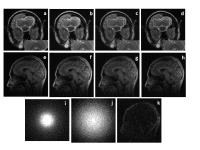
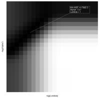
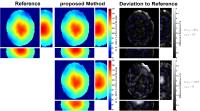
In this study, a new algorithm to better model the receiver coil sensitivities with the purpose of obtaining unbiased proton density maps is proposed. Using optimized orthonormal basis functions for the modeling produces an accurate fit of potential inhomogeneities of the signal due to receiver coil bias. The obtained final image of the proton density has low variance, suitable for quantitative diagnostic information of brain tissue. Results are shown for nine MS patients and one control subject.  


1897.   
Multidimensional Diffusion and Relaxation Data Acquisition for Improved Intravoxel Incoherent Motion Analysis
Anna Scherman Rydhög1, André Ahlgren1, Freddy Ståhlberg1,2,3, Ronnie Wirestam1, and Linda Knutsson1
1Department of Medical Radiation Physics, Lund University, Lund, Sweden, 2Department of Diagnostic Radiology, Lund University, Lund, Sweden, 3Lund Bioimaging Center, Lund University, Lund, Sweden
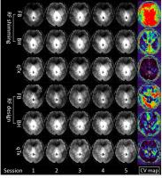
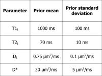
Intravoxel Incoherent Motion (IVIM) is a method for quantification of perfusion parameters, such as the perfusion fraction Fb. Unfortunately, CSF partial volume effects are often seen in the estimated blood compartment. 
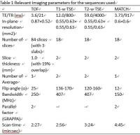
This work introduces a novel version of the IVIM model, containing three compartments (tissue, CSF and blood), where multi-TE and multi-TI data are incorporated to yield a direct relaxation estimate. Using this relaxation-compensated model, results were obtained from in vivo measurements in a volunteer. Compared to a non-relaxation-compensated model, the three-compartment model with relaxation-compensated data reduced the CSF contamination.


1898.   
7TAMIbrainT1w_30 : Whole-brain ultra-high resolution average T1-weighted template at 7 Tesla to improve in vivo depiction of small brain structures
Pierre Besson1,2,3, Arnaud Le Troter1,2, Julien Sein1,2, Gilles Brun1,2, Maxime Guye1,2, and Jean-Philippe Ranjeva1,2
1Aix-Marseille Université, CNRS, Centre de Résonance Magnétique Biologique et Médicale (CRMBM) UMR 7339, Marseilles, France, 2APHM, Timone Hospital, Pôle d’Imagerie, Centre d’Exploration Métabolique par Résonance Magnétique (CEMEREM), Marseilles, France, 3Siemens Healthcare, St Denis, France
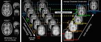
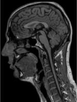
UHF 7T MR scanners offers the possibility to acquire very high resolution in-vivo images, providing a new insight into human brain structural characterization. Nevertheless, in order to obtain highly contrasted and highly spatially resolved atlas, and to compensate for the drop in SNR related to reduction of the voxel size, averaging data among several subjects is needed. We present in this abstract an automatic pipeline that generates a whole brain high-resolution T1-weighted template (called 7TAMIbrainT1w_30) built from MP2RAGE acquisitions obtained in 30 healthy controls at 7T.


1899.   
Complete partial volume solution for ASL brain perfusion data applied to relapsing-remitting multiple sclerosis patients
Ruth Oliver1,2, Linda Ly1,2, Chenyu Wang1,2, Heidi Beadnall2, Ilaria Boscolo Galazzo3,4, Michael Chappell5,6, Xavier Golay7, Enrico De Vita7, David Thomas7, and Michael Barnett1,2
1Sydney Neuroimaging Analysis Centre, Sydney, Australia, 2University of Sydney, Sydney, Australia, 3Institute of Nuclear Medicine, University College London, London, United Kingdom, 4Department of Neuroradiology, University Hospital Verona, Verona, Italy, 5Institute of Biomedical Engineering, University of Oxford, Oxford, United Kingdom, 6FMRIB Centre, University of Oxford, Oxford, United Kingdom, 7Institute of Neurology, University College London, London, United Kingdom
ASL is a low resolution imaging modality that suffers from the partial volume effect, leading to an underestimation of GM perfusion. This effect has two principle causes; blurring from the point spread function in the slice direction, and inadequate resolution due to the need for large voxels to achieve sufficient SNR. Both may act as confounders for measurement of GM CBF abnormalities. Decreased GM perfusion could reflect neuronal loss or metabolic dysfunction; PV correction allows a decoupling of structure and function. We present the first application of a complete PV correction solution for ASL to a cohort of MS patients.


1900.   
Anomalous relaxation in the human brain mapped using ultra-high field magnetic resonance imaging and time-fractional Bloch equation
Shanlin Qin1, Fawang Liu1, Ian William Turner1,2, Qiang Yu3, Qianqian Yang1, and Viktor Vegh3
1School of Mathematical Sciences, Queensland University of Technology, Brisbane, Australia, 2ARC Centre of Excellence for Mathematical and Statistical Frontiers, Melbourne, Australia, 3Centre for Advanced Imaging, University of Queensland, Brisbane, Australia
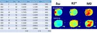
MRI models based on integer order calculus lack the ability to accurately map magnitude signal decay in the human brain, likely due to magnetic susceptibility and microstructure 
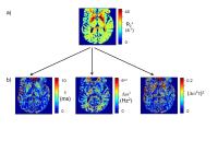
variations in tissues. We applied fractional calculus to the Bloch equation with the aim of developing a model capable of matching experimental findings. Solution of the time-fractional Bloch equation resulted in a new five 
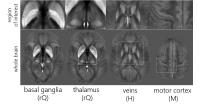
parameter model. We analysed model parameters in nine brain regions using multiple echo gradient recalled echo MRI data from five participants. Time-fractional model parameters may provide new ways of studying microstructure and susceptibility induced changes in the human brain.


1901.   
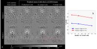
Evaluation the cluster-size inference with random field and permutation methods for group-level MRI analysis
Huanjie Li1, Lisa D. Nickerson2, Yang Fan3, Thomas E. Nichols4, and Jia-Hong Gao5
1Department of Biomedical Engineering, Dalian University of Technology, Dalian, China, People's Republic of, 2McLean Imaging Center, McLean Hospital/Harvard Medical School, Belmont, MA, United States, 3GE Healthcare, MR Research China, Beijing, China, People's Republic of, 4Department of Statistics and Warwick Manufacturing Group, University of Warwick, Coventry, United Kingdom, 5Center for MRI Research, Peking University, Beijing, China, People's Republic of
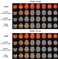
Threshold-free cluster enhancement (TFCE) outperforms the cluster-size test (CST) based on random field theory and our recent papers provide two voxelation-corrected CST (v-CST and vn-CST) which also show the clear advantage over other CST as well. However, it’s not clear which one shows better performance for MRI data analysis. This work provides a very careful, fair and thorough evaluation of the powerful statistical methods,  which may be particularly appealing for group-level MRI data analysis.


1902.   
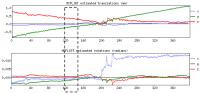
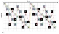
Arterial segmentation and visual stimulus-induced changes in diameter observed in the human brain
Alexandre Bizeau1,2, Guillaume Gilbert3, Minh Tung Huynh4, Michaël Bernier1,2, Christian Bocti5, Maxime Descoteaux2,6, and Kevin Whittingstall1,2,4
1Department of Radiation Sciences and Biomedical imagery, Université de Sherbrooke, Sherbrooke, QC, Canada, 2Centre d’Imagerie Moléculaire de Sherbrooke (CIMS), Centre de Recherche CHUS, Sherbrooke, QC, Canada, 3MR Clinical Science, Philips Healthcare, Markham, ON, Canada, 4Department of Diagnostic Radiology, Université de Sherbrooke, Sherbrooke, QC, Canada, 5Department of Medecine, Université de Sherbrooke, Sherbrooke, QC, Canada, 6Department of Computer Science, Université de Sherbrooke, Sherbrooke, QC, Canada
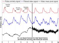
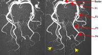
When undergoing stimulation, neurons need to be supplied with oxygen and glucose. This demand then induces vasodilation generated by the astrocytes which act on the muscles of the arteries of the human brain. Using time-of-flight magnetic resonance angiography acquisitions, we extracted the apparent diameter of arterial vessels. We then compared diameter with and without visual stimulation and demonstrated that smaller vessels dilate proportionally more than larger ones in the posterior cerebral arteries. Using this method, the investigation of the coupling between neural activity and regional cerebral vasodilation, also called functional hyperhemia, is now possible. 


1903.   
An Active Learning platform for automatic MR image quality assessment
Thomas Küstner1,2, Martin Schwartz1,2, Annika Kaupp2, Petros Martirosian1, Sergios Gatidis1, Nina F. Schwenzer1, Fritz Schick1, Holger Schmidt1, and Bin Yang2
1University Hospital Tübingen, Tübingen, Germany, 2Institute of Signal Processing and System Theory, University of Stuttgart, Stuttgart, Germany
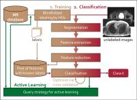
Acquired images are usually analyzed by a human observer (HO) according to a certain diagnostic question. Flexible algorithm parametrization and the enormous amount of data created per patient make this task time-demanding and expensive. Furthermore, definition of objective quality criterion can be very challenging, especially in the context of a missing reference image. In order to support the HO in assessing image quality, we propose a non-reference MR image quality assessment system based on a machine-learning approach with an Active Learning loop to reduce the amount of necessary labeled training data. Labeling is performed via an easy accessible website.


1904.   
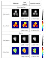
Brain Tissue Clustering Based on Cross-Correlation of Magnetic Resonance Fingerprinting
Mu Lin1, Xiaozhi Cao1, Congyu Liao1, Xu Yan2, and Jianhui Zhong1
1Center for Brain Imaging Science and Technology, Zhejiang University, Hangzhou, China, People's Republic of, 2MR Collaboration NE Asia, Siemens Healthcare, Hangzhou, China, People's Republic of
Multi-component tissue model with priori T1 and T2 have been used to decompose MRF data. We propose that tissue classification can be improved when the selection uses clustering method based on cross-correlation. Our results from phantom and in vivo measurements show that the method successfully separates signal from different tissue types, allows extraction of tissue fractions, and results are more robust with image quality.


1905.   
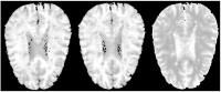
Toward a voxel-based analysis (VBA) of quantitative magnetic susceptibility maps (QSM): Strategies for creating brain susceptibility templates
Jannis Hanspach1, Michael G Dwyer1, Niels P Bergsland1,2, Xiang Feng3, Jesper Hagemeier1, Paul Polak1, Nicola Bertolino1, Jürgen R Reichenbach3,4, Robert Zivadinov1,5, and Ferdinand Schweser1,5
1Buffalo Neuroimaging Analysis Center, Department of Neurology, Jacobs School of Medicine and Biomedical Sciences, The State University of New York at Buffalo, Buffalo, NY, United States, 2MR Research Laboratory, IRCCS Don Gnocchi Foundation ONLUS, Milan, Italy, 3Medical Physics Group, Department of Diagnostic and Interventional Radiology, Jena University Hospital - Friedrich Schiller University Jena, Jena, Germany,4Michael Stifel Center for Data-driven and Simulation Science Jena, Friedrich Schiller University Jena, Jena, Germany, 5MRI Molecular and Translational Research Center, Jacobs School of Medicine and Biomedical Sciences, The State University of New York at Buffalo, Buffalo, NY, United States
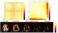
Quantitative susceptibility mapping (QSM) is a recent in vivo magnetic resonance imaging (MRI) technique that provides quantitative information about the bulk magnetic susceptibility distribution in tissues, a promising measure for studying brain iron. A voxel-based analysis (VBA) of susceptibility maps would facilitate a better understanding of the intricate anatomical structure (e.g. sub-nuclear regions) of deep gray matter and its relation to diseases and normal aging.

In the present work, we developed and quantitatively assessed six strategies for creating a susceptibility brain template for VBA based on ANTs, representing the first step toward an understanding of sub-nuclear susceptibility changes without the need for a priori information.


1906.   
Automated multi-parametric segmentation of brain veins from GRE acquisition
Serena Monti1,2, Pasquale Borrelli1, Sirio Cocozza3, Sina Straubb4, Mark Ladd4, Marco Salvatore1, Enrico Tedeschi3, and Giuseppe Palma5
1IRCCS SDN, Naples, Italy, 2Department of Electronics, Information and Bioengineering, Politecnico di Milano, Milan, Italy, 3Department of Advanced Biomedical Sciences, University "Federico II", Naples, Italy,4Department of Medical Physics in Radiology, German Cancer Research Center (DKFZ), Heidelberg, Germany, 5Institute of Biostructure and Bioimaging, National Research Council, Naples, Italy
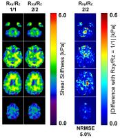
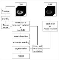
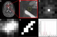
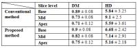
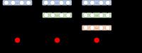
A new fully automated algorithm, based on structural, morphological and relaxometric information, is proposed to segment the entire brain deep venous system from MR images. The method is tested on brain datasets at different magnetic fields and its inter-scan reproducibility is also assessed. The proposed segmentation algorithm shows good accuracy and reproducibility, outperforming previous methods and becoming a promising candidate for the characterization of venous tree topology. 


1907.   
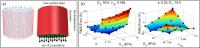
Human Head Models from MRI for Head Impact Analysis
Yash Agarwal1, Philippe Young1, Ross Cotton1, Chris Pearce2, Siddiq Qidwai3, Amit Bagchi3, and Nithyanand Kota3
1Simpleware Ltd., Exeter, United Kingdom, 2Atkins, Epsom, United Kingdom, 3U.S. Naval Research Laboratory, Washington, DC, United States
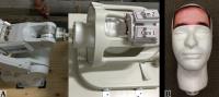
Image-based model generation methods demonstrate the value of creating realistic human head models based on high-resolution MRI data. Head models created by the U.S. Naval Research Laboratory and Simpleware (Exeter, UK) are being used to study head impact and traumatic brain injury; this offers a solution to the problem of limited experimental testing. Results from the modelling methodology and simulation demonstrate a good level of accuracy when compared to experimental benchmarks. The methodology and models have been extended for use in areas such as examining head impact in sports including American football, rugby and cricket.


1908.   
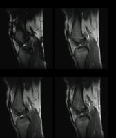
Improve the Detection of Cartilage Degradation by Dividing the Tissue Unequally – A Comparative Study of Two Methods
Farid Badar1, Ji Hyun Lee1, and Yang Xia1
1Department of Physics and Center for Biomedical Research, Oakland University, Rochester, MI, United States
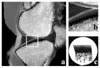
The consequences of two different zone-division methods in MRI T2 of articular cartilage were studied, using an animal model of early osteoarthritis (OA). By dividing the cartilage thickness unequally, significant improvement in OA detection can be achieved – both in the deeper cartilage as well as between the contralateral and normal tissue. This improved detection may become important in the clinical diagnostics of early OA.


1909.   
Accurate Synthetic FLAIR Images Using Partial Volume Corrected MR Fingerprinting
Anagha Deshmane1, Debra McGivney2, Chaitra Badve3, Alice Yu4, Yun Jiang1, Dan Ma2, and Mark Griswold1,2
1Biomedical Engineering, Case Western Reserve University, Cleveland, OH, United States, 2Radiology, Case Western Reserve University, Cleveland, OH, United States, 3Radiology, University Hospitals of Cleveland, Cleveland, OH, United States, 4School of Medicine, Case Western Reserve University, Cleveland, OH, United States
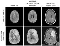
Synthetic weighted images from quantitative parameter maps suffer from partial volume artifacts which can distort contrast.  In this work, Partial Volume MR Fingerprinting is applied to estimate and remove signal due to cerebrospinal fluid (CSF) in the brain, allowing for improved contrast in synthetic FLAIR images generated from MRF relaxation time maps.  


1910.   
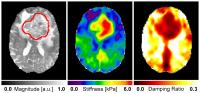
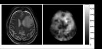
Improvement in Glioma Visualization using Subtraction Maps Derived from Contrast-Enhanced T1- and T2-Weighted MR Images
Mohammed Goryawala1, Bhaswati Roy2, Rakesh K Gupta2, and Andrew A Maudsley1
1Department of Radiology, University of Miami, Miami, FL, United States, 2Department of Radiology, Fortis Memorial Research Institute, Gurgaon, India
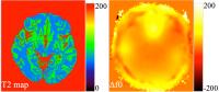
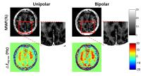
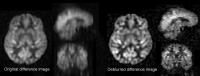
Calculated differences between two images of differing T1 or T2 contrasts, or subtraction images, have been presented as a way to improve image contrast for imaging of brain tumors. In this study the performance of subtraction images for differentiation of tumor, edema, and normal appearing white matter (NAWM) is compared to traditionally acquired anatomic MRIs, diffusion tensor imaging (DTI), perfusion weighted imaging (PWI) and MR spectroscopy imaging (MRSI). Results showed a significant increase in contrast for differentiating between enhancing tumor and edematous regions from NAWM using the ΔT1 map and ΔT2 map, respectively, as compared to other parametric maps. 


1911.   
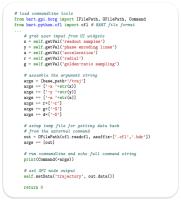
Resource-efficient architecture of FPGA-based 2D FFT processors
Limin Li1 and Alice M Wyrwicz1,2
1Center for Basic MR Research, Northshore University Healthsystem, Evanston, IL, United States, 2Department of Biomedical Engineering, Northwestern University, Evanston, IL, United States
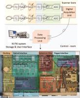
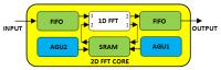
The processing rate for real-time multi-slice image reconstruction on an FPGA can be improved significantly by taking advantage of its parallel processing capability. In particular, multiple 2D FFT processors can be embedded into a single FPGA and run simultaneously. In this abstract, we report a new design of a 2D FFT processor with significant reduced usage of hardware resource. Test results show that an important type of resource, DSP48 slice, can be reduced by up to 50% without degrading processing performance, which implies that more 2D FFT cores can be installed into a single FPGA with a given size.


1912.   
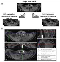
Quantitative image analysis based on Image registration of brain MR and SPECT for dopamine transporter imaging
Takeshi Hara1, Yuta Takeda1, Tetsuro Katafuchi2, Taiki Nozaki3, Masaki Matsusako3, and Hiroshi Fujita1
1Intelligent Image Information, Gifu University Graduate School of Medicine, Gifu, Japan, 2Health Science, Gifu University of Medical Science, Seki, Japan, 3Radiology, St. Luke's International Hospital, Tokyo, Japan
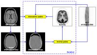
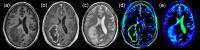
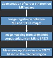
Features in Parkinson's disease (PD) are a degeneration and loss of the dopamine neurons in striatum. 123I-FP-CIT can visualize the distribution by binding to the dopamine neurons. The radioactivated medicine is used for diagnosis of PD and Dementia with Lewy Bodies (DLB). The material can visualize activities in corpus striatum on SPECT images, but the location of the corpus striatum on SPECT images are often lost because of the low uptake. To realize a quantitative image analysis for the SPECT images, image registration technique to determine the region of corpus striatum on SPECT images are required to measure precise uptakes. In this study, we proposed an image fusion technique for SPECT and MR images by intervening CT image taken by SPECT/CT. We employed 30 cases of SPECT/CT and MR cases for the evaluation. 25 of 30 cases were registered correctly with registration errors less than 5mm. These results enable to measure precise uptake on SPECT images based on the segmentation results on MR images.


1913.   
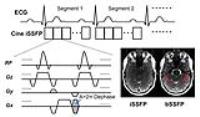
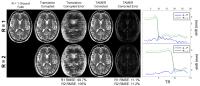
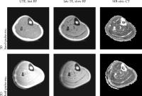
Incorporation of Nonzero Echo Times in the SPGR and bSSFP Signal Models used in mcDESPOT
Mustapha Bouhrara1 and Richard G. Spencer1
1NIA, NIH, Baltimore, MD, United States
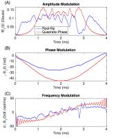
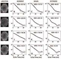
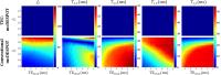
Formulations of the two-component spoiled gradient recalled echo (SPGR) and balanced steady-state free precession (bSSFP) models that incorporate nonzero echo time (TE) effects are presented in the context of mcDESPOT and compared with the conventionally used SPGR and bSSFP models which ignore nonzero TEs. Relative errors in derived parameter estimates from conventional mcDESPOT, omitting TE effects, are assessed using simulations over a wide range of experimental and sample parameters. The neglect of nonzero TE leads to an overestimate of the SPGR and an underestimate of the bSSFP signals. These effects introduce large errors in parameter estimates derived from conventional mcDESPOT.


1914.   
A noise correction model incorporating weighted neighborhood information for liver R2* mapping
Changqing Wang1,2,3, Xinyuan Zhang2, Yanying Ma4, Xiaoyun Liu1, Diego Hernando3, Scott B. Reeder3,5,6,7,8, Wufan Chen1,2, and Yanqiu Feng2
1School of Automation Engineering, University of Electronic Science and Technology of China, Chengdu, China, People's Republic of, 2School of Biomedical Engineering and Guangdong Provincial Key Laboratory of Medical Image Processing, Southern Medical University, Guangzhou, China, People's Republic of, 3Radiology, University of Wisconsin-Madison, Madison, WI, United States, 4School of Mathematical Sciences, University of Electronic Science and Technology of China, Chengdu, China, People's Republic of, 5Medical Physics, University of Wisconsin-Madison, Madison, WI, United States, 6Biomedical Engineering, University of Wisconsin-Madison, Madison, WI, United States, 7Medicine, University of Wisconsin-Madison, Madison, WI, United States, 8Emergency Medicine, University of Wisconsin-Madison, Madison, WI, United States
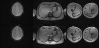
R2* mapping has the potential to provide rapid and accurate quantification of liver iron overload. However, conventional voxelwise liver R2* mapping methods are challenging when using echo images with low signal-noise ratio (SNR). The purpose of this work was to improve liver R2* mapping by a noise correction model incorporating weighted neighborhood information. Simulation and in vivo results demonstrate that the proposed method produces more accurate R2* maps with high spatial resolution compared to two recently proposed R2* mapping methods.


1915.   
Automatic MR-based Skull Segmentation using Local Shape and Global Topology Priors
Max W.K. Law1, Calvin M.H. Lee1, Gladys G. Lo2, Jing Yuan1, Oilei Wong1, Abby Y. Ding1, and Siu Ki Yu1
1Medical Physics and Research Department, Hong Kong Sanatorium & Hospital, Hong Kong, Hong Kong, 2Department of Diagnostic and Interventional Radiology, Hong Kong Sanatorium & Hospital, Hong Kong, Hong Kong
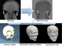
This abstract proposes a new algorithm that automatically segments the skull from gradient echo based magnetic resonance images to facilitate MR-based radiotherapy planning. The proposed algorithm compared the neighboring voxel intensity to capture local structural information of bone. The structural information was incorporated in a topology template which encapsulated global topology prior of skulls to achieve automatic segmentation. With the sequence-independent structural and topology priors, this method is potentially applicable to other scanning sequences. The segmented skull will be helpful for clinical applications such as cephalometry and MR-based radiotherapy planning to reduce ionizing-radiation received by patients.


1916.   
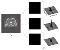
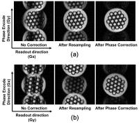
Image-based estimation of point spread function in distorted EPI images
Seiji Kumazawa1, Takashi Yoshiura2, Akihiro Kikuchi1, Go Okuyama1, Daisuke Shimao1, and Masataka Kitama1
1Hokkaido University of Science, Sapporo, Japan, 2Kagoshima University, Kagoshima, Japan
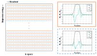
To correct the distortion in EPI due to field inhomogeneity, the information regarding the signal from adjacent points within each voxel is needed. The PSF approach can provide this information. Our purpose was to develop an image-based-method for estimating the PSF images in the distorted EPI image using T1WI. Our method synthesizes the distorted image to match the measured EPI image through the generation process of EPI image according to a single-shot EPI k-space trajectory and field inhomogeneity. The results demonstrate that the PSF image for each voxel in distorted EPI image can be estimated by proposed method using segmented T1WI instead of additional acquisitions for PSF measurement.


1917.   
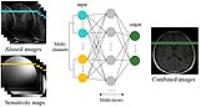
Combining Multi-channel MP2RAGE Images with Minimized Noise
Jing Zhang1, Bruce Bjornson2, and Qing-San Xiang3
1Applied Science Laboratory, GE Healthcare Canada, Vancouver, BC, Canada, 2Department of Pediatrics, University of British Columbia, Vancouver, BC, Canada, 3Department of Radiology, Department of Physics and Astronomy, University of British Columbia, Vancouver, BC, Canada
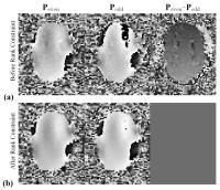
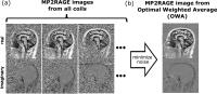
Magnetization-prepared rapid gradient echo (MP-RAGE) has been widely used for T1-weighted imaging. In order to overcome B1 field inhomogeneity effect, the MP2RAGE sequence was introduced, with two complex images, GRETI1 and GRETI2, acquired at two inversion times TI1 and TI2. The MP2RAGE images are usually calculated from all the coils first and combined later into a final result. We propose an algorithm for  multi-channel MP2RAGE image combination with minimized resulting noise.


1918.   
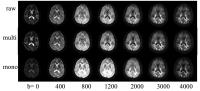
Improving the Quality of the Multi-b Diffusion Weighted Images Using the Intrinsic Multi-Exponential Pattern
He Wang1, Kaining Shi1, Weibo Chen1, and Guilong Wang1
1Philips Healthcare, shanghai, China, People's Republic of
The study developed a methodology to improve the quality of the multi-b DWIs using the intrinsic multi-exponential pattern. It was evaluated on a healthy brain and compared with the mono-exponential model. In addition, its potential value of improving the robustness of IVIM was also evaluated. According to the results, the multi-exponential method can improve the image quality of the multi-b DWIs and may become an effective preprocessing way for the non-monoexponential models.


1919.   
Multi-Inversion EPI-based imaging of T1 distribution within individual voxels
Ville Renvall1 and Jonathan R. Polimeni2
1Department of Neuroscience and Biomedical Engineering, Aalto University School of Science, Espoo, Finland, 2Athinoula A. Martinos Center for Biomedical Imaging, Department of Radiology, Massachusetts General Hospital, Harvard Medical School, Charlestown, MA, United States
T1 mapping using multiple inversion time IR-EPI can provide a large number of different TI values in a short time, which can be utilized to characterize the relaxation time distributions within individual voxels, as an extension to multi-parametric fitting.


1920.   
Generation of hybrid color images from T1 and T2 acquired simultaneously with MRF
Katherine L. Wright1, Peter Schmitt2, Dan Ma1, Anagha Deshmane3, Vikas Gulani1, and Mark Griswold1
1Radiology, Case Western Reserve University, Cleveland, OH, United States, 2Siemens Healthcare, Erlangen, Germany, 3Biomedical Engineering, Case Western Reserve University, Cleveland, OH, United States
This work proposes a method for the calculation of a single color image using quantitative T1 and T2 measurements acquired with Magnetic Resonance Fingerprinting. Quantitative MRF parameters are transformed and scaled with the goal of making normal tissues appear in grayscale and tissues with different T1 and T2 values (lesions) appear in color. 


1921.   
High-SNR susceptibility weighted venography (SWV) for multi-echo magnetic resonance (MR) images based on complex signal modeling
Taejoon Eo1, Dosik Hwang1, and Jinseong Jang1
1Yonsei University, Seoul, Korea, Democratic People's Republic of
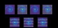
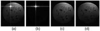
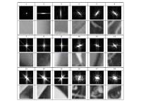
The multi-echo SWV with the proposed complex signal modeling method can provide high-SNR and multi-contrast phase masks and SWV images. The multiplication number of the phase mask for SWV was increased up to 16 without image degradation even at the long TE of 49.8 ms. More detailed vein structures were visualized with higher- and multiple contrasts than the conventional single-echo GRE SWV.


1922.   
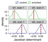
Estimating Registration Variance Using Deformation Field Perturbations
Jan Scholz1, Kaitlyn Easson2, and Jason P Lerch1,3
1Mouse Imaging Centre, Hospital for Sick Children, Toronto, ON, Canada, 2Department of Biomedical and Molecular Sciences, Queen's University, Toronto, ON, Canada, 3Department of Medical Biophysics, Department of Medical Biophysics, Toronto, ON, Canada
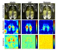
Most image registration algorithms do not output any information about the variance of the transformation estimates. Here we show that by perturbing input files we can recover this information without modifying the underlying algorithms. We demonstrate that local brain volume estimates can be improved by using the determinant of the average across the distribution of transformations. Our methods will improve morphological analyses, registration-based label alignment, and help find optimal registration parameters.


1923.   
Graph-based segmentation of signal voids in time series of diffusion-weighted images of musculature in the human lower leg
Martin Schwartz1,2, Günter Steidle1, Petros Martirosian1, Bin Yang2, and Fritz Schick1
1Section on Experimental Radiology, Department of Radiology, University of Tuebingen, Tuebingen, Germany, 2Institute of Signal Processing and System Theory, University of Stuttgart, Stuttgart, Germany
The segmentation of signal voids, which occur in time-series of single-shot diffusion-weighted images, is important for an accelerated evaluation providing larger studies on this phenomenon. The proposed segmentation is based on a two-stage detection and segmentation approach, which utilizes a graph-based representation with random walker optimization. It was demonstrated that the presented method enables a fast and accurate segmentation of signal voids in time-series of diffusion-weighted images.


1924.   
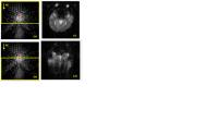
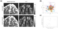
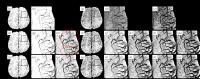
Power spectrum detects corpus callosum directionality using T2-weighted MRI in secondary progressive MS patients and controls
Shrushrita Sharma1 and Yunyan Zhang2
1Biomedical Engineering Program, University of Calgary, Calgary, AB, Canada, 2Departments of Radiology and Clinical Neurosciences, University of Calgary, Calgary, AB, Canada
Standard MRI is routinely collected in patient care but is limited in assessing changes in tissue microstructure. We developed a new method to assess tissue directionality using the power spectrum of T2-weighted MRI and validated it using the highly coherent structure, corpus callosum. In controls, power spectrum-derived angles corresponded exactly with the predicted aligning directions of the corpus callosum, and such aligning patterns were interrupted in advanced MS patients with increased variability and angular entropy. Fourier-based power spectrum may provide advanced measures of tissue directionality following myelin and axonal pathology using clinical scans.


1925.   
The Impact of Polar based initialization and frame time curve selection on Left Ventricle short axis Perfusion MR Segmentation
Doaa Mousa1, Nourhan Zayed1, and Inas Yassine2,3
1Computer and Systems, Electronic Research Institute, Giza, Egypt, 2Systems and Biomedical Engineering, Cairo University, Giza, Egypt, 3Medical Informatics and Image processing Lab, Nile University, Giza, Egypt
Cardiovascular diseases (CVDs) cause 31% of the death rate globally. Automatic accurate segmentation is needed for CVDs early detection. In this paper, we propose a modified workflow to automatically segment the left ventricle (LV) for the short axis cardiac perfusion MRI (perfusion CMR) images using levelset method. We propose mitigating the initial contour extraction, and modify the technique used to initialize the levelset algorithm in order to improve the accuracy of segmentation results. The system workflow consists of five main modules: preprocessing, localization, initial contour extraction, registration, and segmentation. Our results showed enhancement in the segmentation accuracy by 5%.


1926.   
Multi-layered Atlas Registrations for Multi-atlas Segmentation of Brain MRI
Han Sang Lee1 and Junmo Kim1
1School of Electrical Engineering, KAIST, Daejeon, Korea, Republic of
Multi-atlas segmentation has often suffered from the registration error. We propose a novel method for multi-atlas registration for multi-atlas segmentation inspired by the template generation and deep neural network. We first add an intra-atlas registration layer which performs image-based registration between atlas images to duplicate the atlases. We then add a label-wise registration layer which rectifies the registered images by label-based registration. We present preliminary results of our multi-layered atlas registration on brain MRI segmentation.


1927.   
The impact of data analysis method, scanner type and scan session on volume measurements of brain structures
Michael Amann1,2, Pavel Falkovskiy3,4,5, Alain Thoeni1, Tobias Kober3,4,5, Alexis Roche3,4,5, Bénédicte Maréchal3,4,5, Philippe Cattin6, Tobias Heye2, Oliver Bieri2, Till Sprenger7, Christoph Stippich2, Gunnar Krueger4,5,8, Ernst-Wilhelm Radue1, and Jens Wuerfel1
1Medical Image Analysis Center (MIAC), Basel, Switzerland, 2Department of Radiology, University Hospital of Basel, Basel, Switzerland, 3Advanced Clinical Imaging Technology, Siemens Healthcare AG, Lausanne, Switzerland, 4Department of Radiology, University Hospital (CHUV), Lausanne, Switzerland, 5École Polytechnique Fédérale de Lausanne, Lausanne, Switzerland, 6Department of Biomedical Engineering, University of Basel, Basel, Switzerland, 7Department of Neurology, DKD Helios Klinik, Wiesbaden, Germany, 8Siemens Medical Solutions USA, Boston, MA, United States
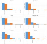
Performance of FreeSurfer and FSL was compared on T1-weighted 3D MRI data of 22 controls as function of scan session, scanner type and segmentation pipeline. Intra-class correlation coefficients and percentage volume differences were calculated for the segmentation results of both pipelines. Strong agreement was found for whole brain, white matter and cortex. For each pipeline, the impact of experimental factors was assessed by linear mixed effects analysis. We found significant scanner effect on the results of both segmentation pipelines. For subcortical structures, segmentation reliability was higher in FSL than in FreeSurfer, whereas for cortex and WM, FreeSurfer was more stable.


1928.   
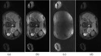
Image Inhomogeneity Correction using Geometric Average of Channels in Sum-of-Squares Multi-channel MR Imaging
Renjie He1, Yu Ding1, and Qi Liu1
1United Imaging Healthcare America, Houston, TX, United States
Geometric average is insensitive to the value variation between components to be averaged, this is used to noticeably reduce the inhomogeneity caused by Sum-of-Squares (SOS) in channel combination in parallel MR imaging.


1929.   
In-vivo characterization of grey matter microstructure at 3T from the transverse component of the MRI signal
Antoine Lutti1
1LREN, Dept. of Clinical Neurosciences, Centre Hospitalier Universitaire Vaudois, Lausanne, Switzerland
The characterization of brain microstructure from MRI data requires the development of specific MRI tissue biomarkers and of advanced models linking microscopic tissue properties to MRI signals. We apply the Anderson-Weiss theory, which describes the transverse relaxation of the MRI signal as a function of tissue microstructure, on in-vivo MRI data acquired at 3T. In grey matter, parameter estimates show a strong correlation with histological measures of iron concentration. The time constants provided by the model yield realistic estimates of microscopic compartment size. These results offer a promising perspective for the histological assessment of brain tissue in-vivo using MRI.


1930.   
Hippocampal subfields segmentation derived from Freesurfer 6.0: a multisite 3T reproducibility study in healthy elderly
Moira Marizzoni1, Daniele Orlandi1, Luigi Antelmi2, Flavio Nobili3, Mira Didic4,5, David Bartrés-Faz6, Ute Fiedler7, Peter Schonknecht8, Pierre Payoux9,10, Andrea Soricelli11,12, Alberto Beltramello13, Lucilla Parnetti14, Magda Tsolaki15, Paolo Maria Rossini16,17, Pieter Jelle Visser18, Regis Bordet19, Oliver Blin20, Giovanni Battista Frisoni1,21, Jorge Jovicich22, and on behalf of the PharmaCog Consortium1
1LENITEM Laboratory of Epidemiology, Neuroimaging, & Telemedicine — IRCCS San Giovanni di Dio-FBF, Brescia, Italy, 2Health Department, Foundation IRCCS Neurological Institute Carlo Besta, Milan, Italy,3Department of Neuroscience, Ophthalmology, Genetics and Mother–Child Health (DINOGMI), University of Genoa, Genoa, Italy, 4APHM, CHU Timone, Service de Neurologie et Neuropsychologie, Marseille, France,5Aix-Marseille Université, INSERM U 1106, Marseille, France, 6Department of Psychiatry and Clinical Psychobiology, Universitat de Barcelona and IDIBAPS, Barcelona, Spain, 7LVR-Clinic for Psychiatry and Psychotherapy, Institutes and Clinics of the University Duisburg-Essen, Essen, Germany, 8Department of Neuroradiology, University Hospital Leipzig, Leipzig, Germany, 9INSERM, Imagerie cérébrale et handicaps neurologiques, UMR 825, Toulouse, France, 10Université de Toulouse, UPS, Imagerie cérébrale et handicaps neurologiques, UMR 825, CHU Purpan, Place du Dr Baylac, Toulouse, France, 11IRCCS SDN, Naples, Italy,12University of Naples Parthenope, Naples, Italy, 13Department of Neuroradiology, General Hospital, Verona, Italy, 14Section of Neurology, Centre for Memory Disturbances, University of Perugia, Perugia, Italy, 153rd Department of Neurology, Aristotle University of Thessaloniki, Thessaloniki, Greece, 16Dept. Geriatrics, Neuroscience & Orthopaedics, Catholic University, Policlinic Gemelli, Rome, Italy, 17IRCSS S.Raffaele Pisana, Rome, Italy, 18Department of Neurology, Alzheimer Centre, VU Medical Centre, Amsterdam, Italy, 19Department of Pharmacology, EA1046, University of Lille Nord de France, Lille, Italy, 20Pharmacology, Assistance Publique-Hôpitaux de Marseille, Aix-Marseille University-CNRS UMR 7289, Marseille, France, 21Memory Clinic and LANVIE - Laboratory of Neuroimaging of Aging, University Hospitals and University of Geneva, Geneva, Switzerland, 22Center for Mind/Brain Sciences, University of Trento, Rovereto, Italy
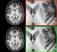
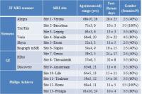
In this study we quantify the across-session reproducibility of hippocampus subfields obtained from the recently proposed ex-vivo atlas tool available in Freesurfer version 6.0. We use structural 3T multisite data from 65 healthy elderly participants scanned twice at least a week apart. We show that several subfields  like Cornu Ammonis (CA) 1, hippocampal tail, molecular layer and subiculum offer, despite being smaller, comparable reliability errors to the whole hippocampus volume (2%). This suggests that these subfields may be valid and more specific markers to test disease progression in longitudinal studies, like for example Alzheimer's disease.


1931.   
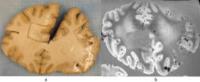
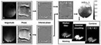
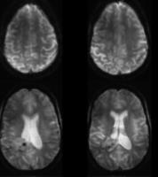
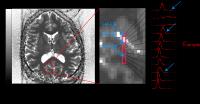
Identification of Microbleeds on Postmortem Brain of Normal Aging Elderly and Dementia Patients
Shunshan Li1, Lily Zhou2, Mark J Fisher3, Ronald C Kim4, Vitaly Vasilevko5, David Cribbs5, Annlia Hill3, and Min-Ying Su6
1Tu & Yuen Center for Functional Onco-Imaging, Department of Radiological Sciences, university of california, irvine, irvine, CA, United States, 2Sun Yat-Sen Memorial Hospital, Sun Yat-Sen University, Guangzhou, China, People's Republic of, 3Department of Neurology, University of California, Irvine, Irvine, CA, United States, 4Department of Pathology, University of California, Irvine, Irvine, CA, United States, 5Institute for Memory Impairments and Neurological Disorders, University of California, Irvine, irvine, CA, United States, 6Tu & Yuen Center for Functional Onco-Imaging, Department of Radiological Sciences, University of California, Irvine, irvine, CA, United States
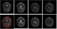
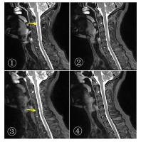
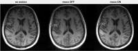
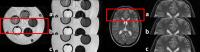
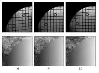
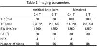
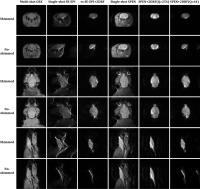
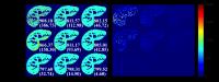
The postmortem brain MR images include air-bubble artifacts and typical microbleeds(MBs) are less than 200 µm which make MBs detection very challenging. In this project we developed an optimization MR imaging method to detect possible MBs on postmortem brains of patients with and without dementia, hoping to provide information to guide neuropathological examination to sample the suspicious MBs areas, and improve the chance of identifying true MBs to better understand its role in normal aging and development/progression of dementia, and further develop streamlined automatic MBs detection software.


1932.   
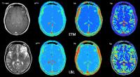
Dynamic Contrast Enhanced MRI Measurements in Glioma: Comparison Between Two Models
Sameeha Fallatah1, Rolf Jäger1, and Xavier Golay1
1Brain Repair and Rehabilitation, UCL, Institute of Neurology, London, United Kingdom
Dynamic Contrast Enhanced MRI is used to assess the integrity of the blood brain barrier. A major difficulty for the method to be accepted in the clinics is the variety of pharmacokinetic models used and their strong dependence on the underlying assumptions and/or acquisition parameters. Thus the far simpler methods based on signal intensity curve characteristics are the most commonly used approaches in clinical practice. In this study we compare two different pharmacokinetic models, the extended Tofts model and Lawrence & Lee model in patients with primary brain tumours. 


1933.   
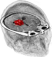
Development and Implementation of a Matlab-based multi-modal 3D visualization, co-registration and quantification platform for assessing brain tumor physiology and metabolism
Gaurav Verma1, Suyash Mohan1, Sanjeev Chawla1, John Y.K. Lee2, Sumei Wang1, Andrew Maudsley3, Steven Brem2, and Harish Poptani4
1Department of Neuroradiology, University of Pennsylvania, Philadelphia, PA, United States, 2Department of Neurosurgery, University of Pennsylvania, Philadelphia, PA, United States, 3Department of Radiology, University of Miami, Miami, FL, United States, 4Department of Cellular and Molecular Physiology, University of Liverpool, Liverpool, United Kingdom
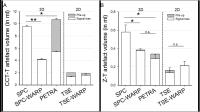
A 3D visualization, co-registration and quantification platform was developed in Matlab to combine anatomical imaging with physiological and metabolic data from diffusion tensor, perfusion-weighted and echo-planar spectroscopic imaging. This data can be co-registered across modalities and imaging time-points to provide detailed information about the spatial extent of a brain tumor. 3D visualization was applied in datasets from patients undergoing neurosurgery and a separate cohort of patients undergoing long-term Tumor Treating Fields (TTFields) therapy. This visualization platform could have an impact in the planning of neurosurgery and the placement and monitoring of location-sensitive techniques like TTFields.


1934.   
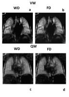
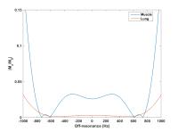
Non-Contrast-Enhanced Perfusion and Ventilation Assessment of the Human Lung by Means Of Wavelet Decomposition in Proton MRI
David Bondesson1,2, Thomas Gaass1,3, Julien Dinkel1,2, and Berthold Kiefer4
1Josef Lissner Laboratory for Biomedical Imaging, Department of Clinical Radiology, Ludwig-Maximilians-University Hospital Munich, Munich, Germany, 2Comprehensive Pneumology Center, German Center for Lung Research, Munich, Germany, 3Comprehensive Pneumology Center,German Center for Lung Research, Munich, Germany, 4Siemens AG Healthcare Sector, Erlangen, Germany
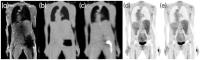
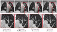
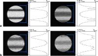
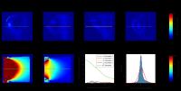
Evaluating regional lung perfusion and ventilation is diagnostically valuable in regards of pulmonary diseases. Standard methods however, expose patients to risks from ionizing radiation and contrast agents. MRI screening is not based on radiation and a new method has previously been presented as a non-contrast-enhanced estimation. This work presents wavelet decomposition as a potential improvement to fourier decomposition for perfusion and ventilation assessment of the human lung in proton MRI.  


1935.   
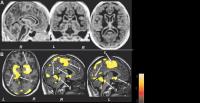
Regional Brain Tissue Entropy Assessment in Patients with Obstructive Sleep Apnea
Sudhakar Tummala1, Bumhee Park1, Ruchi Vig1, Mary A Woo2, Daniel W Kang3, Ronald M Harper4,5, and Rajesh Kumar1,5,6,7
1Anesthesiology, University of California at Los Angeles, Los Angeles, CA, United States, 2UCLA School of Nursing, Los Angeles, CA, United States, 3Medicine, University of California at Los Angeles, Los Angeles, CA, United States, 4Neurobiology, University of California at Los Angeles, Los Angeles, CA, United States, 5Brain Research Institute, University of California at Los Angeles, Los Angeles, CA, United States, 6Radiological Sciences, University of California at Los Angeles, Los Angeles, CA, United States, 7Bioengineering, University of California at Los Angeles, Los Angeles, CA, United States
Obstructive sleep apnea subjects show gray matter volume loss in multiple brain areas, based on voxel-based morphometry procedures, which are less sensitive in detecting subtle chronic/acute gray or white matter changes. We assessed brain injury in recently-diagnosed, treatment naïve OSA subjects by evaluating regional entropy, which measures the extent of homogeneity or randomness in tissue texture, and found significantly decreased regional entropy values in areas regulating autonomic, respiratory, cognitive, and neuropsychologic functions that are deficient in the condition, suggesting predominantly acute tissue pathology in those sites.. The findings suggest that regional entropy can demonstrate acute tissue changes.


1936.   
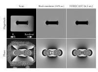
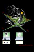
Fast simulation of off-resonance artifacts in MRI using FORECAST (Fourier-based Off-REsonanCe Artifact Simulation in the STeady-State)
Frank Zijlstra1, Job G Bouwman1, Ieva Braškute1, and Peter R Seevinck1
1Image Sciences Institute, UMC Utrecht, Utrecht, Netherlands
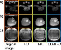
We present a fast alternative to Bloch simulation for simulation of off-resonance artifacts in steady-state imaging. By assuming a steady-state, the signal equation can be quickly evaluated by using multiple Fast Fourier Transforms. We show an acceleration factor of over 350 for a 2D simulation of a titanium cylinder phantom, while the differences with Bloch simulation were minor. The speed of the proposed method enables 3D simulations at high resolution and may benefit various applications.


1937.   
Estimation of voxel-wise phase offsets in a phased array coil using multi-echo GRE data
Minju Jo1, Yoonho Nam2, Jeehun Kim1, Hyeong Geol Shin1, and Jongho Lee1
1Laboratory for Imaging Science and Technology, Department of Electrical and Computer Engineering, Seoul National University, Seoul, Korea, Republic of, 2Department of Radiology, Seoul St. Mary's Hospital, College of Medicine, The Catholic University of Korea, Seoul, Korea, Republic of
In this work, we present a method of estimating the phase offsets in multi-echo GRE data, Multi-Channel Phase Combination using all N echoes (MCPC-N). MCPC-N, which calculates the phase offsets from all echoes, provides more accurate estimation of voxel-wise phase offsets particularly in low SNR.


1939.   
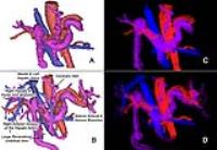
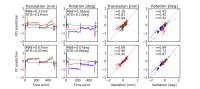
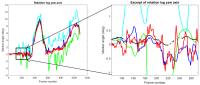
Optimized 4D flow MRI Processing for Evaluation of Abdominal Blood Flow
Eric James Keller1, Jeremy Douglas Collins1, Cynthia K Rigsby2, James C Carr1, Michael Markl1,3, and Susanne Schnell1
1Radiology, Northwestern University, Chicago, IL, United States, 2Radiology, Ann & Robert H. Lurie Children's Hospital of Chicago, Chicago, IL, United States, 3Biomedical Engineering, Northwestern University, Evanston, IL, United States
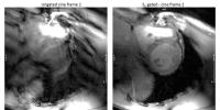
4D flow MRI quantification of abdominal hemodynamics is challenged by a wide range of blood flow velocities and vessel diameters. By adjusting critical pre-processing steps required to analyze 4D flow MRI data, we were able to both recover vessels of interest lost by our previous method and significantly reduce the relative error in flow measurements. We conclude that it is critical to apply background phase error correction prior to any other filters and/or corrections to ensure accurate background offset estimation. Additionally, low venc acquisitions should not be noise corrected to ensure low flow data is not inadvertently deleted.


1938.   
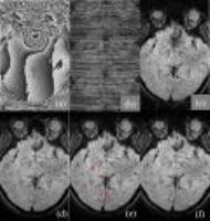
Characterization of atherosclerotic carotid plaque using MATCH with histopathologic validation: initial clinical experience
Lixin Yang1, Wei Yu1, Zhaoyang Fan2, and DeBiao Li3
1Department of Radiology, Beijing AnZhen Hospital, Beijing, China, People's Republic of, 2Biomedical Imaging Research Institute, Cedars-Sinai Medical Center, Los Angeles, CA, United States, 3Biomedical Imaging Research Institute, Cedars-Sinai Medical Center,Department of Bioengineering, University of California, Los Angeles, CA, United States
Purpose: Determine the accuracy of MATCH in the characterization of plaque composition in patients in comparison with the conventional multi-contrast approach, using histopathology as the gold standard.

Methods: Twenty-two patients scheduled for carotid endarterectomy underwent preoperative carotid MRI with MATCH and the conventional protocol, blinded image review for composition identification was performed by 2 radiologists. Carotid histopathological specimens stained with HE and Masson, matched with this two protocol images, Cohen kappa (K) was computed to quantify the agreement in the detection of components among this two protocols and histopathology.

Results: Moderate to good agreement was seen between histopathological specimens and multi-contrast protocol in the detection of plaque components (IH k=0.704 , CA k=0.763, LR/NC k=0.844). Similar results were seen between histopathological specimens and MATCH (IH k=0.703CA k=0.740, LR/NC k=0.850). 


1940.   
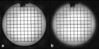
MRI-SPAMM Based Magnetic Resonance Electrical Impedance Tomography
Kemal Sümser1, Nashwan Naji1,2, Mehdi Sadighi1, Hasan Hüseyin Eroglu1,3, and Murat Eyüboglu1
1Electrical and Electronics Engineering Department, Middle East Technical University, Ankara, Turkey, 2On Leave from Ibb University, Ibb, Yemen, 3TSK Rehabilitation and Care Center, Ankara, Turkey
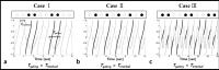
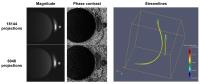
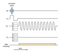
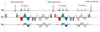
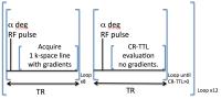
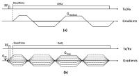
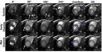
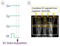
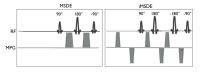
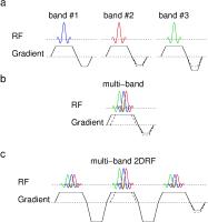
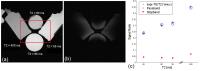
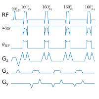
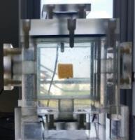
In magnetic resonance electrical impedance tomography (MREIT) currents are injected to the object during MRI imaging sequence. In this study, we propose a new pulse sequence based on the spatial modulation of magnetization (SPAMM) to be used in MREIT applications. In this pulse sequence, the current is injected during a pre SPAMM module which can be followed by any conventional Magnetic Resonance Imaging pulse sequence for data acquisition. Experimental result in comparison with the simulation result shows that this method is an applicable technique for MREIT data acquisition.


1941.   
The Influence of Bolus Arrival Time in Pharmacokinetic Analysis of Dynamic Contrast-Enhanced MRI of Breast Masses
Endre Grøvik1,2, Atle Bjørnerud1,2, Tryggve Holck Storås1, Kjell-Inge Gjesdal3, and Kathinka Dæhli Kurz4
1The Intervention Centre, Oslo University Hospital, Oslo, Norway, 2Department of Physics, University of Oslo, Oslo, Norway, 3Sunnmøre MR klinikk AS, Ålesund, Norway, 4Department of Radiology, Stavanger University Hospital, Stavanger, Norway
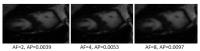
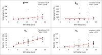
The purpose was to evaluate the influence of BAT in pharmacokinetic analysis of breast masses, by estimating the kinetic parameters both with and without BAT-delay correction. Thirty-nine verified breast masses were examined using a high temporal resolution EPI sequence. The image-data were analyzed using a two-compartment kinetic model with and without BAT-delay correction. The relationship between the relative parametric error and BAT-delay were investigated. The result indicates that neglecting the delayed BAT leads to an overestimation of Ktrans, kep, and, ve, and a underestimation of vp, and that the delayed BAT needs to be accounted for in the model-based analysis.

1942.   
Liver stiffness in pediatric subjects is lower than in adults, and increases with age: a multifrequency MR elastography study
Emily Etchell1, Lauriane Jugé 1,2, Alice Hatt1, Ralph Sinkus3, and Lynne E. Bilston1,4
1Neuroscience Research Australia, Randwick, NSW, Australia, 2School of Medical Sciences, University of New South Wales, Kensington, NSW, Australia, 3BHF Centre of Excellence, Division of Imaging Sciences and Biomedical Engineering, King's College London, London, United Kingdom, 4Prince of Wales Clinical School, University of New South Wales, Kensington, NSW, Australia
Magnetic resonance (MR) elastography provides clinical information for chronic hepatic disorders by quantifying an increase in liver stiffness compared to heathy baseline values. Thus far however, baseline stiffness values have only been reported for adults. We aimed to fill this gap by quantifying healthy liver stiffness of children and adolescents. Results showed that pediatric liver stiffness increases with age during normal development, approaching adult values during adolescence. This implies that comparing pediatric liver stiffness measurements to adult baseline values when using MR elastography may miss disease or underestimate disease severity.


1948.   
Quantification of Breast Stiffness using Magnetic Resonance Elastography at 3T: A Reproducibility Study
Prateek Kalra1, Arunark Kolipaka, PhD1, Jeffrey R. Hawley, MD1, and Brian Raterman1
1Radiology, Ohio State University Wexner Medical Center, Columbus, OH, United States
Magnetic resonance elastography (MRE) is a non-invasive technique to estimate stiffness of soft tissues and has been applied in the breast. However, none of the earlier studies have extensively tested MRE to induce vibrations in the breast using a soft sternum driver at higher field strength and its repeatability of stiffness measurements. The aim of the study is to estimate breast stiffness using MRE by inducing vibrations using a soft sternum driver in normal volunteers at 3T and to determine the reproducibility of stiffness measurements. Preliminary results show that the MRE-derived stiffness values are reproducible in normal volunteers at 3T and can be further extended to detect breast tumors in patients. 


1945.   
Increasing the Spatial Resolution and Sensitivity of High-Resolution Magnetic Resonance Elastography by Correcting for Subject Motion and Susceptibility-Induced Image Distortions
Andreas Fehlner1, Sebastian Hirsch1, Mykola Kadobianskyi2, Patric Birr1, Eric Barnhill1,3, Martin Weygandt2,4, Johannes Bernarding5, Jürgen Braun6, Ingolf Sack1, and Stefan Hetzer2,4
1Department of Radiology, Charité - Universitätsmedizin Berlin, Berlin, Germany, 2Berlin Center for Advanced Neuroimaging, Charité - Universitätsmedizin Berlin, Berlin, Germany, 3Clinical Research Imaging Centre, School of Clinical Sciences and Community Health, College of Medicine and Veterinary Medicine, The University of Edinburgh, Edinburgh, United Kingdom, 4Bernstein Center for Computational Neuroscience Berlin, Berlin, Germany, 5Institut für Biometrie und Medizinische Informatik, Universitätsklinikum Magdeburg, Magdeburg, Germany, 6Institute of Medical Informatics, Charité - Universitätsmedizin Berlin, Berlin, Germany
High-resolution Multifrequency MR Elastography (MMRE) is hampered by susceptibility-induced image distortions. We corrected MMRE data of a 3T and 7T MR scanner for motion and EPI distortion artefacts. The correction of subject motion significantly sharpened the images, which was demonstrated by a decrease of the point-spread function. The improvement was highly correlated with the degree of subject motion. Distortion correction enhanced the accuracy of normalization in the MNI152 space as shown by an increase of the correlation between individual and standard tissue probability maps. This method could help increasing the sensitivity of multi-subject studies exploring |G*| e.g. in small subcortical areas.


1946.   
In vivo multifrequency MR elastography of the human prostate using a surface-based compressed air driver operated in the lower frequency regime
Florian Dittmann1, Heiko Tzschätzsch1, Jing Guo1, Sebastian Hirsch1, Jürgen Braun2, and Ingolf Sack1
1Institute of Radiology, Charité, Berlin, Germany, 2Department of Medical Informatics, Charité, Berlin, Germany
We demonstrate the feasibility of in vivo prostate exam utilizing shear waves induced by pressurized-air actuators previously developed for abdominal MRE.  High wave amplitudes throughout the prostate were achieved in the lower frequency regime from 30 to 50 Hz. Using a 2D multifrequency wave number inversion algorithm, wave speed maps with sufficiently high resolution are obtained to discriminate between the central zone and peripheral zone despite longer wavelengths pertaining to lower vibration frequencies. The proposed MRE setup promises robust and easy-to-use applications in the clinic without the need of specialized hardware in addition to the abdominal MRE setup.


1962.   
Robust Harmonic Estimation for MR Elastography: Application to Brain
Joshua D. Trzasko1, Arvin Arani1, Armando Manduca1, Kevin J. Glaser1, Richard L. Ehman1, Philip A . Araoz1, and John Huston III1
1Mayo Clinic, Rochester, MN, United States
In this work, we adapt the previously-described robust harmonic estimation (RHE) strategy for magnetic resonance elastography (MRE) to brain imaging, and demonstrate that use of this novel signal processing tool improves the accuracy of estimated stiffness information both in a geometrically-accurate phantom and in vivo.


1943.   
MR elastography of intracranial tumors: Initial experience with high-resolution imaging and nonlinear inversion
Curtis L Johnson1, Emily S Matijevich1,2, Emily D Cullum1,2, Matthew DJ McGarry3, Keith D Paulsen3, Bradley P Sutton1,4, Tracey M Wszalek1,2, and William C Olivero1,2
1Beckman Institute for Advanced Science and Technology, University of Illinois at Urbana-Champaign, Urbana, IL, United States, 2Carle Neuroscience Institute, Carle Foundation Hospital, Urbana, IL, United States,3Thayer School of Engineering, Dartmouth College, Hanover, NH, United States, 4Department of Bioengineering, University of Illinois at Urbana-Champaign, Urbana, IL, United States
MR elastography has emerged as an important tool for presurgical evaluation of intracranial tumors. Due to the localized nature of the lesion properties of interest, there is a need for high-resolution MRE methods for characterizing tumors. Here we present our initial experience with MRE of intracranial tumors using a protocol based on high-resolution imaging and nonlinear inversion. We found that glial tumors are soft and have a generally low viscosity, while meningeal tumors are stiff and have a very low viscosity.


1947.   
Reproducibility of low-frequency MR elastography of the human brain
Florian Dittmann1, Sebastian Hirsch1, Jing Guo1, Jürgen Braun2, and Ingolf Sack1
1Institute of Radiology, Charité, Berlin, Germany, 2Department of Medical Informatics, Charité, Berlin, Germany
Since shear waves at low drive frequencies are nearly unaffected by attenuation, we introduce a brain MRE setup, which is based on remote excitation of intracranial shear waves by a pressurized-air actuator in the regime of 20 Hz. MRE-scans, which were repeated 27 times on three different days for each of six healthy volunteers, show differences between individuals as well as from day-to-day for the same individual. The investigation demonstrates that cerebral low frequency MRE provides a fast and reproducible novel source of mechanical information of brain tissue with less onerous head stimulation as required by conventional MRE.


1944.   
Cross vendor comparison of gradient recalled echo (GRE) and spin echo-echo planar imaging (SE-EPI) based MR elastography of the liver at 3T.
Suraj D Serai1, Jonathan R Dillman1, Hui Wang2, and Andrew T Trout1
1Radiology, Cincinnati Children's Hospital, Cincinnati, OH, United States, 2Philips Healthcare, Cincinnati, OH, United States
MR elastography (MRE) allows non-invasive evaluation of hepatic stiffness and samples a larger area of the liver than liver biopsy. The high accuracy of MRE for liver fibrosis staging suggests that MRE could potentially replace liver biopsy. MRE has traditionally been performed using a GRE sequence. GRE, however, has SNR limitations at higher field strengths that can result in under-sampling potentially leading to erroneous stiffness values. SE-EPI is an alternative means of performing MRE that has higher SNR, lower susceptibility related signal loss and increased speed. In this work, we compared GRE and SE-EPI MRE across two vendor platforms.  


1949.   
Assessing the viscoelastic properties of abdominal tumour models in vivo using MRE
Jin Li1, Lisa Asher1, Filipa Lopes2, Craig Cummings1, Alexander Koers2,3, Laura S. Danielson2,3, Louis Chesler2,3, Caroline J. Springer2, Jeffrey C. Bamber1, Ralph Sinkus4, Yann Jamin1, and Simon P. Robinson1
1Division of Radiotherapy & Imaging, The Institute of Cancer Research, London, United Kingdom, 2Division of Cancer Therapeutics, The Institute of Cancer Research, London, United Kingdom, 3Division of Clinical Studies, The Institute of Cancer Research, London, United Kingdom, 4Division of Imaging Sciences and Biomedical Engineering, King’s College London, King’s Health Partners, St. Thomas’ Hospital, London, United Kingdom
MRE was applied to assess the viscoelastic properties of orthotopic pancreatic ductal adenocarcinoma (PDAC) xenografts, and tumours arising in a transgenic mouse model of MYCN-amplified neuroblastoma, within the mouse abdomen. The stromal-rich PDAC tumours were quantified with markedly elevated elasticity (Gd) and viscosity (Gl), whilst the pathologically diverse neuroblastomas exhibit more heterogeneity in their biomechanical properties and were relatively soft. MRE can non-invasively assess the viscoelastic properties of deep-seated tumours arising within the abdomen of mice in vivo


1950.   
Comparison of breath-hold, respiratory navigated and free-breathing MR Elastography
Ian Gavin Murphy1, Martin Graves2, Scott Reid3, Andrew Patterson2, Ilse Gavin Joubert1, Andrew N Priest2, and David J Lomas2
1Radiology, Cambridge University Hospitals NHS Foundation Trust, Cambridge, United Kingdom, 2Radiology, Cambridge University NHS Foundation Trust, Cambridge, United Kingdom, 3GE, Little Chafont, United Kingdom
In patients with liver disease, MR elastography (MRE) is a non-invasive method for evaluating fibrosis. MRE is phase-based and sensitive to motion artefact, and is typically performed in end expiration. We found that navigator timed MRE shows no statistical difference to breath-held techniques for stiffness and reproducibility in 6 healthy volunteers, and may prove superior in patients unable to adequately hold their breath 


1951.   
Brain MR elastography with multiband excitation and nonlinear motion-induced phase error correction
Curtis L Johnson1, Joseph L Holtrop1,2, Aaron T Anderson3, and Bradley P Sutton1,2
1Beckman Institute for Advanced Science and Technology, University of Illinois at Urbana-Champaign, Urbana, IL, United States, 2Department of Bioengineering, University of Illinois at Urbana-Champaign, Urbana, IL, United States, 3Department of Mechanical Science and Engineering, University of Illinois at Urbana-Champaign, Urbana, IL, United States
We propose a novel sequence for magnetic resonance elastography (MRE) of the brain based on multiband excitation and 3D encoding of the distributed slab with multishot spirals. This sequence allows access to optimal SNR efficiency and reduced distortions from field inhomogeneity, but also parallel imaging acceleration both in-plane and thru-plane without onerous artifacts and g-factor penalties. We also incorporate correction for nonlinear motion-induced phase errors through a kz-blipped spiral-in 3D navigator. In this abstract we demonstrate the performance of the sequence and its ability to capture whole-brain MRE data at 2x2x2 mm3 resolution in 3 minutes.


1952.   
Impact of Field Strength and Image Resolution on MRE Stiffness Estimation
Eric Barnhill1, Jing Guo2, Florian Dittmann2, Sebastian Hirsch2, Michael Perrins3, Lucy Hiscox3, Tim Herrmann4, Johannes Bernarding4, Neil Roberts3, Jürgen Braun1, and Ingolf Sack2
1Institute of Medical Informatics, Charité Universitätsmedizin Berlin, Berlin, Germany, 2Department of Radiology, Charité Universitätsmedizin Berlin, Berlin, Germany, 3Clinical Research Imaging Centre, The University of Edinburgh, Edinburgh, United Kingdom, 4Institute for Biometrics and Medical Informatics, Otto von Guericke University Magdeburg, Magdeburg, Germany
We investigated the impact of field strength and image resolution on brain MRE stiffness results.  A cohort of 18 healthy volunteer subjects was scanned at 1.5T (2mm isotropic voxels), 3T (2mm) and 7T (1mm), with a fourth set downsampling the 7T to 2mm.  Means were 1634 Pa (+/-613) for 1.5T, 1743 Pa (+/-811) for 3T, 1786 (+/-634) Pa for 7T 2mm, and 927 (+/-364) Pa for 7T 1mm. In the paired sign-rank tests, there were no significant effects for field strength. Examination of histograms of example slices suggests that a different distribution of features is being captured at the higher resolution.


1953.   
Investigation Of The Relationship Between Feature Detail And Stiffness Estimate In Magnetic Resonance Elastography (MRE) Elastograms
Eric Barnhill1, Florian Dittmann2, Sebastian Hirsch2, Jing Guo2, Jürgen Braun1, and Ingolf Sack2
1Institute of Medical Informatics, Charité Universitätsmedizin Berlin, Berlin, Germany, 2Department of Radiology, Charité Universitätsmedizin Berlin, Berlin, Germany
Magnetic Resonance Elastography (MRE) stiffness estimates show differentiated results by feature scale. Here progressive denoising was applied to study the relation between image sharpness (as meaured by Reduced Energy Ratio) and image stiffness estimate (as measured by complex shear modulus magnitude |G*|). Progressive complex-wavelet-based denoising appears to reach stable stiffness estimates in phantom and brain acquisitions. Images of maximum sharpness result in lower overall stiffness estimates than the stable global estimate, suggesting that coarse elasticity estimates do not average fine feature results, but measure a different stiffness scale.


1954.   
Reducing Time Samples Needed for MR Elastography
Roger Grimm1, Jun Chen1, and Richard Ehman1
1Mayo Clinic, Rochester, MN, United States
A 3D gradient recalled echo sequence has been developed that samples the three shear wave displacement polarization at 3 time points for a total of 9 image samples. A multi-coil recon generates phase difference images and then uses a 3 point discrete Fourier transform to provide the complex displacement fields. The sequence is shown in breast and head applications.


1955.   
MR Elastography using SS-SE-EPI with reduced FOV: phantom study and preliminary volunteer study for the pancreas
Yohei Itoh1, Yasuo Takehara2, Naoki Ooishi2, Masanori Kawade2, Tetsuya Wakayama3, Mikio Suga4, Takasuke Ushio1, Yuki Hirai1, Nobuko Yoshizawa1, Shuhei Yamashita1, Hatsuko Nasu1, and Harumi Sakahara1
1Diagnostic Radiology & Nuclear Medicine, Hamamatsu University school of medicine, Hamamatsu, Shizuoka, Japan, 2Department of Radiology, Hamamatsu University Hospital, Hamamatsu, Shizuoka, Japan,3GEHCJ, Hino-shi, Tokyo, Japan, 4Center for Frontier Medical Engineering, Chiba University, Chiba, Japan
To achieve the high spatial resolution MR elastography(MRE), it has been reported that combining SS-SE-EPI with a spatially selective excitation provides an efficient way of reducing FOV. Using this technique, we performed phantom study and pancreatic MRE. We report the result of our study and the point we found when performing the reduced-FOV MRE.


1956.   
Noise-robust multifrequency wave number inversion for high-resolution MR elastography in the abdomen
Heiko Tzschätzsch1, Jing Guo1, Florian Dittmann1, Sebastian Hirsch1, Eric Barnhill1, Jürgen Braun2, and Ingolf Sack1
1Department of Radiology, Charité - University Medicine Berlin, Berlin, Germany, 2Institute of Medical Informatics, Charité - University Medicine Berlin, Berlin, Germany
Elastography often suffers from limited anatomical resolution due to noise and insufficient elastic deformation. We here introduce noise-robust multifrequency wave number inversion for multifrequency MR elastography. Compound maps of wave speed are obtained, which reveal variations in tissue elasticity in a tomographic fashion, i.e. an unmasked, slice-wise display of anatomical details at pixel-wise resolution. The method is demonstrated using data from the literature including abdominal and pelvic organs such as the liver, spleen, uterus and cervix. Elastic parameters consistent with literature values were obtained even in small regions with low wave amplitudes such as nucleus pulposus and spinal cord.


1958.   
Compact and fully automated 3D multifrequency tabletop MR elastography for the measurement of viscoelastic parameters in small tissue samples
Navid Samavati1,2, Clara Körting1, Toni Drießle3, Stefan Wintzheimer3, Jing Guo1, Florian Dittmann1, Ingolf Sack1, and Jürgen Braun2
1Department of Radiology, Charité University Medicine, Berlin, Germany, 2Department of Medical Informatics, Charité University Medicine, Berlin, Germany, 3Pure Devices GmbH, Würzburg, Germany
A fully integrated tabletop MR elastography (MRE) system based on a 0.5-T permanent magnet for investigations of small tissue samples is introduced. A 3D spin echo MRE sequence allows control of all MRE parameters including frequency and amplitude of a piezoelectric actuator. The device enables fully automated measurements of maps of viscoelastic parameters in soft tissue samples by 3D multifrequency MRE. Initial results are in good agreement to published data and demonstrate the great potential of the system as a preclinical research unit in histopathological laboratories and operating rooms.


1957.   
Development of a novel phantom for routine quality assurance of an MR elastography system
Lumeng Cui1,2, Conrad Yuen3, Ted Lynch4, Paul Babyn5, Francis M. Bui6, and Niranjan Venugopal1,5
1Department of Medical Physics, Saskatchewan Cancer Agency, Saskatoon, SK, Canada, 2Division of Biomedical Engineering, University of Saskatchewan, Saskatoon, SK, Canada, 3Department of Medical Physics, BC Cancer Agency, Vancouver, BC, Canada, 4Non-ionizing Radiation, CIRS Inc., Norfolk, VA, United States, 5Department of Medical Imaging, University of Saskatchewan, Saskatoon, SK, Canada, 6Department of Electrical & Computer Engineering, University of Saskatchewan, Saskatoon, SK, Canada
Magnetic Resonance Elastography (MRE) is a new imaging technique that combines the acoustic waves and MRI to retrieve elastic properties of tissue. Because MRE is non-invasive, there is great clinical interest for its use in the detection of cancer. In this work, we focus on the design of an MRE phantom to be used in the clinical commissioning of an MRE System. With the aid of newly designed pulse sequences and inversion algorithms we have developed a quality assurance process to validate the efficacy of MRE for applications to many clinical sites (i.e. prostate, cervix, uterus).


1959.   
Identification of Myocardial Anisotropic Material Properties using Magnetic Resonance Elastography and the Finite Element Method
Renee Miller1, Arunark Kolipaka2, Vicky Wang3, Martyn Nash3, and Alistair Young1
1Anatomy with Radiology, University of Auckland, Auckland, New Zealand, 2Radiology, Biomedical Engineering and Internal Medicine, The Ohio State University Wexner Medical Center, Columbus, OH, United States,3Auckland Bioengineering Institute, University of Auckland, Auckland, New Zealand
In this study, we examined the determinability of anisotropic stiffness parameters using finite element analysis simulations of harmonic steady-state wave behaviour. Two simulation experiments, of cylindrical phantom and left ventricular geometries, and one phantom experiment using magnetic resonance elastography (MRE) were carried out. The transversely isotropic material properties were determined using a least-squares optimisation algorithm by matching a modelled displacement field to the reference, or MRE, displacement field. The results showed that the parameters were uniquely identifiable even in the presence of noise.


1960.   
Experimental Validation of High Shear Wave Displacement at Mode Frequencies in MR Elastography
Cemre Ariyurek1,2, Safa Ozdemir1,2, Arif Sanli Ergun3, Yusuf Ziya Ider1, and Ergin Atalar1,2
1Department of Electrical and Electronics Engineering, Bilkent University, Ankara, Turkey, 2National Magnetic Resonance Research Center (UMRAM), Ankara, Turkey, 3Department of Electrical and Electronics Engineering, TOBB-University of Economics and Technology, Ankara, Turkey
Experimental validation of modes of shear waves in MR elastography (MRE) is demonstrated. For the first time, frequency response of the actuator is investigated and actuator displacement is measured. Normalizing shear wave displacement to the actuator displacement removes the effect of actuation system and isolates shear wave resonance. It is demonstrated that 10-20 times greater shear wave displacement than applied displacement by the actuator can be observed at resonance. Thus, safety issues in MRE should be reconsidered. Presenting repeatability of determining mode frequency validates feasibility of detecting stiffness changes by observing any shift in mode frequency.


1961.   
Poroelastic mechanical properties of brain tumors using intrinsic actuation MR elastography
Ligin Solamen1, Matthew McGarry1, Elijah Van Houten2, Jennifer Hong3, John Weaver1,4, and Keith Paulsen1,5
1Thayer School of Engineering, Dartmouth College, Hanover, NH, United States, 2Department of Mechanical Engineering, University of Sherbrooke, Sherbrooke, QC, Canada, 3Department of Neurosurgery, Dartmouth-Hitchcock Medical Center, Lebanon, NH, United States, 4Department of Radioogy, Dartmouth-Hitchcock Medical Center, Lebanon, NH, United States, 5Norris Cotton Cancer Center, Dartmouth-Hitchcock Medical Center, Lebanon, NH, United States
Intrinsically actuated poroelastic MR elastography (IA-pMRE) is a technique which estimates tissue mechanical and hydrodynamic properties using measurements of displacement during the cardiac cycle, and does not require external vibration as in traditional MRE. Compared to conventional MRE, which obtains displacements in the range of 25-100Hz, IA-pMRE uses intrinsically generated low frequency (1-2Hz) displacements for elastography reconstruction. IA-pMRE was applied to 7 brain tumor patients and showed a significant difference in both the shear modulus and hydraulic conductivity of brain tissue compared to healthy tissue.
 

The International Society for Magnetic Resonance in Medicine is accredited by the Accreditation Council for
Continuing Medical Education to provide continuing medical education for physicians.