|
2129.
 |
Traveling wave MR using an array of regular RF resonators
Xinqiang Yan1,2 and
Xiaoliang Zhang3
1Institute of Imaging Science, Vanderbilt
University, Nashville, TN, United States, 2Radiology,
Vanderbilt University, Nashville, TN, United States, 3Department
of Radiology and Biomedical Imaging, University of
California San Francisco, San Francisco, CA, United States
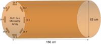
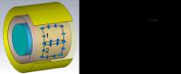
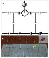
In this study, we investigate the feasibility of using
regular microstrip resonators as RF array elements for
traveling wave parallel imaging. In the proposed microstrip
array, electromagnetic decoupling between the array elements
is sufficient for the practical use. Additionally, geometric
factors and diverse B1 fields from individual array elements
can be obtained in a relatively large area in the magnet
bore. Furthermore, in non-accelerated imaging applications,
this decoupled multi-channel traveling wave method could
improve sensitivity of traveling wave MRI, which is
currently a main issue for traveling wave MRI.
|
|
2130.
 |
Slotted-tube-resonator design for whole-body MR imaging at 14T
Jérémie Daniel Clément1, Arthur Magill2,
Hongxia Lei3, Özlem Ipek3, and Rolf
Gruetter4,5,6
1CIBM-LIFMET, Ecole Polytechnique Fédérale de
Lausanne, Lausanne, Switzerland, 2Forschungszentrum
Jülich, Jülich, Germany, 3CIBM-AIT,
Ecole Polytechnique Fédérale de Lausanne, Lausanne,
Switzerland, 4LIFMET,
Ecole Polytechnique Fédérale de Lausanne, Lausanne,
Switzerland, 5Department
of Radiology, University of Geneva, Geneva, Switzerland, 6Department
of Radiology, University of Lausanne, Lausanne, Switzerland
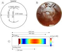
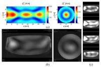
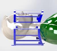
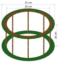
The purpose of the study was to build a slotted-tube
resonator for whole-body MR imaging at 14T. Flip angle maps
were computed to assess the transmit field distribution in a
phantom. A longitudinal coverage of 8 cm and flip angle
homogeneity are observed and spin-echo images were acquired.
|
|
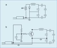
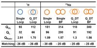
2135.
 |
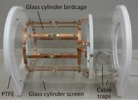
Design and construction of a triple-tuned RF probe for
23Na/31P/1H using traps
Arthur W. Magill1, Chang-Hoon Choi1,
Yonghyun Ha1, and N. Jon Shah1,2
1Institute of Neuroscience and Medicine - 4,
Forschungszentrum Juelich GmbH, Juelich, Germany, 2Department
of Neurology, JARA, RWTH Aachen University, Aachen, Germany
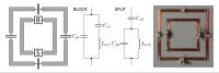
Traps may be used to dual-tune an RF probe, either by
splitting the resonance of a single tuned circuit, or by
blocking coupling at the higher frequency when using a pair
of resonant circuits. This work combines both methods to
construct a triple-tuned probe consisting of a nested pair
of loops. The inner loop incorporates two traps, one to
prevent coupling to the outer loop, which is tuned to 1H,
and a second to simultaneously tune the loop to 23Na
and 31P.
The probe is designed for use at 4T, with resonances at
45MHz (23Na), 69MHz (31P) and 170MHz (1H).
|
|
2152.
 |
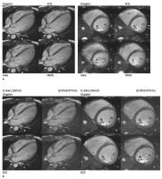
13C RF coil combination for cardiac and abdominal human and pig
studies
Steffen Ringgaard1, Rolf F Schulte2,
James Tropp3, Carsten Kögler4, Titus
Lanz4, Miguel A Navarro5, Jan Henrik
Ardenkjaer-Larsen6,7, Fraser J Robb5,
Hans Stødkilde-Jørgensen1, and Christoffer
Laustsen1
1MR Research Centre, Aarhus University, Aarhus,
Denmark, 2GE
Global Research, Munich, Germany, 3GE
Healthcare, Fremont, CA, United States, 4Rapid
Biomedical, Rimpar, Germany, 5GE
Healthcare, Cleveland, OH, United States, 6GE
Healthcare, Copenhagen, Denmark, 7DTU,
Copenhagen, Denmark
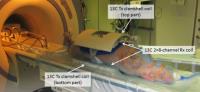
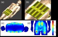
We have developed and validated a dedicated coil system for
human and large animal hyperpolarised 13C measurements. The
system consists of an outer two-element transmit coil and an
inner 16-element receive coil. It was validated by
hyperpolarised experiments in two healthy pigs using a
multi-echo spiral CSI sequence. The 13C metabolic images
showed good SNR and there was low noise correlation between
the receive elements. Hence, the coil system is promising
for future human hyperpolarised examinations.
|
|
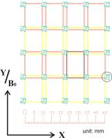
2159.
 |
Effect of the RF Shield on the Mutual Coupling Between Adjacent
and Non-Adjacent Array Elements 
Andreas Pfrommer1, Nikolai I Avdievich1,
and Anke Henning1,2
1Max Planck Institute for Biological Cybernetics,
Tuebingen, Germany, 2Institute
for Biomedical Engineering, UZH and ETH Zurich, Zurich,
Switzerland
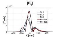
In this study we investigated the effect of an RF shield on
the mutual coupling between adjacent and non-adjacent array
elements in a simple model mimicking our previously
developed cylindrical eight channel transceiver head array.
Both numerical EM simulations and experimental measurements
suggest that at 124 MHz and 400 MHz an RF shield can
substantially decrease S12 for
non-adjacent-array elements.
|
|
2161.
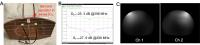 |
Tunable Defected Ground Structure for Decoupling Monopole
Antenna Transmit/Receive Arrays in 7T MRI
Xinqiang Yan1,2 and
William A. Grissom1,2,3
1Institute of Imaging Science, Vanderbilt
University, Nashville, TN, United States, 2Radiology,
Vanderbilt University, Nashville, TN, United States, 3Biomedical
Engineering, Vanderbilt University, Nashville, TN, United
States
Radiative dipole and monopole coil arrays are increasingly
used for ultrahigh ?eld MRI, but few decoupling methods have
been proposed for radiative arrays. To overcome this
problem, we propose a Tunable-Defected-Ground-Structure
(TDGS) method to decouple monopole arrays at 7T. This
concept was successfully validated by EM simulation, bench
test and MR experiments. By using the TDGS method, the
cross-talk between two closely-spaced monopoles was reduced
from -7 dB to -25 dB. It was also found that the TDGS method
had little effect on the original B1 fields
of the individual monopole elements.
|
|
2158.
 |
New decoupling method for receiver arrays with small coils
Xueming Cao1, Elmar Fischer1, Oliver
Gruschke2, Jan Korvink2, Jürgen Hennig1,
and Maxim Zaitsev1
1University Medical Center Freiburg, Freiburg,
Germany, 2Karlsruher
Institut für Technologie, Karlsruher, Germany
In receiver coil arrays, the most commonly used decoupling
methods are overlap together with low-input-impedance
preamplifiers. But very small receiver coils can not be
decoupled effectively with these two methods. A new
decoupling method, which is helpful for receiver coil arrays
with very small coils, is developed here. The coil arrays
decoupled with this method have less noise correlation and
better performance in highly accelerated imaging in the
sample periphery.
|
|
2126.
 |
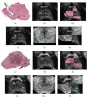
Design of a forward view antenna for prostate imaging at 7 Tesla
Bart Steensma1, Dennis Klomp1, Nico
van den Berg1, Peter Luijten1, Abe van
der Werf2, and Alexander Raaijmakers1
1University Medical Centre Utrecht, Utrecht,
Netherlands, 2Machnet
B.V., Maarn, Netherlands
The forward view antenna has been introduced as a novel
antenna for ultrahigh field imaging. This study has
investigated its potential for prostate imaging where the
antenna is placed between the legs, to contribute as an
additional element of an existing dipole antenna transceiver
array. A significant increase in signal-to-noise ratio is
expected because of the generally smaller distance towards
the prostate from this side. Numerical simulations and in
vivo scans show that signal-to-noise ratio in the prostate
region increases as a result of adding the forward view
antenna to the dipole antenna array.
|
|
2127.
 |
Multi-Channel Helical-Antenna Inner-Volume RF Coils for
Ultra-High-Field MR Scanners
Pranav S. Athalye1, Milan M. Ilic1,2,
Pierre-Francois Van de Moortele3, Andrew J. M.
Kiruluta4, and Branislav M. Notaros1
1Department of Electrical and Computer
Engineering, Colorado State University, Fort Collins, CO,
United States, 2School
of Electrical Engineering, University of Belgrade, Belgrade,
Yugoslavia, 3Center
for Magnetic Resonance Research, Department of Radiology,
University of Minnesota, Minneapolis, MN, United States, 4Radiology
Department, Massachusetts General Hospital, Harvard Medical
School, Boston, MA, United States
RF coil design for human ultra-high-field scanners is an
area of intense development, to address difficult challenges
including RF excitation spatial heterogeneity and low RF
efficiency. We present the development and testing of a
novel category of multi-channel RF volume coil structures at
both 7T and 10.5T based on a subject-loaded multifilar
helical-antenna RF coil. Phantom data show excellent
consistency between numerical simulations and experimental
results with 4- and 8-channel helical-antenna coil
prototypes. This design shows capability for multi-channel
RF-transmit technology and parallel imaging. This work may
help decide which coil structure should be used for future
studies at 10.5T.
|
|
2128.
 |
A proton-free birdcage coil to enable zero-echo-time MRI without
background signal
Markus Weiger1, David Otto Brunner1,
Thomas Schmid1, Romain Froidevaux1,
Manuela Barbara Rösler1, Simon Gross1,
and Klaas Paul Pruessmann1
1Institute for Biomedical Engineering, University
and ETH Zurich, Zurich, Switzerland
MRI of tissues with very short T2s below 1 ms, such as bone,
lung, or myelin is usually performed with 3D radial
sequences with ultra-short or even zero TE. However, with
these techniques also signals from hardware parts are
detected, in particular from the RF coils. Especially the
ZTE method is highly sensitive also to materials with
extremely short T2 of tens of us. In this work, it is
demonstrated how the undesired signal is avoided during coil
design and production, presenting for the first time a
birdcage coil which is virtually free of proton signal.
|
|
2154.
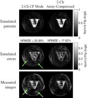 |
Experimental Implementation of Array-compressed Parallel
Transmission at 7T
Zhipeng Cao1,2, Xinqiang Yan1,3, and
William A. Grissom1,2,3
1Vanderbilt University Institue of Imaging
Science, Vanderbilt University, Nashville, TN, United
States, 2Biomedical
Engineering, Vanderbilt University, Nashville, TN, United
States, 3Radiology,
Vanderbilt University, Nashville, TN, United States
With a constructed 8 channel transmit array and a tunable 2
channel-to-8 coil compression matrix, the array-compressed
parallel transmit pulse design is demonstrated on 7T MRI
through B1+ mapping and accelerated spiral excitation.
Results showed more accurate excitation pattern can be
achieved with the compression matrix hardware and compressed
parallel transmit pulses than two-channel CP-mode pulses.
|
|
2155.
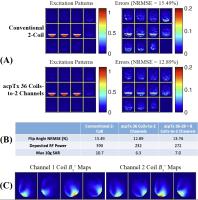 |
Array-compressed parallel transmit pulse design with optimized
coil-channel assignments and coil pruning for simultaneous
multislice and 3D reduced-field-of-view excitations
Zhipeng Cao1,2, Xinqiang Yan1,3, and
William A. Grissom1,2,3
1Vanderbilt University Insitute of Imaging
Science, Vanderbilt University, Nashville, TN, United
States, 2Biomedical
Engineering, Vanderbilt University, Nashville, TN, United
States, 3Radiology,
Vanderbilt University, Nashville, TN, United States
An improved array-compressed parallel transmit pulse design
is proposed and validated to optimally connect transmit
arrays with a large number of elements to a few transmit
channels. It is further demonstrated to achieve
better performance with array-compressed coil designs than
conventional designs for multiband RF shimming for human
brain imaging and 3D spatially selective excitation for
human occipital lobe imaging.
|
|
2144.
 |
A Cervical Spine Array Coil with Volume Transmitter at 7 Tesla
Tsinghua Zheng1, Matthew Finnerty1,
Xiaoyu Yang1, Matthew Diprimio1, Luke
Beery1, Paul Taylor1, Johanna Vannesjo2,
Stuart Clare2, and Hiroyuki Fujita1,3,4,5
1Quality Electrodynamics, LLC, Mayfield Village,
OH, United States, 2FMRIB
Centre, Oxford University, Oxford, United Kingdom, 3Physics,
Case Western Reserve University, Cleveland, OH, United
States, 4Radiology,
University Hospital of Cleveland, Cleveland, OH, United
States, 55School
of Information and Electrical Engineering, the University of
Queensland, Brisbane, Australia
A cervical spine array coil with a volume transmit coil for
7.0 Tesla was constructed and tested. The coil uses one
partially shielded birdcage volume transmit coil for
generating uniform excitation throughout the cervical spine
region and an array of sixteen loop coils for receiving.
Initial volunteer imaging demonstrated good coverage and
uniformity along cervical spine.
|
|
2139.
 |
Transceive surface array of dipole antennas for multi-transmit
imaging at 3T
Aidin Ali Haghnejad1, Shaihan J. Malik2,
Francesco Padormo2, Cornelis A.T. van den Berg1,
Peter R. Luijten1, Dennis W.J. Klomp1,
Joseph V. Hajnal 2,
and Alexander J.E. Raaijmakers1
1UMC Utrecht, Utrecht, Netherlands, 2King's
College London, London, United Kingdom
The birdcage body coil at 3T has some considerable
disadvantages. Most of all it has very large power
requirements. The use of local transmit arrays severely
reduces these power requirements. In this study, we intend
to explore the use of dipole antennas as transceive surface
array elements at 3T. Three designs are investigated after
which a strongly meandering dipole antenna is selected. An
array of eight of these element is used for prostate imaging
at 3T in a 8ch. multi-transmit MRI system. Using 8x200W
input power, 12 µT is achieved inside the prostate.
Relatively homogeneous T2w images have been acquired
|
|
2140.
 |
A Mixed Dipole and Microstrip Transmit/Receive Array
Xinqiang Yan1,2, John C. Gore1,2,3,
and William A. Grissom1,2,3
1Institute of Imaging Science, Vanderbilt
University, Nashville, TN, United States, 2Radiology,
Vanderbilt University, Nashville, TN, United States, 3Biomedical
Engineering, Vanderbilt University, Nashville, TN, United
States
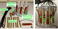
Dipole and microstrip coils produce different and somewhat
complementary B1 patterns and hybrid E-field
distributions. Based this observation, we developed a
16-channel transmit/receive array for 7T head imaging by
interleaving dipole and microstrip elements. Mutual coupling
among any elements is <-14 dB without including any other
decoupling. Compared with 8-channel microstrip-only and
dipole-only arrays, the proposed 16-ch dipole+microstrip
array has a higher SNR gain and lower g-factor. No
decoupling treatment is needed for the mixed dipole and
microstrip array, so it can be used as a flexible
transceiver array at ultrahigh field.
|
|
2133.
 |
A 7-Tesla Transmit with 32-Channel Receive-Only Array Head Coil
for fMRI
Matthew Finnerty1, Derick Petrey1,
Paul Taylor1, Luke Beery1, Tsinghua
Zheng1, Xiaoyu Yang1, Hiroyuki Fujita1,2,3,4,
Se-Hong Oh5, Ken Sakaie5, and Mark
Lowe5
1Quality Electrodynamics, LLC, Mayfield Village,
OH, United States, 2Department
of Physics, Case Western Reserve University, Cleveland, OH,
United States, 3Department
of Radiology, University Hospitals of Cleveland, Cleveland,
OH, United States, 4School
of Information Technology and Electrical Engineering, The
University of Queensland, Brisbane, Australia, 5Imaging
Institute, Cleveland Clinic, Cleveland, OH, United States
While fMRI at 7-Tesla can provide clinically relevant
increases in functional sensitivity over 3-Tesla, it also
typically uses visual and audio stimulation devices that
require additional space accommodations inside the RF coil.
In order to accommodate a wider range of stimulus devices
than possible with high filling factor designs, a head array
coil utilizing a volume transmitter and 32 receive elements
for 7-Tesla was constructed inside a versatile mechanical
package to support fMRI and other applications.
|
|
2142.
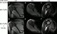 |
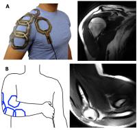
Cost-Efficient 7ch Rx Shoulder Array for 7T UHF MRI Featuring
External Switchbox Detuning
Stefan HG Rietsch1,2, Oliver Kraff1,
Stephan Orzada1, Andrea Lazik3, and
Harald H Quick1,2
1Erwin L. Hahn Institute for MR Imaging,
University of Duisburg-Essen, Essen, Germany, 2High
Field and Hybrid MR Imaging, University Hospital Essen,
Essen, Germany, 3Department
of Diagnostic and Interventional Radiology and
Neuroradiology, University Hospital Essen, Essen, Germany
MRI at 7T and above opens the field for high resolution
human imaging for example in the shoulder. In order to
improve a present setup consisting of an 8ch Tx/Rx shoulder
coil using microstrip line elements with meanders, we
present an additional low cost 7ch Rx loop coil and utilize
a simple approach for detuning of this coil during transmit
via a custom built 8ch Tx/Rx switchbox. With the additional
7ch Rx coil a factor of 2 in SNR can be achieved in the
center of the humeral head in proton-density weighted images
with a spatial resolution of 0.4x0.4x2.5 mm3.
|
|
2141.
 |
Design of RF Coils Mixing Elements of Dissimilar Radiation
Pattern
Ian RO Connell1,2 and
Ravi S Menon1,2
1Centre for Functional and Metabolic Mapping,
Robarts Research Institute, London, ON, Canada, 2Department
of Medical Biophysics, University of Western Ontario,
London, ON, Canada
At ultra-high field (UHF), multi-channel radio-frequency
(RF) arrays have found increasing utility in mitigating
wave-like behaviour during transmission (1), while
continuing to provide increases in sensitivity to MRI signal
with densely filled conformal receive arrays (2). In an
effort to more efficiently excite spin populations, and
increase sensitivity to the transverse magnetization during
relaxation, work into mixing array elements of dissimilar
radiation pattern has been demonstrated to better
encapsulate UHF ideal current patterns (3). Application of
our method - coupling matrix synthesis - is used to robustly
decouple a sample of these array-types.
|
|
2134.
 |
A Hybrid 8 channel TR Dipole and 8 channel Rx Birdcage Body Coil
Array for 7T
Jan Paska1,2, Martijn Cloos1,2,
Gillian Haemer1,2,3, Bei Zhang1,2, and
Graham C Wiggins1
1Center for Biomedical Imaging, Department of
Radiology, NYU School of Medicine, Newyork, NY, United
States, 2Center
for Advanced Imaging Innovation and Research (CAI2R), NYU
School of Medicine, Newyork, NY, United States, 3The
Sackler Institute of Graduate Biomedical Sciences, NYU
School of Medicine, Newyork, NY, United States
A body array at 7T was optimized in simulation for potential
hybrid elements, including dipoles, loops, and birdcage
arrays. The optimal coil, consisting of 8 transmit/receive
dipoles and an 8ch birdcage receive coil, was built and
tested as proof of principle.
|
|
2164.
 |
Analytical theory, circuit and numerical simulations to design a
splittable degenerate birdcage for MSK applications.
Riccardo Stara1,2,3, Fabio Morsani2,
Gianluigi Tiberi4,5, Maria Evelina Fantacci2,3,
Massimo Marletta6, Virna Zampa6, Brian
Rutt1, Alessandra Retico2, and Michela
Tosetti5
1Stanford University, Stanford, CA, United
States, 2Istituto
Nazionale di Fisica Nucleare (Pisa), Pisa, Italy, 3Dipartimento
di Fisica, Universita' di Pisa, Pisa, Italy, 4IMAGO7,
Pisa, Italy, 5IRCCS
Stella maris, Calambrone (Pisa), Italy, 6Dipartimento
di radiologia diagnostica ed interventistica AOU, Pisa,
Italy
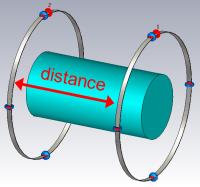
The degenerate birdcage is not a common design for
ultra-high field transmit array due to the technical
difficulties in its construction, such as the
interdependence of tuning and degeneracy on the value of
capacitors. We present here a combination of an analytical
theory, circuit simulations and numerical simulations to be
used for an efficient design and construction of the
degenerate birdcage at 7T. We demonstrate satisfactory
performance in terms of decoupling, B1+ homogeneity
and B1+ efficiency
on the workbench and with scanner measurements on phantoms
and human volunteers.
|
|
2168.
 |
Optimized MRI RF Body Coil for Integration with In-bore Therapy
or Biopsy System
Jiaqi Li1, Masahiro Fujimoto1, Amy Sue
Meyers1, Qiong Zhang2, and Huaiyu Dong2
1GE Healthcare, Waukesha, WI, United States, 2GE
Healthcare, Beijing, China, People's Republic of
An optimized MRI RF coil for integration with in-bore
therapy or biopsy system is discussed. The RF coil is
optimally designed into an open Ω shape to allow a much
bigger room for therapy or biopsy system. Horizontal rails
as well as coil support brackets are integrated with body
coil. Such that, the in-bore treatment system can have
bigger space and more power. The optimized design also
separated HIFU or SWL sub-assembly from high voltage RF
parts, which reduces EMI between those two, and safety issue
due to liquid leakage from HIFU or SWL sub-assembly is also
greatly reduced.
|
|
2153.
 |
A new monopole intravascular coil with three parasitic elements
optimized for MRI 1.5 T
mohammad mohammadzadeh1,2 and
alireza ghasempour Shirazi1
1ICT, University of applied science and
technology, Tehran, Iran, 2Shahid
Beheshti, Tehran, Iran
Monopole coil has a thin and flexible structure provides
high-resolution MR images from the internal vessels such as
aorta and coronary arteries. However, its SNR homogeneity
decreases at higher Tesla MRI systems, leading to increasing
the image artifact caused by the wall movement which is not
fully compensated using the post processing algorithms. In
this study, we introduced a monopole coil with tree
parasitic elements and compared its ISNR (Intrinsic SNR)
magnitude and distribution homogeneity to a conventional
monopole coil with one parasitic element for MRI 1.5 T at
64MHz . We optimized the coil geometry using a fast genetic
algorithm written in MATLAB and performed the simulation of
the ISNR indices by Involving HFSS and MATLAB inside a
saline phantom.
|
|
2165.
 |
Simulation, measurement, and optimization of a microcoil design
for MR Microscopy at 9.4 T
Mohammad Mohammadzadeh1,2 and
Mohammad Mohammadi2
1ICT, University of Applied Science and
Technology, Tehran, Iran, 2Nuclear
Engineering, Shahid Beheshti, Tehran, Iran
MR micro coils provide high SNR images of the mass limited
samples. To increase the coil sensitivity and then the image
SNR, microcoils geometries are adapted to the sample
dimension. However, differences between magnetic
susceptibility of the coil conductor and its surrounding
materials distorts the B0 magnetic fields homogeneity across
the sample. In this study, we measured 2D maps of a
solenoid of 1mm diameter and compared them with the
simulated results at 9.4 T. Considering the good agreement
of the computed and measured maps, effects of the shimming
and susceptibility matching processes were assessed in
removing the B0 fields inhomogeneities. Simulated results
verify that shimming coils are not able to fully cancel the
B0 field inhemogenities but embedding the micro coils in
susceptible materials will remove the B0 inhomogeneity
completely.
|
|
2156.
 |
Increasing transmit coil efficiency without local transmit
coils: a novel device for locally concentrating B1
Tracy Wynn1, Olli Friman1, and Randy
Duensing1
1Technology Architecture, Philips/Invivo,
Gainesville, FL, United States
Bore size increases can contribute to decreased efficiency
of transmit body coils, but modern protocols often increase
the requirements for B1 power.
This paper describes a novel solution for concentrating B1 power
without the use of a traditional local transmit coil, based
on the observation that roughly half of the transmit field
from a traditional birdcage coil comes from the end rings. A
dual-ring structure, modeled on a birdcage without rungs,
was built and shown to enable head imaging with up to a 30%
reduction in required RF input power. Uniformity was
maintained.
|
|
2137.
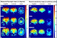 |
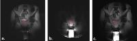
Evaluation of Spiral Extended Monopole Antenna Array with
Individual Whields (SEMAS) at 7T
Myung Kyun Woo1, Chang-Ki Kang2, and
Zang-Hee Cho3
1Electrical and Computer Engineering, Seoul
National University, Seoul, Korea, Republic of, 2Neuroscience
Research Institute, Incheon, Korea, Republic of, 3Seoul
National University, Seoul, Korea, Republic of
This abstract is to propose and evaluate the Spiral Extended
Monopole antenna Array with individual Shield (SEMAS) coil.
This coil was compared with the original Monopole antenna
Array (MA) coil and an Spiral Monopole antenna Array coil
with no shield (SMA) coil. The SEMAS coil showed larger flip
angle than the MA and SMA coils in the inferior areas of the
brain and relatively uniform flip angles across the brain.
|
|
2146.
 |
Printed Receive Coil Arrays with High SNR
Joseph Corea1, Balthazar P. Lechene1,
Thomas Grafendorfer2, Fraser Robb3,
Ana Claudia Arias1, and Michael Lustig1
1UC Berkeley, Berkeley, CA, United States, 2GE
Healthcare, Stanford, CA, United States, 3GE
Healthcare, Aurora, OH, United States
Extremely thin, lightweight, and flexible receive arrays can
be achieved by the use of printed electronics. Coil arrays
printed layer-by-layer from solution have shown potential to
deliver a comfortable customized fit for many patients.
However, relatively low SNR and poor mechanical robustness
prevented these devices from performing to their full
potential. Here we offer SNR within 3% of a traditionally
made coil by using high quality polymeric films as
dielectric layers in capacitors, high conductivity inks, and
a mechanically robust fabrication processes using fewer
printed layers and stronger connections. Using these
techniques shoulder and elbow images of a volunteer were
obtained.
|
|
2131.
 |
An 8Tx/32Rx RF Coil for 7T UHF Body MRI
Stefan HG Rietsch1,2, Stephan Orzada1,
and Harald H Quick1,2
1Erwin L. Hahn Institute for MR Imaging,
University of Duisburg-Essen, Essen, Germany, 2High
Field and Hybrid MR Imaging, University Hospital Essen,
Essen, Germany
In order to allow for improved SNR and higher acceleration
during image acquisition in the body at 7T, we present a
coil with 8Tx/Rx microstrip line elements with meanders and
24Rx loop elements. This coil comprises 8 building blocks
each consisting of one Tx/Rx element and 3 overlapping loops
which are actively detuned during transmit. With about
-19 dB reflection and an average decoupling of more than
-30 dB, the SNR can be boosted by about 21% in the abdomen.
Evaluation of g-factors as well as in vivo images of healthy
volunteers in both abdomen and heart show promising results.
|
|
2132.
 |
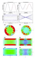
A 7T head coil with 16-channel dual-row transmit and 32-channel
receive array for pTx applications and high SNR
Shajan Gunamony1, Jens Hoffmann1,
Gregor Adriany2, Kamil Ugurbil2, and
Klaus Scheffler1
1Max Planck Institute for Biological Cybernetics,
Tuebingen, Germany, 2Center
for Magnetic Resonance Research, University of Minnesota,
Minneapolis, MN, United States
Transmit elements arranged in multiple rows are beneficial
in extending longitudinal coverage and achieve whole brain
excitation at ultra-high field strengths. Furthermore,
studies have shown that dual-row arrays produce less local
SAR. Receive arrays shaped to the contours of the anatomy
improves the signal-to-noise ratio (SNR) of the image. In
this work, we develop a 2x8 transmit array for spin
excitation in combination with a 32-channel high sensitive
receive array for human brain imaging at 7T. Critical coil
performance parameters like transmit efficiency and SNR were
evaluated.
|
|
2138.
 |
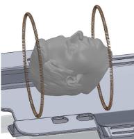
End-Loaded Dipole Array for 10.5T Head Imaging
Russell Luke Lagore1, Lance DelaBarre1,
Jinfeng Tian1, Gregor Adriany1,
Yigitcan Eryaman1, and J. Thomas Vaughan1
1Center for Magnetic Resonance Research,
University of Minnesota, Minneapolis, MN, United States
The feasibility of human head imaging at 10.5T is
demonstrated by the successful acquisition of in
vivo porcine
head images. This is achieved with an 8-element end-loaded
dipole array resonant at 10.5T (447MHz). This dipole array
is compared in terms of transmit efficiency and
signal-to-noise ratio to a high-pass birdcage coil and loop
array at 3T, 7T, and 10.5T. All coils share identical
dimensions and element count. While both transmit arrays
have comparable SNR performance at 7T, the dipole array is
inferior in terms of transmit efficiency compared to the
loop array and birdcage coil at all field strengths
examined.
|
|
2143.
 |
Interchangeable Patient-Specific Receive-Only Carotid Coils for
Simultaneous Imaging with Radio Frequency Head Coils at 3 Tesla
Michael J Beck1, Dennis L Parker1,
Bradley D Bolster, Jr.2, Seong-Eun Kim1,
J Scott McNally1,3, Gerald S Treiman1,4,5,
and J Rock Hadley1
1Utah Center for Advanced Imaging Research, Salt
Lake City, UT, United States, 2Siemens
Healthcare, Salt Lake City, UT, United States, 3University
of Utah Department of Radiology, Salt Lake City, UT, United
States,4University of Utah Department of Surgery,
Salt Lake City, UT, United States, 5Veterans
Affairs Department of Surgery (VASLCHCS), Salt Lake City,
UT, United States
We developed interchangeable carotid coils that can image
simultaneously with clinical head coils. Both 7 and 9
channel carotid coils were built to demonstrate the
interchangeability concept. SNR results show that the 7
channel coil has ~4x the SNR and the 9 channel coil has ~3x
the SNR of the commercial neck coil at the carotids. The
carotid coils image simultaneously with a head coil
providing greater coil sensitivity at the carotid
bifurcation and extending total coverage from the carotid
bifurcation to the circle of Willis.
|
|
2162.
 |
Electric-LC resonators decoupling approach for monopole antenna
arrays at 7T
Xinqiang Yan1 and
Xiaoliang Zhang2
1Key Laboratory of Nuclear Analysis Techniques,
Institute of High Energy Physics, Chinese Academy of
Sciences, Beijing, China, People's Republic of, 2Department
of Radiology and Biomedical Imaging, University of
California San Francisco, San Francisco, CA, United States
Induced current elimination (ICE) method could efficiently
reduce the element coupling in monopole and dipole arrays,
and ultimately improve their SNR and parallel imaging
performance. Nevertheless, in current ICE method, the
decoupling element has possible effect on the original B1 field,
leading to dark spots at the areas near decoupling elements.
To address such effects, we introduce a new structure,
electric-LC (ELC) resonator, for decoupling monopole arrays.
Based on the simulation and experimental results, ELC
resonators could also effectively reduce the coupling of
monopoles and meanwhile have less influence on the original
B1 fields of the elements.
|
|
2167.
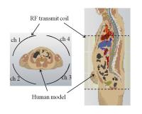 |
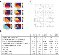
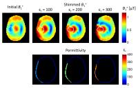
Improvement of B1+ Homogeneity and Reduction of Transmit RF
Power Using 4-channel Regional RF Shimming in L-spine Imaging at
3T
Yukio Kaneko1, Kosuke Ito2, Masahiro
Takizawa2, Yoshihisa Soutome1,2,
Hideta Habara1,2, Yusuke Seki1,
Tetsuhiko Takahashi2, Yoshitaka Bito2,
and Hisaaki Ochi1
1Research and Development Group, Hitachi Ltd.,
Tokyo, Japan, 2Healthcare
Company, Hitachi, Ltd., Chiba, Japan
The B1+ inhomogeneity
in a human body increases as the strength of a static
magnetic field increases. Previous studies showed the effect
of the number of RF transmit channels in RF shimming.
However, the effect for a partial region of the lumbar spine
in a sagittal plane has not yet been investigated. In this
study, the effect of the number of RF transmit channels for
regional RF shimming in the lumbar spine region was
investigated. The results show that 4-channel RF shimming
can contribute to improving B1+ homogeneity
and reducing the transmit RF power more than 2-channel RF
shimming.
|
|
2145.
 |
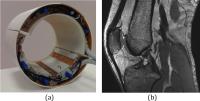
A 6 Channel Transmit-Receive Coil Array for 7T Cervical Spine
Imaging
Zidan Yu1,2, Bei Zhang1, Jerzy Walczyk1,
Gang Chen1,2, and Graham Wiggins1
1The Bernard and Irene Schwartz Center for
Biomedical Imaging, Department of Radiology, New York
University School of Medicine, New York, NY, United States, 2The
Sackler Institute of Graduate Biomedical Sciences, New York
University School of Medicine, New York, NY, United States
The cervical spine presents a challenging target for 7T RF
coils. In this work, we describe a 6 channel
transmit-receive cervical spine coil constructed like a
cervical collar, wrapping around the back of the neck.
In-vivo experiments demonstrate higher transmit efficiency,
better B1+ uniformity in the
transverse plane and equivalent SNR compared to a RAPID
Biomedical cervical spine coil.
|
|
2163.
 |
RF coil design using circulant and block circulant matrix
algebra
Sasidhar Tadanki1
1Electrical and Computer Engineering, Worcester
Polytechnic Institute, Worcester, MA, United States
In this work a simple, efficient method to designing a
transmission line volume resonator coil for MR applications
is presented. A multiconductor transmission line is
represented as a multiport network using its port admittance
matrix. Closed form solutions for port resonant mode
frequencies are calculated by solving the eigenfunctions of
the port admittance matrix using block matrix and circulant
block matrix algebra. Detailed analysis and simulated
results are presented and compared with standard published
results. A dual-tuned surface coil is developed to
demonstrate the efficacy of the proposed method.
|
|
2148.
 |
Inverse Design of Dielectric Pads based on Contrast Source
Inversion
Wyger Brink1, Jeroen van Gemert2, Rob
Remis2, and Andrew Webb1
1Radiology, Leiden University Medical Center,
Leiden, Netherlands, 2Circuits
and Systems, Delft University of Technology, Delft,
Netherlands
The design of passive dielectric pads can be an exhaustive
procedure with many degrees of freedom to address. In this
study we developed a constrained inverse design approach
based on the contrast source inversion method. The procedure
can yield design guidelines efficiently, enabling automated
design of dielectric pads.
|
|
2149.
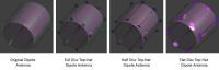 |
Improvement of B1+ Homogeneity along Z-Direction Using Top-Hat
Dipole-Antenna pTX Array for Body Imaging at 7 Tesla
Suchit Kumar1, Joshua Haekyun Park2,3,
Young-Seung Jo2,4, Jeong-Hee Kim2,3,
Chulhyun Lee2, and Chang-Hyun Oh1,4,5
1Department of Biomicrosystem Technology, Korea
University, Seoul, Korea, Republic of, 2Korea
Basic Science Institute, Cheongju, Chungcheongbuk-do, Korea,
Republic of, 3Industrial
Technology Institute, Korea University, Sejong City, Korea,
Republic of, 4Department
of Electronics and Information Engineering, Korea
University, Seoul, Korea, Republic of, 5ICT
Convergence Technology Team for Health&Safety, Korea
University, Seoul, Korea, Republic of
In ultra-high field (UHF), body imaging suffers from B1
inhomogeneity due to shorter wavelength. A range of new RF
coil designs has been proposed to overcome this problem.
But, B1 inhomogeneity in the coronal plane still exists due
to limited coverage. In this work, a novel design of an
8-channel top-hat dipole antenna with parallel transmission
is proposed to improve B1+ homogeneity along Z-direction. B1+ field
distribution and SAR field were simulated in FDTD solver.
Comparison with original dipole antenna array confirms the
improved B1+ homogeneity
in proposed design.
|
|
2147.
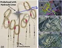 |
3D-printed RF Probeheads for Low-cost, High-throughput NMR
R. Adam Horch1,2 and
John C. Gore1,2
1Vanderbilt University Institute of Imaging
Science, Vanderbilt University, Nashville, TN, United
States, 2Department
of Radiology & Radiological Sciences, Vanderbilt University,
Nashville, TN, United States
3D printing is demonstrated as a new means to fabricate
complete RF probeheads for solution-state NMR. Current 3D
printing methods yield mm-scale RF coils with integral
sample chambers for self-contained NMR probes, and
3D-printed microcoils are imminent given ongoing advances in
technology. The unique properties of 3D printing enable
facile construction of potentially thousands of coils at low
cost, giving way to dense coil arrays for high-throughput
NMR and novel coil geometries.
|
|
2166.
 |
Numerical Comparison of Stacked and Planar Coil Reception Arrays
for Prostate MRI at 3 T
Jorge Chacon-Caldera1, Javier Uranga Solchanga1,
Paulina Koziol1,2, and Lothar R Schad1
1Computer Assisted Medical Medicine, Medical
Faculty Mannheim, Heidelberg University, Mannheim, Germany, 2Department
of Medical Physics and Biophysics, Faculty of Physics and
Applied Computer Science, AGH University of Science and
Technology, Krakow, Poland
Prostate MRI is commonly performed using endorectal coils
which are invasive. This is done since body planar arrays
are not sensitive enough for prostate imaging. Increasing
sensitivity of an array for deep structures in the body is
not trivial. In this study, we extended the traditional
stacked figure-8 and single loop quadrature pair to add more
single loop coils and enhanced the sensitivity at the depth
of the prostate without increasing the field over a larger
lateral area. We compared these arrays to classical planar
approaches and found a factor 1.35 increase in maximum
localized |B1-| using numerical
simulations.
|
|
2157.
 |
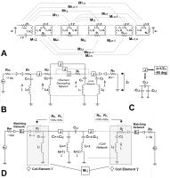
SNR simulations including coupled preamplifier noise
Matthias Malzacher1,2, Markus Vester3,
Robert Rehner3, Christopher Stumpf1,
and Patrick Korf1
1Friedrich-Alexander University
Erlangen-Nuremberg, Erlangen, Germany, 2Computer
Assisted Clinical Medicine, University Heidelberg, Medical
Faculty Mannheim, Mannheim, Germany, 3Siemens
Healthcare GmbH, Erlangen, Germany
Using reciprocity, available SNR from a receive coil array
can be calculated by maximizing B1- at the target
voxel for unit input power. However, for strongly coupled or
lightly loaded coil elements, the noise figure degradation
due to coupled preamplifier noise becomes significant. It is
shown here that this effect can be modeled by power loss in
a resistive attenuator at each coil port. Thus, it is now
possible to simulate any coil configuration, including those
where coil coupling cannot be neglected.
|
|
2160.
 |
Transmission Line Resonator Segmented with Series Capacitors
Vitaliy Zhurbenko1, Vincent Boer 2,
and Esben Thade Petersen 2
1Technical University of Denmark, Kgs. Lyngby,
Denmark, 2Danish
Research Centre for Magnetic Resonance, Centre for
Functional and Diagnostic Imaging and Research, Copenhagen
University Hospital Hvidovre, Hvidovre, Denmark
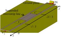
Transmission line resonators are often used as coils in high
field MRI. Due to distributed nature of such resonators,
coils based on them produce inhomogeneous field. This work
investigates application of series capacitors to improve
field homogeneity along the resonator. The equations for
optimal values of evenly distributed capacitors are
presented. The performances of the segmented resonator and a
regular transmission line resonator are compared.
|
|
2136.
 |
Design of quadrature-compensated double-tuned RF surface coil
using trap circuits
Chang-Hoon Choi 1,
YongHyun Ha1, Arthur W. Magill1, and
N. Jon Shah1,2
1Institute of Neuroscience and Medicine-4,
Research Centre Juelich, Juelich, Germany, 2Faculty
of Medicine, Department of Neurology, JARA, RWTH Aachen
University, Aachen, Germany
A novel double tuned (1H/23Na)
butterfly/loop surface coil using LCC traps was designed
whereby the sodium mode was operated in a quadrature. The
performance of this coil was evaluated on a 4T whole-body
scanner and compared with a single-tuned butterfly and a
loop coil. Images obtained by the quadrature-compensated
double-tuned RF coil were more uniform in each slice and the
SNRs were slightly higher over the selected ROIs compared to
those from the reference coils.
|
|
2151.
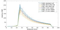 |
Copper plating of conductive silver ink coils for improved SNR
performance
J. Rock Hadley1, Emilee Minalga1, and
Dennis L. Parker1
1Radiology, University of Utah, Salt Lake City,
UT, United States
This work tests how much loop conductivity and SNR is
improved with copper plating of the silver ink trace. Coils
made with a silver ink base and different amounts of copper
plating were compared against solid copper. This work
demonstrates that copper plating of silver ink coils is
possible and it indicates that significant improvements in
coil trace conductivity can be achieved. Consequently, the
SNR performance of silver ink coils that have been plated
with copper improves over silver ink coils without plating.
|
|
2150.
 |
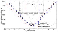
TMS positioning in MRI using NMR probes
Yi-Cheng Hsu1, Ying-Hua Chu1, Pu-Yeh
Wu1, Shang-Yueh Tsai2, and Fa-Hsuan
Lin1
1Institute of Biomedical Engineering, National
Taiwan University, Taipei, Taiwan, 2Institute
of Applied Physic, National Chengchi University, Taipei,
Taiwan
We propose a method and a system to precisely place the TMS
coil inside the MRI using NMR probes.The positioning can be
completed in 0.1 s with high translation (0.015 mm) and
rotation precision (0.0047°) as well as low bias (~0.8 mm in
50 mm FOV).
|
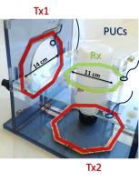
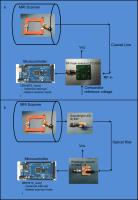
|