|
Interventional
 |
2091.
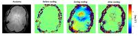 |
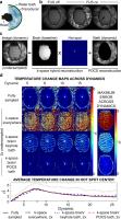
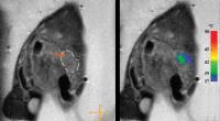
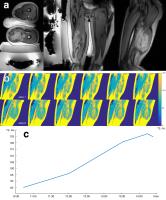
Monitoring temperature changes in the brain during high flow
cold air cooling 
Åsmund Kjørstad1, Fabian Temme1, Jens
Fiehler1, and Jan Sedlacik1
1Neuroradiology, University Medical Center
Hamburg-Eppendorf, Hamburg, Germany
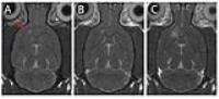
Targeted temperature management is a treatment that seeks to
reduce and control the body temperature. We demonstrate a
novel localized cooling technique using high flow cold air
applied nasally and orally to the airways by monitoring the
brain temperature using gradient echo phase imaging at 3T. 2
healthy volunteers were investigated, with one subject being
scanned twice and the other once. A significant temperature
reduction (p<0.05) was seen in the inferior frontal lobe in
all three experiments with an average cooling effect of
-0.33°C. This demonstrates the feasibility of our proposed
high flow cold air system.
|
|
2092.
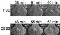 |
Early Assessment of MRgFUS Thalamotomy Using a Diffusion
Weighted Steady State MRI Sequence in an In-vivo Porcine Model
Juan Camilo Plata1, Sam Fielden2,
Bragi Sveinsson3, Brian Hargreaves4,
and Craig Meyer2
1Bioengineering, Stanford University, Las Vegas,
NV, United States, 2Biomedical
Engineering, University of Virginia, Charlottesville, VA,
United States, 3Electrical
Engineering, Stanford University, Palo Alto, CA, United
States, 4Stanford
University, Palo Alto, CA, United States
Early detection of thermal lesions generated using MR-guided
focused ultrasound systems is critical for treatment
feedback. Irreversible changes in the apparent diffusion
coefficient (ADC) have been previously shown to be an early
indicator for loss of viability in the prostate. Due to poor
image quality using standard diffusion weighted imaging
strategies inside the focused ultrasound system,
radiologists rely on T2-weighted fast spin echoes (FSE) for
lesion detection. T2-weighted changes due to lesion
formation develop more slowly than ADC changes. We propose
using a diffusion-weighted steady state sequence for early
detection of thermal lesions inside the focused ultrasound
system.
|
|
2093.
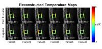 |
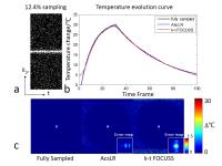
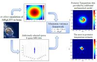
Fast Temperature Estimation from Undersampled k-Space with Fully
Sampled Center for Real Time MR Guided Microwave Ablations
Fuyixue Wang1, Zijing Dong1, Shuo Chen2,
Bingyao Chen3, Jiafei Yang3, Xing Wei3,
Shi Wang2, and Kui Ying2
1Department of Biomedical Engineering, Tsinghua
University, Beijing, China, People's Republic of, 2Key
Laboratory of Particle and Radiation Imaging, Ministry of
Education, Medical Engineering and Institute, Department of
Engineering Physics, Tsinghua University, Beijing, China,
People's Republic of, 3Department
of Orthopedics, First Affiliated Hospital of PLA General
Hospital, Beijing, China, People's Republic of
Real time thermometry is desirable for thermal therapy such
as microwave ablation to ensure patient safety. MR
temperature imaging using proton resonance frequency (PRF)
shift technique can provide temperature maps during the
treatment. In this work, we proposed a novel reconstruction
framework that estimates temperature changes from
undersampled k-space with a few fully sampled k-space
points. Simulation studies, phantom heating experiments and
human experiments were performed to validate the proposed
method. The proposed method can provide temperature images
with relatively high accuracy and short reconstruction time
at a reduction factor of 4 in presence of motion.
|
|
2094.
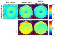 |
Temporal Weighted Sliding Window SPIRiT with Golden Angle Radial
Sampling for Real Time MR Temperature Imaging
Fuyixue Wang1, Zijing Dong1, Haikun Qi2,
Shi Wang3, Huijun Chen2, and Kui Ying3
1Department of Biomedical Engineering, Tsinghua
University, Beijing, China, People's Republic of, 2Center
for Biomedical Imaging Research, Department of Biomedical
Engineering, School of Medicine, Tsinghua University,
Beijing, China, People's Republic of, 3Key
Laboratory of Particle and Radiation Imaging, Ministry of
Education, Medical Engineering and Institute, Department of
Engineering Physics, Tsinghua University, Beijing, China,
People's Republic of
Real time MR temperature imaging during thermal therapy is
beneficial for monitoring and controlling the treatment in
clinical applications. In this work, we explored
correlations in the temporal dimension of temperature
imaging and proposed a novel method, temporal weighted
sliding window SPIRiT using motion-insensitive golden angle
radial sampling, to achieve real time temperature imaging.
Through simulation studies and phantom heating experiments,
we validated the ability of the proposed method to obtain
temperature images with relatively high temperature accuracy
at a reduction factor of 8.
|
|
2095.
 |
Spatially-segmented undersampled temperature map reconstruction
for transcranial MR-guided focused ultrasound
Pooja Gaur1, Xue Feng2, Samuel Fielden2,
Craig H Meyer2, Beat Werner3, and
William A Grissom1
1Vanderbilt University, Nashville, TN, United
States, 2University
of Virginia, Charlottesville, VA, United States, 3University
Children's Hospital, Zurich, Switzerland
Accelerated temperature imaging is desirable to improve
spatiotemporal coverage during MR-guided focused ultrasound
procedures in the brain. Circulating water prevents skull
overheating, but also creates signal variations that disrupt
correlations between images collected before and during
treatment (which are relied on to overcome undersampling
artifacts), leading to errors in temperature measurements.
We propose a spatially-segmented iterative reconstruction
method, which applies the k-space hybrid model to
reconstruct temperature changes in the brain and a POCS
method to reconstruct the image in the water bath.
Separately reconstructing brain and water bath signal
results in lower temperature error when undersampling
k-space.
|
|
2096.
 |
3D UTE MR thermometry of frozen tissue: feasibility and accuracy
during cryoablation at 3T
Christiaan G. Overduin1, Jurgen J. Fütterer1,2,
and Tom W.J. Scheenen1
1Radiology, Radboud University Medical Centre,
Nijmegen, Netherlands, 2MIRA
Institute for Biomedical Engineering and Technical Medicine,
University of Twente, Enschede, Netherlands
Our study assessed the feasibility and accuracy of 3D
ultrashort TE (UTE) MR thermometry to dynamically track
temperatures across frozen tissue during cryoablation on a
clinical MR system at 3T. We demonstrated 3D UTE imaging to
achieve measurable MR signal from frozen tissue down to
temperatures as low as -40°C within a clinically realistic
time-frame (~1min) and with sufficient spatial resolution
(1.63mm isotropic). Using a calibration curve, we could
derive 3D MR-estimated temperature maps of the frozen
tissue, which showed good agreement with matched temperature
sensor readings on statistical analysis.
|
|
2097.
 |
Acceleration of Temperature Mapping with an Ascending Threshold
Low Rank Constraint (AscLR)
Fuyixue Wang1, Zijing Dong1, Bingyao
Chen2, Jiafei Yang2, Xing Wei2,
Shi Wang3, and Kui Ying3
1Department of Biomedical Engineering, Tsinghua
University, Beijing, China, People's Republic of, 2Department
of Orthopedics, First Affiliated Hospital of PLA General
Hospital, Beijing, China, People's Republic of, 3Key
Laboratory of Particle and Radiation Imaging, Ministry of
Education, Medical Engineering and Institute, Department of
Engineering Physics, Tsinghua University, Beijing, China,
People's Republic of
Thermal therapies such as microwave ablation require
temperature imaging with high temporal resolution to
calculate thermal absorption and evaluate the curative
effects of the ablation. Thus, acceleration techniques of
data acquisition for MR temperature imaging using PRF shift
technique are desirable. In this work, we explored the low
rank property of k-t space in dynamic MR temperature imaging
and proposed a novel fast reconstruction method AscLR with
an ascending-threshold low rank constraint. Through
simulation studies and microwave heating experiments, we
validated the ability of the proposed method to provide
relatively accurate temperature estimation at a reduction
factor of 8.
|
|
2098.
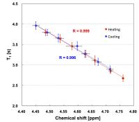 |
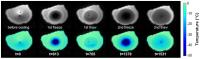
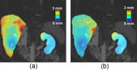
Feasibility of Absolute Thermometry of Knee Joint Cartilage
using Spin-lattice Relaxation Time
Tomoya Kimura1, Atsushi Shiina1, Kenji
Takahashi2, and Kagayaki Kuroda1,3
1Course of Electrical and Electronic Engineering,
Graduate School of Engineering, Tokai University, Hiratsuka,
Japan, 2Department
of Orthopaedic Surgery, Nippon Medical School, Tokyo, Japan, 3Center
for Frontier Medical Engineering, Chiba University, Chiba,
Japan
Temperature dependence of T1 of tissue water in the porcine
knee joint cartilage in vitro was examined at 9.4T in
comparison with that of the water proton resonance
frequency. The absolute value of T1 at each temperature
between room temperature and 60 oC was
reproducible. Hysteresis was negligible during heating and
cooling processes. The correlation coefficient with
temperature was higher than 0.998, and hence that with water
proton chemical shift was also high (>= 0.996). The
temperature coefficient was 1.28%/ oC at 30 oC
for heating and 1.24%/ oC for cooling. These
results suggested that T1 is a favorable index for
thermometry of the knee joint cartilage under thermal
therapies.
|
|
2104.
 |
High Speed, High Sensitivity MR-ARFI Using a Balanced
Steady-State Free Precession Pulse Sequence
Yuan Zheng1, Michael Marx1, G. Wilson
Miller2, and Kim Butts Pauly1
1Radiology, Stanford University, Stanford, CA,
United States, 2Radiology
and Medical Imaging, University of Virginia,
Charlottesville, VA, United States
We have developed a novel MR-ARFI technique that makes use
of transition band balanced steady-state free precession (bSSFP).
Due to the strong dependence of image phase on the
motion-encoded phase, this technique improves the
sensitivity of MR-ARFI measurements over commonly used
spoiled sequences. The proposed technique also features high
speed, as an ARFI contrast image can be acquired in a few
seconds. With its high speed and high sensitivity, the
bSSFP-ARFI technique could be useful in
confirming/calibrating the HIFU focal spot before thermal
ablation treatment.
|
|
2099.
 |
A Hybrid Model Integrated with Correction of Susceptibility
Induced Phase Error in Magnetic Resonance Thermometry
Kexin Deng1, Yuxin Zhang1, Yu Wang1,
Bingyao Chen2, Xing Wei2, Jiafei Yang2,
Shi Wang3, and Kui Ying3
1Biomedical Engineering, Tsinghua University,
Beijing, China, People's Republic of, 2Department
of Orthopedics, First Affiliated Hospital of PLA General
Hospital, Beijing, China, People's Republic of, 3Key
Laboratory of Particle and Radiation Imaging, Ministry of
Education, Department of Engineering Physics, Tsinghua
University, Beijing, China, People's Republic of
The temperature dependency of susceptibility, especially for
fat, could introduce errors in temperature estimation. To
address this problem, a hybrid model integrated with
susceptibility change induced phase is proposed to reduce
the phase error. Simulation was conducted to validate the
proposed model and a water-fat phantom was made and heated
to illustrate the effect of susceptibility-induced phase
error correction. The proposed model shows more accurate
temperature estimation near the water-fat interface both in
simulation and phantom heating experiment.
|
|
2100.
 |
Patient preparation by oral fluid intake for proton resonance
frequency shift based MR thermometry in the pancreas
Cyril J Ferrer1, Lambertus W Bartels1,
Marijn van Stralen1, Chrit T.W Moonen1,
and Clemens Bos1
1University Medical Center Utrecht, Utrecht,
Netherlands
Magnetic Resonance Imaging-guided High Intensity Focused
Ultrasound has recently been suggested as an alternative
treatment modality for pancreatic cancer that is
non-invasive, and may be suited for treatment in cases where
surgery is not an option. However, using proton resonance
frequency shift based thermometry in this area is highly
challenging, because of motion and air in the digestive
tract near the pancreas. We have shown experimentally that
patient preparation by filling the stomach and duodenum with
juice can be a pragmatic solution for more precise
temperature monitoring during MR-HIFU therapy particularly
in the head of the pancreas.
|
|
2103.
 |
Rapid HIFU refocusing based on MR-ARFI
Charles Mougenot1, Samuel Pichardo2,3,
Steven Engler2,4, Adam Waspe5,6,
Elodie Constanciel5, and James Drake5,6
1Philips Healthcare, Toronto, ON, Canada, 2Thunder
Bay Regional Research Institute, Thunder Bay, ON, Canada, 3Electrical
Engineering, Lakehead University, Thunder Bay, ON, Canada, 4Computer
Science, Lakehead University, Thunder Bay, ON, Canada, 5Hospital
for Sick Children, Toronto, ON, Canada, 6University
of Toronto, Toronto, ON, Canada
Algorithms have been developed that use Magnetic Resonance
Acoustic Radiation Force Imaging (MR-ARFI) to maximize the
intensity at the focal point of a high intensity focused
ultrasound beam in order to compensate for tissue related
phase aberrations. A combination of two methods is proposed
to achieve refocusing using a clinically acceptable
acquisition time at 3T. Compensation of three aberrators
inducing a relative intensity of 95%, 67.4% and 25.3% were
successfully evaluated in a phantom to retrieve a relative
intensity of 101.6%, 91.3% and 93.3% in 10 minutes or
103.9%, 94.3% and 101% in 25 minutes.
|
|
2101.
 |
Magnetic Resonance Acoustic Radiation Force Imaging for
interventional planning of HIFU therapy in the kidney
Johanna Maria Mijntje van Breugel1, Martijn de
Greef2, Charles Mougenot3, Maurice AAJ
van den Bosch2, Chrit CW Moonen2, and
Mario AAJ Ries2
1Radiology, University Medical Center Utrehct,
Utrecht, Netherlands, 2University
Medical Center Utrehct, Utrecht, Netherlands, 3Torontp,
Canada
Hypothesis: MR-ARFI can be deployed in the kidney as an
alternative for the thermal test shot at low power. The
employed respiratory gated MR-ARFI sequence in combination
with a 450 W excitation tone-burst is sensitive enough to
exceed the noise level and to clearly display the focal
point of the HIFU beam. Both at 450W and at 1000W the
displacement due to the radiation force coincided with the
location of the temperature rise due to thermal ablation at
equivalent power. Hence, radiation force in combination with
a pencil beam navigator to compensate for respiratory motion
is a reliable indicator of the location of the thermal
lesion and might be an alternative to the low power thermal
test shot in highly perfused organs such as the kidney.
|
|
2102.
 |
MR-Shear Wave Elasticity Imaging (SWEI) with Bipolar
Motion-Encoding Gradients
Yuan Zheng1, Michael Marx1, Rachelle
R. Bitton1, and Kim Butts Pauly1
1Radiology, Stanford University, Stanford, CA,
United States
We have demonstrated a method for shear wave elasticity
imaging (SWEI). A shear wave was generated by a short
focused ultrasound (FUS) pulse, and was tracked by
collecting images with different delays (tdelay)
between the FUS pulse and bipolar motion-encoding gradients
(MEG). The time-of-flight (TOF) at each pixel was determined
by the zero-crossing of the image phase as a function of tdelay.
Based on the TOF map, a shear wave velocity map was
generated in polar coordinates.
|
|
2105.
 |
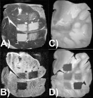
A Novel method for developing clinical grade active devices
dedicated to interventional MRI procedures
Korel Dursun Yildirim1, Engin Baysoy1,
Zahid Sagiroglu2, Çagla Özsoy1, Ozgur
Kocatürk1, and Senol Mutlu2
1Biomedical Engineering, Bogaziçi University,
Institute of Biomedical Engineering, Istanbul, Turkey, 2Electrical
and Electronics Engineering, Bogaziçi University, Institute
of Graduate Studies in Science and Engineering, Istanbul,
Turkey
In this study, A Novel method and system were devoloped for
developing clinical grade active devices dedicated to
interventional MRI procedures. Before prototype fabrication,
according to desired component dimensions, component values
were simulated. With the exact dimensions used in
simulations, component prototypes were fabricated via
conductive ink as component material. Finally, simulation
results and banch top measurements of component values were
compared and reliability of simulation results were
comfirmed.
|
|
2106.
 |
Real-Time Hemodynamic Monitoring during MR Imaging and
Interventional Procedures derived from induced
Magnetohydrodynamic Voltages
T. Stan Gregory1, Ehud Schmidt2, John
Oshinski3, and Zion Tsz Ho Tse1
1College of Engineering, The University of
Georgia, Athens, GA, United States, 2Radiology,
Brigham and Women's Hospital, Boston, MA, United States, 3Radiology,
Emory University Hospital, Atlanta, GA, United States
Magnetic Resonance Imaging (MRI) is increasingly becoming
the preferred diagnostic and interventional imaging modality
for a variety of diseases. Despite the increasing clinical
merit, practical implementation of these procedures in the
clinic is oftentimes limited due to the high risk associated
with these patient groups and the subsequent need for
advanced physiological monitoring for each patient to be
cleared for MRI imaging and interventional workflows. The
presented method for beat-to-beat SV and continuous aortic
flow monitoring within the MRI bore based on
Magnetohydrodynamic Voltages (VMHD) induced onto 12-lead
Electrocardiograms (ECG), enables MR imaging and MRI-guided
interventional procedures for these patients.
|
|
2107.
 |
Accelerated MR Thermometry in the Presence of Uncertainties
Reza Madankan1, Wolfgang Stefan1,
Christopher MacLellan1, Samuel Fahrenholtz1,
Drew Mitchell1, R.J. Stafford1, John
Hazle1, and David Fuentes1
1Imaging Physics, MD Anderson Cancer Center,
Houston, TX, United States
Compressive sensing and sparse image reconstruction has
received significant attention and has demonstrated
potential in reduction of acquisition times. However, in
many methods, under-sampling strategies are heuristically
chosen and empirically validated. This often leads to a
relatively larger number of k-space
samples than needed for a particular application. The
presented work develops a mathematically rigorous and
quantitative methodology for k-space under-sampling with
respect to model-based reconstruction of MR thermometry. The
key idea of the proposed approach is to detect the useful
samples of k-space
in order torefine the
model, and then the refined mathematical model is utilized
to reconstruct the image.
|
|
2108.
 |
Kalman Filtered Bio Heat Transfer Model Based Self-adaptive
Hybrid Magnetic Resonance Thermometry
Yuxin Zhang1, Kexin Deng1, Shuo Chen2,
Bingyao Chen3, Xing Wei3, Jiafei Yang3,
Shi Wang2, and Kui Ying2
1Biomedical Engineering, Tsinghua University,
Beijing, China, People's Republic of, 2Key
Laboratory of Particle and Radiation Imaging, Ministry of
Education, Department of Engineering Physics, Tsinghua
University, Beijing, China, People's Republic of, 3Department
of Orthopedics, First Affiliated Hospital of PLA General
Hospital, Beijing, China, People's Republic of
The proposed Kalman ?ltered Bio Heat Transfer Model Based
Self-adaptive Hybrid MR Thermometry, abbreviated as KalBHT
hybrid algorithm, introduced the BHTE model to synthesize a
window on the regularization term of the hybrid algorithm,
which leads to a self-adaptive regularization both spatially
and temporally with change of temperature. Further, to
decrease the sensitivity to accuracy of the BHTE model,
Kalman ?lter is utilized to update the window at each
iteration time. Besides, the BHTE model is able to
interpolate temperature maps during the acquisition and
reconstruction of the next MR image to make real time
temperature monitoring possible. To investigate the effect
of the proposed model, phantom microwave heating experiment
and in-vivo experiment
with heating simulation were conducted in this study.
|
|
2109.
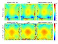 |
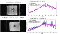
Evaluation of the Effect of Trajectory Correction with Radial
Sampling on Temperature Imaging
Tongxin Chen1, Fuyixue Wang1, Zijing
Dong1, Haikun Qi2, Shi Wang3,
Huijun Chen2, and Kui Ying3
1Department of Biomedical Engineering, Tsinghua
University, Beijing, China, People's Republic of, 2Center
for Biomedical Imaging Research, Department of Biomedical
Engineering, School of Medicine, Tsinghua University,
Beijing, China, People's Republic of, 3Key
Laboratory of Particle and Radiation Imaging, Ministry of
Education, Medical Engineering and Institute, Department of
Engineering Physics, Tsinghua University, Beijing, China,
People's Republic of
Radial sampling is sensitive to trajectory errors and can
cause image distortions. To investigate the effect of
trajectory errors on temperature imaging, we first evaluated
the use of Trajectory Auto-Corrected Image Reconstruction
(TrACR), a method to reconstruct radial images without
trajectory errors, for radial temperature imaging. Then, we
examined the feasibility of TrACR with only one calibration
on dynamic temperature imaging based on the assumption that
gradient errors are time-invariant. Through phantom heating
experiments, we validated that both of the TrACR and the
single-calibration TrACR can correct the errors of normal
and golden angle radial sampling and provide improved
temperature accuracy.
|
|
2110.
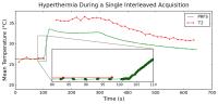 |
On-Demand Dynamic Updating of the Temporal Resolution of
Interleaved PRFS and T2 Temperature Mapping Methods for MR-HIFU
Steven Engler1,2, Charles Mougenot3,
Jochen Keupp4, Steffen Weiss4, Edwin
Heijman5, and Samuel Pichardo1,6
1Thunder Bay Regional Research Institute, Thunder
Bay, ON, Canada, 2Lakehead
University, Computer Science, Thunder Bay, ON, Canada, 3Philips
Healthcare, Toronto, ON, Canada, 4Philips
Research, Hamburg, Germany, 5Philips
Research, Eindhoven, Netherlands, 6Lakehead
University, Electrical Engineering, Thunder Bay, ON, Canada
Temperature changes can be assessed in non-adipose tissue
using proton resonance frequency shift MR-thermometry
methods based on gradient-echo sequences, and in adipose
tissue using apparent T2-mapping MR-thermometry methods
based on multi-echo fast spin-echo sequences. It has been
previously demonstrated that these sequences can be
interleaved to simultaneously monitor temperature in all
tissues. In this study we show the feasibility of
controlling the sequence duty-cycle of the aforementioned
interleaved scanning technique on-demand in order to
dynamically change the temporal resolution of the two
interleaved scans in response to actual temperature changes
and the stage of the hyperthermia application.
|
|
2111.
 |
Model predictive filtering MR thermometry utilizing ultrasound
beam modeling SAR predictions
Henrik Odéen1, Scott Almquist2, Joshua
de Bever1, and Dennis L Parker1
1Utah Center for Advanced Imaging Research,
Department of Radiology, University of Utah, Salt Lake City,
UT, United States, 2School
of Computing, University of Utah, Salt Lake City, UT, United
States
Thermal model based reconstruction of subsampled MR
temperature data for focused ultrasound applications rely on
acoustic and thermal parameters that are often analytically
determined from a pre-treatment sonication. In this work we
combine a thermal model based reconstruction method with
ultrasound beam simulations to determine the specific
absorption rate in order to avoid potentially damaging the
tissue during a pre-treatment sonication. Proof-of-concept
experiments are performed in a homogenous gelatin phantom
and a gelatin phantom embedded with a plastic skull. The
temperature estimations using US modeling show the same
accuracy as those using a pre-treatment sonication.
|
|
2112.
 |
Investigation of temperature dependent changes in signal
intensity, T1 and T2* in cortical bone
Henrik Odéen1, Bradley Bolster2, Eun
Kee Jeong1, and Dennis L Parker1
1Utah Center for Advanced Imaging Research,
Department of Radiology, University of Utah, Salt Lake City,
UT, United States, 2Siemens
Healthcare, Salt Lake City, UT, United States
Measurements of changes in signal intensity and T1
relaxation time with temperature has been suggested for
temperature monitoring in cortical bone during MR guided
focused ultrasound treatments. In this study we compare
changes in signal intensity, T1, and T2* with temperature
using a 3D ultrashort echo time pulse sequence and a 2D
gradient recalled echo pulse sequence with short TE. The
effects of T1 and T2* change with temperature counteract
each other making the change in signal intensity small, and
therefore T1 and T2* appears to have the greatest
sensitivity to changes in temperature.
|
|
2113.
 |
Simultaneous PRFS and T1 quantification using bSSFP for
Temperature Monitoring
Mingming Wu1, Matthew Tarasek2, Axel
Haase3, and Silke Lechner-Greite4
1IMETUM, Technische Universität München,
Garching, Germany, 2GE
Global Research, Niskayuna, NY, United States, 3Technische
Universität München, Garching, Germany, 4GE
Global Research, Garching, Germany
Inversion Recovery prepared bSSFP sequence is used to
quantify T1 and PRFS simultaneously based on a phase
sensitive bSSFP readout. This technique allows for
temperature mapping in both adipose and aqueous tissues at
the same time. The feasibility of this method is shown with
means of a cooling down experiment of a heterogeneous
phantom. B0 drift correction is performed based on
neighboring voxels in the fatty tissue.
|
|
2114.
 |
MRI-guided robotic arm (MgRA) to target deep brain nuclei in
vitro
Yi Chen1,2, Filip Sobczak1, and Xin Yu1,2
1Research Group of Translational Neuroimaging and
Neural Control, High-Field Magnetic Resonance, Max Planck
Institute for Biological Cybernetics, Tuebingen, Germany, 2Graduate
School of Neural Information Processing, University of
Tuebingen, Tuebingen, Germany
A key challenge of the fiber optic-mediated multi-model fMRI
methodologies is locating the fiber tip accurately and
precisely to target deep brain nuclei. The requirement of
precision is only several hundreds of microns in the animal
brains. In this work, a multi degree of freedom robotic arm
was developed with the use of step motors. The setup is in
compatible with 14.1T MRI scanner. This MRI-guided robotic
arm provides visually monitored fiber insertion to reduce
the position error significantly in the perfused rat brain.
|
|
2117.
 |
MR-guided focused ultrasound for antibody delivery in a brain
metastasis model
Thiele Kobus1,2, Yongzhi Zhang2,
Natalia Vykhodtseva2, and Nathan McDannold2
1Radiology and Nuclear Medicine, Radboud
University Medical Center, Nijmegen, Netherlands, 2Radiology,
Brigham and Women's Hospital, Boston, MA, United States
We studied the treatment effect of HER2-targeting antibodies
in combination with MR-guided focused ultrasound (FUS) to
disrupt the blood-brain barrier in a breast cancer brain
metastasis model. Tumors were implanted in rats and animals
either received no treatment, six weekly treatments with
antibodies, or six treatments of the antibodies combined
with FUS-mediated BBB disruption. MR was used to guide the
treatments and monitor tumor volume. 4/10 animals in the
FUS+antibody-group responded to the treatment, but none of
the other animals did. We could not explain with our results
why only some of the FUS+antibody-animals responded and this
requires further investigation.
|
|
2115.
 |
Fast generation of pseudo-CT in the Head and Neck for MR guided
Radiotherapy: Comparison of different UTE readout strategies
Michaela A U Hoesl1, Peter R Seevinck1,
Matteo Maspero1, Gert J Meijer2, Jan J
W Lagendijk1, Bas W Raaymakers1, and
Cornelis A T van den Berg1
1Center of Image Sciences, University Medical
Center Utrecht, Utrecht, Netherlands, 2Radiotherapy,
University Medical Center Utrecht, Utrecht, Netherlands
Pseudo-CT (pCT) generation for Head and Neck region based on
ultrashort echo time and radial under sampling is
investigated in order to reach a clinical acceptable time
frame for image acquisition. Two different UTE sequences, a
3D radial “kooshball” and a 3D radial “stack-of-stars”
k-space acquisition are compared for image acquisition and
pCT result using tissue classification and bulk density
assignment. The results suggest that radial undersampling is
feasible and thus results in a time frame of clinical
relevance of 3 min for image acquisition plus 1 min for
post-processing pCT generation.
|
|
2116.
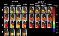 |
Iron-based T1 MRI contrast agent for MR-guided drug delivery
from temperature sensitive liposomes
Esther Kneepkens1, Adriana Fernandes2,
Klaas Nicolay3, and Holger Grüll3,4
1Biomedical NMR, Biomedical Engineering,
Eindhoven University of Technology, Eindhoven, Netherlands, 2Universidade
de Lisboa, Lisbon, Portugal, 3Eindhoven
University of Technology, Eindhoven, Netherlands,4Philips
Research, Eindhoven, Netherlands
The aim of this study was to investigate the potential of
Fe(III) N-succinyl deferoxamine (Fe-SDFO) as a safe T1 contrast
agent for encapsulation in temperature sensitive liposomes
(TSLs) in order to visualize drug release from TSLs when
using Magnetic Resonance-guided High Intensity Focused
Ultrasound (MR-HIFU). Two TSLs were developed that
contained either Fe-SDFO or doxorubicin. Both TSLs showed
suitable release and stability characteristics in vitro. An
in vivo proof-of-concept study was carried out in
tumor-bearing rats treated with MR-HIFU. Treated tumors
showed an increase in R1 and
future work aims to correlate the R1change with
tumor drug concentrations.
|
|
2118.
 |
An ultrasound compatible rat RF array for MRI guided high
intensity focused ultrasound
Xiao Chen1, Rou Li1, Changjun Tie1,
Xiaoqing Hu1, Xiaoliang Zhang2,3, Chao
Zou1, Xin Liu1, Hairong Zheng1,
and Ye Li1
1Paul C. Lauterbur Research Center for Biomedical
Imaging, Shenzhen Institutes of Advanced Technology, CAS,
Shenzhen, China, People's Republic of, 2Department
of Radiology and Biomedical Imaging, University of
California San Francisco, San Francisco, CA, United States, 3UCSF/UC
Berkeley Joint Graduate Group in Bioengineering, San
Francisco, CA, United States
Due to MRI’s unique capability of providing accurate,
non-invasive and real-time target localization and
temperature monitoring, MRI guided high intensity focused
ultrasound (HIFU) has been a critical modality for imaged
guided thermal therapy. We propose a 3 channel ultrasound
compatible rat array to obtain high resolution and
homogeneous rat brain images at 3T for temperature
monitoring. Phantom and in-vivo imaging
experiments in temperature mapping demonstrate the
capability of the proposed array to provide homogenous and
high SNR images and temperature map in the whole rat brain
at 3T, which provides the possibility to perform MRI guided
HIFU treatment in-vivo.
|
|
2119.
 |
MR-guided high intensity focused ultrasound mediated
hyperthermia for targeted drug delivery to treat pancreatic
cancer
Navid Farr1, Yak-Nam Wang2, Samantha
D’Andrea3, Frank Starr2, Ari Partanen4,
Kayla Gravelle3, Donghoon Lee5, and
Joo Ha Hwang1,3
1Department of Bioengineering, University of
Washington, Seattle, WA, United States, 2Applied
Physics Laboratory, University of Washington, Seattle, WA,
United States, 3Department
of Medicine, University of Washington, Seattle, WA, United
States, 4Philips
Healthcare, Andover, MA, United States, 5Department
of Radiology, University of Washington, Seattle, WA, United
States
Pancreatic cancer has one of the lowest survival rates
because current therapies are ineffective. Dense stromal
tissue and poor vascular perfusion limits drug penetration
and uptake into the tumor. Growing evidence suggests that
hyperthermia in combination with temperature sensitive
liposomal drug delivery can lead to increased organ
perfusion and drug extravasation resulting in high local
drug concentration. We performed MR-guided heating methods
that enable accurate and precise spatial and temporal
control of heating. Enhanced drug delivery was achieved to
treat pancreatic tumors using Magnetic Resonance-guided High
Intensity Focused Ultrasound (MR-HIFU) in conjunction with a
heat triggered drug delivery system.
|
|
2120.
 |
An improved tracking technique for real-time MR-guided beam
therapies in moving organs
Cornel Zachiu1, Nicolas Papadakis2,
Mario Ries1, Chrit Moonen1, and
Baudouin Denis de Senneville1,2
1Imaging Division, University Medical Center
Utrecht, Utrecht, Netherlands, 2Institut
de Mathématiques de Bordeaux, Bordeaux, France
Current methods for real-time MR-guided HIFU and EBRT
interventions in moving organs rely on an algorithm that is
sensitive to gray-level intensity variations from other
sources than motion. In this work, an improved real-time
tracking algorithm with increased robustness to such effects
is proposed and experimentally compared to the existing
methods. Results have shown a notable improvement in the
quality of the motion estimates when the proposed method was
used, while maintaining real-time capabilities. Our method
was shown to be potentially beneficial for MR-guided HIFU
and EBRT interventions in the abdomen, where cardiac
activity might become problematic for current approaches.
|
|
2121.
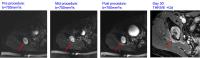 |
Monitoring tissue damage during MRgHIFU of bone metastases:
relating intra-procedural DWI changes to post-procedural
appearances
Sharon L Giles1, Matthew Brown2,
Jessica M Winfield1, David J Collins3,
Ian Rivens4, John Civale4, Gail R ter
Haar4, and Nandita M deSouza1
1CRUK Cancer Imaging Centre, The Royal Marsden
Hospital NHS Foundation Trust and The Institute of Cancer
Research, London, United Kingdom, 2Anaesthetic
Department, The Royal Marsden Hospital NHS Foundation Trust,
London, United Kingdom, 3CRUK
Cancer Imaging Centre, The Institute of Cancer Research,
London, United Kingdom, 4Therapeutic
Ultrasound, The Institute of Cancer Research, London, United
Kingdom
This study assessed intraprocedural DWI for detecting extra-
and intra-osseous tissue change during MRgHIFU treatment of
bone metastases by comparing appearances with
post-procedural and Day-30 DWI and T1-W contrast-enhanced
image appearances. Change in image appearances for n=9
patients was assessed by 2 observers assigning a consensus
score where 0=no, 1=mild, 2=moderate and 3=striking change.
Extra-osseous DWI changes were more conspicuous than
intra-osseous DWI changes, but were less striking than
immediate post-procedural contrast-enhanced changes.
However, intra-procedural DWI changes significantly
correlated with post-procedural and Day-30 DWI and
contrast-enhanced changes, suggesting that intra-procedural
DWI can provide an indicator of subsequent extra-osseous
tissue damage.
|
|
2122.
 |
Response of MR Contrast Parameters in Tissues and Tissue
Mimicking Phantoms to Histotripsy
Steven P Allen1, Luis Hernandez-Garcia2,
Charles A Cain1, and Timothy L Hall1
1Biomedical Engineering, University of Michigan,
Ann Arbor, MI, United States, 2fMRI
Lab, University of Michigan, Ann Arbor, MI, United States
We estimate the R2 relaxation rate and the apparent
diffusion coefficient at 7T in a variety of in vitro tissues
and tissue mimicking phantoms after they have been subjected
to homogenization by ultrasonic cavitation (histotripsy).
The estimated R2 rate of these lesions decreases with
increased treatment so long as the lesions are made in
materials with high iron content. When lesions are made in
brain tissue or phantoms with low iron content, the R2 rate
remains unperturbed by homogenization. The apparent
diffusion coefficient increases with increasing treatment
for all tissues and phantoms.
|
|
2123.
 |
T2-Mapping as a Predictor for Non-Perfused Volume in MRgFUS
Treatments of Desmoid Tumors
Eugene Ozhinsky1, Matthew D. Bucknor1,
and Viola Rieke1
1Radiology and Biomedical Imaging, University of
California San Francisco, San Francisco, CA, United States
Desmoid tumors are benign but locally aggressive soft tissue
tumors that arise from fibroblast cells. Focused ultrasound
has shown promising results in reduction of tumor volume
without significant side effects. Post-treatment contrast
enhanced MR imaging allows assessment of the non-perfused
volume (NPV), the gold standard assessment of the quantity
of tumor ablation. However, safety concerns regarding
heating of tissue after gadolinium injection prevent further
treatment following the NPV assessment. We have shown that
T2 mapping can be used to visualize the extent of ablation
with focused ultrasound and be used as a predictor of NPV
without the need for contrast injections.
|
|
2124.
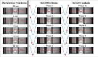 |
Validation of a 4D-MRI motion framework using an MRI-compatible
motion phantom
Bjorn Stemkens1, Rob HN Tijssen1, Jan
JW Lagendijk1, and Cornelis AT van den Berg1
1Department of Radiotherapy, University Medical
Center Utrecht, Utrecht, Netherlands
Geometric accuracy is vital for MR-guided radiotherapy. In
this study we quantify the geometric fidelity of a
retrospectively sorted 4D-MRI and 2D MS cine-MR acquisition,
which serve as input for a motion model for dose
accumulation mapping and tumor tracking. A linearly moving
MRI-compatible motion phantom was used to quantify the
positional error in the 4D-MRI and 2D MS acquisitions using
a range of user-defined motion trajectories. Geometrical
errors were found to be smaller than the voxel or pixel
size.
|
|
2125.
 |
Robust and flexible real-time MRI-guided interventions using
coRASOR-mediated passive device tracking
Peter Roland Seevinck1, Frank Zijlstra1,
Jouke Smink2, Sascha Krueger3, Frebus
Jan van Slochteren4,5, Steven A.J. Chamuleau4,
Max A Viergever1, and Marinus Adriaan Moerland1
1Center for Image Sciences, University Medical
Center Utrecht, Utrecht, Netherlands, 2Philips
Healthcare, Best, Netherlands, 3Innovative
Technologies, Philips Research Laboratories, Hamburg,
Germany, 4Department
of Cardiology, University Medical Center Utrecht, utrecht,
Netherlands, 5ICIN,
Utrecht, Netherlands
The Co-RASOR imaging technique for high temporal resolution
passive device visualization was implemented in the
interventional Suite software package. This facilitates
MRI-guided device tracking by combining high temporal
resolution color overlays on top of high spatial resolution
3D roadmaps. Titanium needles were accurately depicted in
two orthogonal planes with 2.5Hz framerate, facilitating
easy freehand needle targeting. The ability to adapt
crucial Co-RASOR reconstruction parameters, including the
off-resonance value, during the intervention was
demonstrated to provide unprecedented flexibility and
robustness in device visualization.
|
|