|
MSK
|
2241.
 |
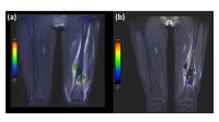
Multi-parametric assessment of thigh muscles in patients with
limb girdle muscular dystrophies (LGMD): preliminary results.
Alberto De Luca1,2, Maria Grazia D'Angelo3,
Denis Peruzzo2, Fabio Triulzi4,
Alessandra Bertoldo1, and Filippo Arrigoni2
1Department of Information Engineering,
University of Padova, Padova, Italy, 2Neuroimaging
Lab, Scientific Institute IRCCS Eugenio Medea, Bosisio
Parini (LC), Italy, 3Functional
Rehabilitation Unit, Neuromuscular Disorders, Scientific
Institute IRCCS Eugenio Medea, Bosisio Parini (LC), Italy, 4Department
of Neuroradiology, Scientific Institute IRCCS Ca Granda
Foundation - Ospedale Maggiore Policlinico, Milan, Italy
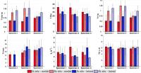
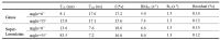
Limb girdle muscular dystrophies (LGMD) are a heterogeneous
family of disorders characterized by the substitution of
muscles with fat and fibrotic tissue. In this work we show
the initial results of our acquisition protocol, that
included DW-MRI, T2 mapping and DIXON imaging, on
two subtypes of LGMD (type 2A and 2B). Statistical tests and
Pearson’s correlation were performed on parametric maps at
single muscle level. Preliminary results show that
multi-parametric MRI is promising in the characterization of
LGMD subtypes on the thigh. Considered MRI techniques show
different sensibilities to damages induced by muscular
dystrophies and can be considered complimentary.
|
|
2242.
 |
Multiparametric voxel-based analysis of standardized uptake
values and apparent diffusion coefficients in soft-tissue tumors
with a positron emission tomography-magnetic resonance system:
Application for evaluation of treatment effect
Koji Sagiyama1, Yuji Watanabe2,
Ryotaro Kamei1, Sungtak Hong3, Satoshi
Kawanami2, Yoshihiro Matsumoto4, and
Hiroshi Honda1
1Department of Clinical Radiology, Graduate
School of Medical Sciences, Kyushu University, Fukuoka,
Japan, 2Department
of Molecular Imaging and Diagnosis, Graduate School of
Medical Sciences, Kyushu University, Fukuoka, Japan, 3Healthcare,
Philips Electronics Japan, Fukuoka, Japan, 4Department
of Orthopaedic Surgery, Graduate School of Medical Sciences,
Kyushu University, Fukuoka, Japan
A combination of single measurements would be necessary to
improve the efficacy of evaluating the treatment effect in
heterogeneous soft-tissue tumors. This study aimed to
investigate the feasibility of direct voxel-by-voxel
comparison of SUVs and ADCs with the PET/MR system in the
evaluation of the treatment effect in soft-tissue tumors.
The ADCs and SUVs were recorded on a voxel-by-voxel basis
for all slices. The scatter plots clearly demonstrated
significant difference between pre- and post-treatment.
Multiparametric voxel-based analysis of SUVs and ADCs could
be a promising tool for evaluating the treatment effect in
soft-tissue tumors.
|
|
2243.
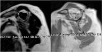 |
Predicting re-tear after repair of full-thickness rotator cuff
tear: 2-Point Dixon MR quantification of fatty muscle
degeneration – Initial experience with 1-year follow-up
Taiki Nozaki1, Atsushi Tasaki2, Saya
Horiuchi1, Junko Ochi1, Jay Starkey1,
Takeshi Hara3, Yukihisa Saida1,
Yasuyuki Kurihara1, and Hiroshi Yoshioka4
1Radiology, St.Luke's International Hospital,
Tokyo, Japan, 2Orthopaedic
Surgery, St.Luke's International Hospital, Tokyo, Japan, 3Intelligent
Image Information, Gifu University, Gifu, Japan, 4Radiological
Sciences, University of California, Irvine, Orange, CA,
United States
Rotator cuff tear is a common cause of shoulder pain and
disability. Minimally-invasive arthroscopic rotator cuff
repair is increasingly popular for treatment of
full-thickness rotator cuff tear. However, operative
outcomes are far from perfect. Postoperative re-tears are
associated with greater fatty degeneration. The purpose of
this study was to quantify the pre- and post-operative
muscular fatty degeneration using a 2-Point Dixon sequence
in patients with rotator cuff tears treated by arthroscopic
rotator cuff repair. Further, we aim to assess the
relationship of preoperative fat fraction values within
rotator cuff muscles between patients who experience re-tear
and those who do not.
|
|
2244.
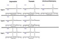 |
In vivo 1H MRS using 3 Tesla to investigate the metabolic
profiles of joint fluids in different types of knee diseases 
Geon-Ho Jahng1, Wook Jin1, Dong-Cheol
Woo2, Chanhee Lee1, Chang-Woo Ryu1,
and Dal-Mo Yang1
1Department of Radiology, Kyung Hee University
Hospital at Gangdong, Kyung Hee University, Seoul, Korea,
Republic of, 2Biomedical
Research Center, Asan Institute for Life Sciences, Asan
Medical Center, Seoul, Korea, Republic of
To assess the ability of proton MR spectroscopy to identify
the apparent heterogeneous characteristics of metabolic
spectra in effusion regions in human knees using a
high-field MRI system, 84 patients with effusion lesions
underwent proton MRS with PRESS single-voxel MRS using a
clinical 3.0 Tesla MRI system. Nonparametric statistical
comparisons were performed to investigate any differences in
metabolites among the degenerative osteoarthritis, traumatic
diseases, infectious and an inflammatory disease groups.
There were no significant differences among the three groups
for the CH3 (p=0.9019), CH2 (p=0.6406), and CH=CH lipids
(p=0.5467) and water (p=0.2853).
|
|
2245.
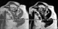 |
Reliability of MR quantification of rotator cuff muscle fatty
degeneration using a 2-point Dixon technique in comparison with
the qualitative modified-Goutallier classification
Saya Horiuchi1, Taiki Nozaki1, Atsushi
Tasaki2, Akira Yamakawa2, Yasuhito
Kaneko3, Takeshi Hara4, Yasuyuki
Kurihara1, and Hiroshi Yoshioka3
1Radiology, St Luke's International Hospital,
Tokyo, Japan, 2Orthopedics,
St Luke's International Hospital, Tokyo, Japan, 3Radiological
Sciences, University of California, Irvine, Orange, CA,
United States, 4Department
of Intelligent Image Information, Gifu University, Gifu,
Japan
The assessment of presurgical rotator cuff muscle fatty
degeneration is a main determinant of management in patients
with rotator cuff tears. The modified-Goutallier
classification has been widely accepted as a qualitative
method for evaluation of fatty degeneration in current
practice. However, reproducibility is insufficient because
it is shown to be highly observer-dependent. The objective
of this study was to quantify fatty degeneration of the
supraspinatus muscle by using 2-point Dixon technique, and
to evaluate the inter- and intra-observer reliability of
quantitative analysis of fatty degeneration in comparison
with the qualitative modified-Goutallier classification.
|
|
2298.
 |
Bone marrow perfusion study on different BMD groups in elderly
female
Chaoyang Zhang1, Hu Xianghui2, Heather
T. Ma2, Li Liang2, and Chenfei Ye2
1Harbin Institute of Technology Shenzhen Graduate
School, Shenzhen, China, People's Republic of, 2Shenzhen,
China, People's Republic of
This study utilized dynamic contrast enhanced (DCE) MRI and
blood oxygen level dependent (BOLD) MRI as imaging method,
using half quantitative analysis of two kinds of methods to
study the relationship between the marrow blood perfusion,
oxygen metabolism and bone mineral density. The research
found that significant differences were observed in A, MaxEn
and Halflife parameters among different BMD groups
(p<0.05).In conclusion, different BMD groups has significant
difference in perfusion ability on marrow. There is a link
between the bone mineral density and marrow blood
circulation and metabolism of oxygen. The changed blood
circulation may be one of the reasons induced osteoporosis.
|
|
2299.
 |
Qualitative and Quantitative Diagnosis of Meniscal Tears Using
SWI Compared with T2mapping at 3-Tesla MRI
Jun Zhao1, Wei Chen1, Jian Wang1,
Shuai Li2, and Wen-Jing Hou1
1Radiology, Southwest Hospital, Third Military
Medical University, Chongqing, China, People's Republic of, 2MR
Collaborations NE Asia, Siemens Healthcare, Beijing, China,
People's Republic of
In the past reports, invariably irregularity and
high-signal-intensity changes of the free edge of meniscus
may lead to a false-positive MR imaging, in addition, MR
imaging of the knee invariably missed small meniscal tears,
tears and abnormalities of the meniscal free edge, and at
times large, unstable tears, result in false-negative. In
recent decades, new MR image of water-tissues and
collagen-rich tissues, including cartilage, menisci and
tendon, has undergone significant progress, which are
biological MR image techniques for the characterization
tissues .This study was to compare the diagnostic
performance of SWI (Susceptibility Weighted Imaging) in the
evaluation of meniscal tears at 3T MR with those of a T2
Mapping sequence, using phase value and T2 value as
the quantitative parameters. The phase value was a
good predictor to diagnose meniscal tears.
|
|
2246.
 |
T2 and T1rho values of grade 1 early degenerative cartilage in
the distal femur using angle/layer dependent approach
Yasuhito Kaneko1,2, Taiki Nozaki1,3,
Hon Yu1,4, Kayleigh Kaneshiro1, Ran
Schwarzkopf5, and Hiroshi Yoshioka1
1Radiological Sciences, University of California,
Irvine, Orange, CA, United States, 2Orthopaedic
Surgery, Saitama City Hospital, Saitama, Japan, 3Radiology,
St. Luke's International Hospital, Tokyo, Japan, 4John
Tu and Thomas Yuen Center for Functional Onco-Imaging,
University of California, Irvine, Orange, CA, United States, 5Orthopaedic
Surgery, University of California, Irvine, Orange, CA,
United States
We assessed patterns of T2 and T1rho value change with
Outerbridge grade 1 lesions in OA patients compared to
healthy control cartilage utilizing angle and layer
dependent approach. T1rho values were more sensitive than T2
values to detect early cartilage degeneration with higher
values in OA cartilage than in healthy control. However, T2
and T1rho values in grade 1 cartilage degeneration with
signal heterogeneity can be lower compared to those in
healthy cartilage.
|
|
2247.
 |
Morphological, Compositional, and Fiber Architectural Changes in
from Unilateral Limb Suspension Induced Acute Atrophy Model in
the Medial Gastrocnemius Muscle.
Shantanu Sinha1, Vadim Malis2, Robert
Csapo1, Jiang Du1, and Usha Sinha3
1Radiology, University of California at San
Diego, San Diego, CA, United States, 2Physics,
University of California at San Diego, San Diego, CA, United
States, 3Physics,
San Diego State University, San Diego, CA, United States
Acute muscle atrophy is characterized by a loss of muscle
mass and muscle force. Changes are likely to occur in
muscle composition, microenvironment, and fiber architecture
which could impact muscle function. This study focuses on
the changes in these parameters using MR based fat and
connective tissue quantification and DTI in a model of acute
atrophy induced by Unilateral limb suspension (ULLS). The %
changes in fat and connective tissue were minimal while
significant decreases were found in fiber diameter
(decrease) and in the pennation angle. These changes could
be primarily responsible for muscle force loss in acute
atrophy.
|
|
2248.
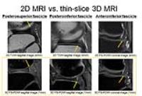 |
Usefulness of thin-slice 3D MR imaging using 3D FSE sequence
with variable flip-angle refocusing RF pulses for assessing the
popliteomeniscal fascicles of the lateral meniscus in knee MR
imaging at 3T
Masayuki Odashima1, Tsutomu Inaoka1,
Hideyasu Kudo1, Tomoya Nakatsuka1,
Rumiko Ishikawa1, Shusuke Kasuya1,
Noriko Kitamura1, Hiroyuki Nakazawa1,
Koichi Nakagawa2, and Hitoshi Terada1
1Radiology, Toho University Sakura Medical
Center, Sakura, Japan, 2Orthopedic
Surgery, Toho University Sakura Medical Center, Sakura,
Japan
Thin-slice 3D MR imaging of the knee joint using 3D FSE
sequence with variable flip-angle refocusing RF pulses may
improve the visualization of the three popliteomeniscal
fascicles of the lateral meniscus in comparison with
conventional 2D MR imaging of the knee joint.
|
|
2249.
 |
Quantitative knee cartilage T2 mapping with in situ mechanical
loading using prospective motion correction
Thomas Lange1, Benjamin R. Knowles1,
Michael Herbst1,2, Kaywan Izadpanah3,
and Maxim Zaitsev1
1Department of Radiology, University Medical
Center Freiburg, Freiburg, Germany, 2John
A. Burns School of Medicine, University of Hawaii, Honolulu,
HI, United States, 3Department
of Orthopedic and Trauma Surgery, University Medical Center
Freiburg, Freiburg, Germany
Robust T2 mapping of knee cartilage with in situ mechanical
loading using prospective motion correction is demonstrated
for the patellofemoral and tibiofemoral knee compartments.
T2 maps are reconstructed from multiple spin-echo data
acquired with slice position updates before every
excitation. While T2 maps of the tibiofemoral joint do not
show significant changes in response to loading, maps of the
patellofemoral joint show a substantial load-induced T2
reduction in the superficial cartilage layers. In
particular, the T2 of tangential fibers at the cartilage
surface appears to undergo a strong reduction due to a
load-induced increase of tissue anisotropy.
|
 |
2250.
 |
CSF-Free Imaging of the Lumbar Plexus using Sub-Millimeter
Resolutions with 3D TSE
Barbara Cervantes1, Houchun Harry Hu2,
Amber Pokorney2, Dominik Weidlich1,
Hendrik Kooijman3, Ernst Rummeny1,
Axel Haase4, Jan S Kirschke5, and
Dimitrios C Karampinos1
1Diagnostic and Interventional Radiology,
Technische Universität München, Munich, Germany, 2Radiology,
Phoenix Children’s Hospital, Phoenix, AZ, United States, 3Philips
Healthcare, Hamburg, Germany, 4Zentralinstitut
für Medizintechnik, Garching, Germany, 5Neuroradiology,
Technische Universität München, Munich, Germany
High-resolution MRI with 3D turbo spin echo (TSE) is arising
as an accurate, non-invasive method for detecting disease
and injury in the nerves of the lumbar plexus. Imaging of
the lumbar plexus with 3D TSE frequently faces signal
contamination of the cerebrospinal fluid (CSF) within the
spine. Increasing spatial resolution in 3D TSE can affect
flowing signal. The present study describes numerically the
effects of the imaging gradients in 3D TSE on flowing CSF
and demonstrates in
vivo that
CSF can be completely suppressed without modifications to
refocusing angle modulation when sub-millimeter voxel sizes
are used with 3D TSE.
|
|
2251.
 |
An assessment of the repeatability and sensitivity of T2 mapping
in low-grade cartilage lesions at 3 and 7 Tesla
Vladimir Juras1,2, Laurent Didier3,
Vladimir Mlynarik1, Pavol Szomolanyi1,
Stefan Zbyn1, Nicole Getzmann3, Joerg
Goldhahn3, Stefan Marlovits4, and
Siegfried Trattnig1,5
1Department of Biomedical Imaging and
Image-Guided Therapy, High Field MR Centre, Medical
University of Vienna, Vienna, Austria, 2Department
of Imaging Methods, Institute for Measurement Science,
Bratislava, Slovakia, 3Novartis
Institutes for Biomedical Research, Basel, Switzerland, 4Department
of Traumatology, Medical University of Vienna, Vienna,
Austria, 5Christian
Doppler Laboratory for Clinical Molecular MR Imaging,
Vienna, Austria
An assessment of the reliability of T2 mapping was achieved
with a 3D-TESS sequence in patients with cartilage lesions
ICRS Grade I-II. Since low-grade cartilage lesions are not
usually accompanied by collagen matrix remodeling, we tested
the sensitivity of T2 to detect these lesions at 3 and 7T.
It seems that the reproducibility of 3T T2 mapping is higher
than that of 7T; however, the sensitivity of T2 mapping for
the detection of low-grade cartilage lesions was greater at
the ultra-high field. T2 mapping could be used in the future
as a good alternative to cartilage biopsies in future
clinical trials on new therapies aimed at cartilage
regeneration.
|
|
2252.
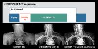 |
Non-Contrast, Flow-Independent, Relaxation-Enhanced Subclavian
MR Angiography Using Inversion Recovery and T2 Prepared 3D
Gradient-Echo DIXON Sequence
Masami Yoneyama1, Nobuyuki Toyonari2,
Seiichiro Noda2, Yukari Horino2,
Kazuhiro Katahira2, and Marc Van Cauteren3
1Philips Electronics Japan, Tokyo, Japan, 2Kumamoto
Chuo Hospital, Kumamoto, Japan, 3Philips
Healthcare Asia Pacific, Tokyo, Japan
This study showed a novel non-contrast MR angiography
sequence based on gradient echo DIXON sequence with
flow-independent relaxation-enhanced method
(Relaxation-Enhanced Angiography without Contrast and
Triggering: REACT) for evaluating thoracic outlet syndrome.
This could provide high-quality MRA with robust fat
suppression entire the subclavian area with/without arm
abduction.
|
|
2253.
 |
Toward a 7T MRI protocol for the evaluation of early
osteoarthritis in knee cartilage
Daniel J. Park1, Neal K. Bangerter2,3,
Antony J. R. Palmer1, Haonan Wang2,
Bragi Sveinsson4, Brian Hargreaves4,
and Siôn Glyn-Jones1
1Nuffield Department of Orthopaedics,
Rheumotology, and Musculoskeletal Sciences, University of
Oxford, Oxford, United Kingdom, 2Department
of Electrical and Computer Engineering, Brigham Young
University, Provo, UT, United States, 3Department
of Radiology, Univerisity of Utah, Salt Lake City, UT,
United States, 4Radiology,
Stanford University, Stanford, CA, United States
Osteoarthritis, a disease that is a burden to society and
individuals, has 3 major stages of progression in cartilage:
(1) glycosaminoglycan loss; (2) collagen matrix
degeneration; and (3) fissures and volume and thickness
loss. A protocol is proposed to measure the progression of
each of these stages of OA at 7 Tesla in about 30 minutes:
(1) T1ρ to measure glycosaminoglycan changes; (2) modified
DESS measurements of T2 and ADC to measure collagen matrix
integrity; and (3) high resolution phase cycled bSSFP images
to measure changes in morphology.
|
|
2300.
 |
Magnetization transfer MRI Evaluation of Autologous Chondrocyte
Membrane Transplantation in The Knee Joint
Yi-Bin Xi1, Fan Guo1, Chun-Li Zhang1,
Hu Xu1, Long-Biao Cui1, Chen Li1,
Ping Tian1, Wei-Guo LI2, and Hong Yin1
1Xijing Hospital, Fourth Mililtary Medical
University, Xi'an, China, People's Republic of, 2Bioengineering,
University of Illinois at Chicago, Chicago, IL, United
States
Magnetization transfer MRI Evaluation of Autologous
Chondrocyte Membrane Transplantation in The Knee Joint
|
|
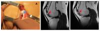
2254.
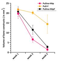 |
3D Longitudinal MRI studies on novel tissue-engineered bone
constructs in living rats : Volume & Perfusion assessments
Neha KOONJOO1,2, Clément Tournier3,
Aurélien Trotier1,2, Didier Wecker4,
William Lefrançois1,2, Didier Letourneur5,
Joëlle Amédée Vilamitjana3, Sylvain Miraux1,2,
and Emeline J Ribot1,2
1CNRS-UMR 5536, Centre de Résonance Magnetique
des Systèmes Biologiques, Bordeaux, France, Metropolitan, 2University
of Bordeaux, Bordeaux, France, Metropolitan, 3U1026,
Bioingénierie Tissulaire (BioTis), Bordeaux, France,
Metropolitan, 4Bruker
Biospin MRI GMBH, Ettlingen, Germany, 5INSERM
U 1148, Cardiovascular Bio-engineering Laboratory, Paris,
France, Metropolitan
In tissue engineering, correct bone regeneration in large
bone defects is a major issue. MRI has revealed its high
potential to assess continuous tracking of three differently
conditioned bone constructs implanted in the rats’ femoral
condyles. These constructs aimed at evaluating cumulative
effects of hydroxyapatite and/or fucoidan in osteogenesis
and vascularization. A water-selective bSSFP sequence with
fat suppression and banding artifacts correction was
implemented for volumetric measurements. 3D Dynamic-contrast
enhanced MRI was applied and pixel-wise analysis resulted in
fairly good constructs perfusion evaluation. 3D images
spotted distinct volume changes and promising area under
curve evolution.
|
|
2255.
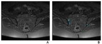 |
Quantifying bone marrow inflammatory edema in psoriatic
arthritis using pixel-based morphometry
Ioanna Chronaiou1,2, Ruth Stoklund Thomsen 3,
Else-Marie Huuse-Røneid 2,3,
and Beathe Sitter1
1Department of Radiography, Sør-Trøndelag
University College, Trondheim, Norway, 2Department
of Circulation and Medical Imaging, Norwegian University of
Science and Technology, Trondheim, Norway, 3St
Olav's University Hospital, Trondheim, Norway
Psoriatic arthritis (PsA) is a highly heterogeneous
inflammatory disease that manifests with inflammation in
sacroiliac (SI) joints and spine among other symptoms. PsA
patients (N=12) underwent magnetic resonance (MR) imaging
examinations to assess the extent of SI joint inflammation.
A pixel-based morphometric method for accurate
quantification of bone marrow inflammatory edema was
compared to SPARCC assessment in MR images of
psoriatic arthritis patients with low or very low
inflammatory activity. A significant correlation was found,
suggesting pixel-based morphometry as a reliable and
sensitive quantitative method for measuring inflammation in
bone marrow.
|
|
2256.
 |
Statistical Comparison of Commonly Used Kinetic Models for
Dynamic Contrast Enhanced Magnetic Resonance Imaging of
Rheumatoid Arthritis in the Wrist
Sameer Khanna1,2, Nicolas Pannetier1,
Jing Liu1, and Xiaojuan Li1
1Radiology, University of California, San
Francisco, San Francisco, CA, United States, 2University
of California, Berkeley, Berkeley, CA, United States
There has been a lack of statistical analysis to determine
which kinetic model is best suited for analysis of the
wrist. This study aims to rectify this by comparing the most
commonly used models: Modified Tofts (MT), Two Compartment
Uptake (2CU), and Two Compartment Exchange (2CX). Goodness
of fit is analyzed by reduced chi squared, while statistical
signifance between models is determined by wilcoxon
signed-rank.
|
|
2257.
 |
Reduced Field of View Multi-Spectral Imaging through Coupled
Coil and Frequency Bin Encoding
Andrew S. Nencka1, Shiv S. Kaushik1,
and Kevin M. Koch1
1Radiology, Medical College of Wisconsin,
Milwaukee, WI, United States
Advanced methods for imaging around metallic implants yield
most benefit in the neighborhood around the implant.
However, due to the extent of the anatomy in the region of
the implant, large field of view acquisitions are often
required. In this work, it is shown that a low-resolution
acquisition can be used to inform a subsequent reduced field
of view acquisition. Significant reductions in the imaged
field of view are possible due to the combination of both
spatially varying coil sensitivity profiles along with
spatially varying resonance frequency bins. Artifact free
regions in the neighborhood of the implant are possible with
extreme field of view reductions because of the rapid
spatial variability of the imaged resonance frequency bins.
|
|
2258.
 |
Cortical bone quality as a biomarker for diabetes risk in
post-menopausal Chinese-Singaporean women: a preliminary study
Francesca A. A. Leek1, Anna Therese Sjoholm1,
Christiani Jeyakumar Henry2, Xiaodi Su3,
Marlena C. Kruger4, and John J. Totman1
1A*STAR-NUS Clinical Imaging Research Centre,
Singapore, Singapore, 2A*STAR
Clinical Nutrition Research Centre, Singapore, Singapore, 3A*STAR
Institute of Materials Research and Engineering, Singapore,
Singapore, 4School
of Food and Nutrition, College of Health, Massey University,
Palmerston North, New Zealand
The feasibility of utilising proximal femur cortical bone
quality as a biomarker for diabetes risk in post-menopausal
Chinese-Singaporean women was investigated. Non-dominant
proximal femurs were imaged with quantitative CT (QCT) and
MR for the assessment of volumetric bone mineral density (vBMD)
and cortical bone porosity. A significant (p<0.01; n=8)
positive correlation between MRI vBMD and QCT vBMD for the
region of maximum cortical thickness was shown. Whether MRI
vBMD is associated with fracture risk and if it is sensitive
to changes due to dietary or drug intervention needs to be
investigated to fully assess the clinical potential of this
method.
|
|
2259.
 |
Assessment of trabecular bone quality of the proximal femur in
vivo: A Preliminary Study
Maria Kalimeri1, Christiani Jeyakumar Henry2,
Xiao Di Su3, Marlena C. Kruger4, and
John J. Totman1
1A*STAR-NUS Clinical Imaging Research Centre,
Singapore, Singapore, 2Clinical
Nutrition Research Centre, Singapore, Singapore, 3Institute
of Materials Research and Engineering, Singapore, Singapore, 4School
of Food and Nutrition, College of Health, Massey University,
Palmerston North, New Zealand
Osteoporosis is a skeletal disorder that affects
predominantly postmenopausal women. The screening method for
osteoporosis is Dual X-ray Absorptiometry (DXA), which has
several limitations, including the inability to
differentiate between trabecular and cortical bone. 3D
imaging modalities can give information about each bone
component, which contribute to bone strength in different
ways. MRI is an attractive alternative due to lack of
ionising radiation. In this abstract, we present a method
for bone density assessment of trabecular bone in the
proximal femur using MRI. Strong correlations with both DXA
and Quantitative Computed Tomography (QCT) measurements of
similar regions were observed.
|
|
2260.
 |
T2-weighted Multispectral Imaging for Postoperative Imaging of
Patients with Lumbar Spinal Fusion
Daehyun Yoon1, Kathryn Stevens1, and
Brian Hargreaves1
1Radiology, Stanford University, Palo Alto, CA,
United States
T2-weighted MRI is essential to detect neural compression in
the lumbar spine after spinal fusion surgery in patients
with recurrent radicular symptoms. Unfortunately,
off-resonance artifacts induced from lumbar fusion devices
make the conventional T2-weighted MR images extremely
challenging or impossible to interpret. We present a
modified version of MAVRIC-SL, an MR sequence designed to
correct for metal-induced artifacts, to allow T2 contrast,
significantly improving diagnostic capabilities in the
postoperative lumbar spine.
|
|
2261.
 |
Evaluation of Chronic Inflammatory Demyelinating Polyneuropathy:
New Simultaneous T2 mapping and neurography method with 3D
Nerve-Sheath Signal Increased with Inked Rest-Tissue Rapid
Acquisition of Relaxation Enhancement Imaging (SHINKEI Quant)
Akio Hiwatashi1, Osamu Togao1, Koji
Yamashita1, Kazufumi Kikuchi1, Masami
Yoneyama2, and Hiroshi Honda1
1Clinical Radiology, Kyushu University, Fukuoka,
Japan, 2Philips
Electronics Japan, Tokyo, Japan
MR neurography (MRN) is a useful technique with which to
evaluate abnormal conditions of the peripheral nerves such
as chronic inflammatory demyelinating polyradiculoneuropathy
(CIDP). We have developed a new simultaneous T2 mapping and
MRN method called SHINKEI Quant. Patients with CIDP could be
distinguished from normal subjects in size and T2 value of
the peripheral nerves with SHINKEI Quant.
|
|
2262.
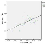 |
Vertebral Bone Marrow Fat Quantification and its Relationship
with Bone Mineral Density: Using Multi-Echo MRS and Multi-Echo
Dxion
Na Chai1, Panli Zuo2, Stephan
Kannengiesser3, Andre De Oliveira3,
Shun Qi1, and Hong Yin1
1Department of Radiology, Xijing Hospital, Xi'an,
China, People's Republic of, 2Siemens
Healthcare, MR Collaborations NE Asia, Beijing, China,
People's Republic of, 3Application
Predevelopment, Siemens Healthcare, Erlangen, Germany
Using the multi-echo 1H-MRS
and multi-echo Dixon VIBE, we measured the proton density
fat fraction (PDFF) using MR imaging in the bone marrow
of L2-L4 vertebra, and compared with the bone mineral
density (BMD) measured using computed tomography (CT). The
resutls showed a significant correlation between PDFF
measured using the two methods, and also PDFF with BMD.
|
|
2263.
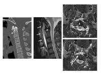 |
Calcific Longus Colli Tendinitis: Emphasis on MRI Appearance
with Variations in Anatomical Correlation
Tamami Shirakawa1, Kazutoshi Inamura2,
Yasuhisa Tanaka3, Takeshi Hoshikawa3,
Megumi Kuchiki1, and Atsuko Oda1
1Radiology, Tohoku Central Hospital, Yamagata,
Japan, 2Otolaryngology,
Tohoku Central Hospital, Yamagata, Japan, 3Orthopaedic
Surgery, Tohoku Central Hospital, Yamagata, Japan
Calcific longus colli tendinitis is an inflammatory lesion
in the prevertebral space. When prevertebral effusion is
observed on MRI, awareness of the prevertebral muscle
swelling with signal change and the associated mass effect
would suggest that the main site of the lesion is the
prevertebral space, not the retropharyngeal space and may
thus prevent both misdiagnosis as a retropharyngeal abscess
and unnecessary treatment. The variability in the level of
calcification and prevertebral effusion is highlighted in
the present study in order to assist in the establishment of
the correct radiological diagnosis.
|
 |
2264.
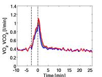 |
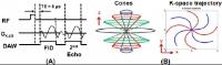
Combined Spiroergometry and 31P MRS in human calf muscle during
high intense exercise
Kevin Tschiesche1, Alexander Gussew1,
Christian Hein2, and Jürgen Rainer Reichenbach1
1Medical Physics Group, Institute of Diagnostic
and Interventional Radiology, Jena University Hospital -
Friedrich Schiller University Jena, Jena, Germany, 2Ganshorn
Medizin Electronic GmbH, Niederlauer, Germany
The aim of this work was the implementation of combined
spirometric and 31P MRS measurements. We adapted
a commercial gas exchange system by extending the gas
sampling line from 3 m to 5 m to perform acquisitions of
pulmonary ventilation in a MR scanner. Calibration
measurements showed changes in an appropriate range in the
delay- and response time.
|
|
2301.
 |
Structural and Biomechanical Properties of Hypertrophic
Articular Cartilage Using Microscopic Magnetic Resonance Imaging
David J Kahn1, Daniel Mittelstaedt1,
and Yang Xia1
1Physics and Center for Biomedical Research,
Oakland University, Rochester, MI, United States
High-resolution T2 imaging of AC is able to quantitatively
measure depth-dependent features of articular cartilage
(AC). When the cartilage articular surface (AS) is oriented
normal (0°) to the external magnetic field, healthy AC takes
on a laminar appearance that indicates the superficial zone
(SZ), transitional zone (TZ), and radial zone (RZ), where
collagen fibers are oriented parallel, random, and
perpendicular to the AS [1]. When the AS is oriented at the
magic angle (55°), the nuclear dipolar interaction is
minimized and the tissue appears homogeneous. Compression of
AC has effects that change many zonal properties [2,3], and
hypertrophy may alter the biomechanical function and
depth-dependent collagen ultra-structure of AC.
|
|
2265.
 |
Observation of in vivo lactate metabolism in skeletal muscle
using hyperpolarized 13C MRS
JAE MO PARK1, Sonal Josan1, Dirk Mayer2,
Ralph E Hurd3, Youngran Chung4, David
Bendahan5, Daniel M Spielman1, and
Thomas Jue4
1Radiology, Stanford University, Stanford, CA,
United States, 2Diagnostic
Radiology and Nuclear Medicine, University of Maryland,
Baltimore, MD, United States, 3Applied
Sciences Laboratory, GE Healthcare, Menlo Park, CA, United
States, 4Biochemistry
and Molecular Medicine, University of California - Davis,
Davis, CA, United States, 5Centre
de Resonance Magnetique Biologique et Medicale,
Aix-Marseille University, Marseille, France
The present study reports the use of hyperpolarized [1-13C]lactate
and [2-13C]pyruvate to measure the rapid pyruvate
and lactate kinetics in rat skeletal muscle. The results
provide support for a critical underpinning of both the
glycogen shunt model and the intracellular lactate shuttle
hypothesis, and cautions against an overly simplistic view
of glycolytic end products as merely hypoxia biomarkers.
|
|
2266.
 |
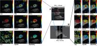
Quantitative magnetization transfer MRI of in-situ and ex-situ
meniscus
Mikaël Simard1, Emily J. McWalter2,
Garry E. Gold2, and Ives R. Levesque1,3
1Medical Physics, McGill University, Montreal,
QC, Canada, 2Radiology,
Stanford University, Stanford, CA, United States, 3Research
Institute of the McGill University Health Centre, Montreal,
QC, Canada
Quantitative magnetization transfer (QMT) probes
macromolecular content in tissue and may be a useful tool in
the early detection of meniscal degeneration. QMT mapping of
the meniscus was performed in 3 cadaver knee specimens in
situ, and repeated ex situ following dissection and
immersion in perflubron. After extraction, a decrease in the
restricted pool fraction f was noted, while T1obs and T1f
increased. A trend towards lower values of the exchange rate
kf was noted after excision. T2 and T2r were relatively
constant. The variation in QMT parameters may be caused by
the diffusion of perflubron into the ex situ samples.
|
|
2267.
 |
Ultrashort echo time magnetization transfer (UTE-MT) imaging and
modeling – magic angle independent biomarkers of tissue
properties
Yajun Ma1, Hongda Shao1, Michael Carl2,
Eric Chang1, and Jiang Du1
1Department of Radiology, UCSD, San Diego, CA,
United States, 2Global
MR Application & Workflow, General Electric, San Diego, CA,
United States
Magnetic resonance imaging biomarkers such as T2 and
T1rho have
been widely used in the evaluation of osteoarthritis (OA).
The principal confounding factor for T2 and
T1rho measures
is the magic angle effect, which may result in a several
fold increase in T2 and
T1rho values
when the fibers are oriented near 55° (the magic angle)
relative to the B0 field. This often far exceeds the changes
produced by OA, and may make definitive interpretation of
elevated T1rho and
T2 values
difficult or impossible. Magic angle independent MR
biomarkers are highly desirable for more accurate assessment
of OA. In this study we report the use of two-dimensional
ultrashort echo time magnetization transfer (UTE-MT) imaging
and modeling for magic angle independent assessment of the
tissue properties.
|
|
2268.
 |
NEW MR PARAMETERS TO ASSESS AND MONITOR TENDON XANTHOMAS
James F Griffith1, Teresa M Hu2, David
KW Yeung1, D F Wang1, Fan Xiao1,
and Brian Tomlinson2
1Imaging and Interventional Radiology, The
Chinese University of Hong Kong, Hong Kong SAR, Hong Kong, 2Medicine,
The Chinese University of Hong Kong, Hong Kong SAR, Hong
Kong
Achilles tendon xanthoma is a key clinical indicator of
familial hypercholesteraemia (FH) and associated
cardiovascular disease. Treatment that reduces the size of
tendon xanthoma also benefits the arterial manifestations of
FH. Ultrasound and MRI are often used to detect and monitor
treatment response of tendon xanthomas using parameters such
as tendon thickness, width and cross-sectional area.
However, MR-based parameters derived from the DIXON
technique to determine tendon volume and intratendinous
percentage fat fraction may be more sensitive
than traditional US and conventional MRI.
|
|
2269.
 |
Magnetic resonance imaging evaluation of acetabular morphology
and long-term prognosis in developmental dysplasia of the hip in
childhood
Mingming Lu1, Peng Peng1, Yu Zhang2,
and Fei Yuan1
1Affiliated Hospital of Logistics University of
Chinese People's Armed Police Forces, Tianjin, China,
People's Republic of, 2Philips
Healthcare, Beijing, China, People's Republic of
This study aimed to investigate the efficacy of MRI for
evaluating morphology and long-term prognosis of acetabulum
in pediatric patients with DDH. The bony acetabular index (BAI),
cartilaginous acetabular index (CAI), acetabular anteversion
index of bone (BAAV) and cartilage (CAAV) were measured and
cartilaginous index (CI=(BAI-CAI) / BAI) was computed. There
was obvious differences with statistical significance in the
CI between non-reduced group and reduced group (t=-2.315,
P=0.24), and age was also negatively correlated with the CI
(r = -0.345, P =0.01) . The CI can preliminarily predict the
long-term prognosis of DDH after reduction.
|
|
2302.
 |
quantitative UTE imaging of the Achilles tendon enthesis of PsA
patients and healthy volunteers
Bimin Chen1,2, Hongda Shao1, Michael
Carl3, Arthur Kavanaugh4, Graeme M
Bydder1, and Jiang Du1
1Radiology Department, UCSD, San Diego, CA,
United States, 2Radiology
Department, The first affiliated hospital of Jinan
University, Guangzhou, China, People's Republic of, 3GE
Healthcare, San Diego, CA, United States, 4Center
for Innovative Therapy Division of Rheumatology, Allergy,
and Immunology, UCSD, San Diego, CA, United States
Achilles tendon enthesitis is the source of the the heel
pain of PsA patients. The current measures based on pressure
being placed on various entheses during physical examination
are both insensitive and non-specific.Also it’s very time
consuming and poorly reproducible.MR imaging with ultrashort
echo time (UTE) sequences provides a good option for
assessing entheses, which has a relatively short T2 and
largely “invisible” with clinical MR sequence.
|
|
2295.
 |
Mitochondrial function as measured by 31P Magnetic Resonance
Spectroscopy between lean Chinese and Asian-Indian males
Ivan P.W. Teng1, Jamie X.M. Ho1, Trina
Kok1, Philip Lee2, Melvin K.S. Leow3,
Hong Chang Tan4, Chin Meng Khoo5,
George K Radda6, and Mary C Stephenson1,5
1Clinical Imaging Research Centre, A*STAR-NUS,
Singapore, Singapore, 2SBIC,
A*STAR, Singapore, Singapore, 3SICS,
A*STAR, Singapore, Singapore, 4Department
of Endocrinology, SGH, Singapore, Singapore, 5Department
of Medicine, NUS, Singapore, Singapore, 6Biomedical
Research Council, A*STAR, Singapore, Singapore
Previous studies have indicated differences in insulin
sensitivity between lean Indian and Chinese men. In this
study we used 31P
MRS and a dorsiflexion task to assess muscle mitochondrial
function, thought to be associated with insulin
sensitivity, via PCr recovery rates. No inter-ethnic group
differences were observed in measured blood parameters
(HbA1c, fasting blood glucose level and M-value) between
groups. However, positive correlations were observed
between τPCr and both HbA1c and fasting blood glucose
levels suggesting poorer mitochondrial function. No
correlation was observed with M-value. Larger sampling
sizes are necessary for these correlations and group
differences to reach statistically-significant conclusions.
|
|
2294.
 |
The changes in vertebra subchondral bone and cartilage endplate
perfusion of degenerated intervertebral disks :a quantitative
DCE-MRI study
Jiao WANG1, Yun fei ZHA1, Dong XING1,
Lei HU1, Chang sheng LIU1, Hui LIN2,
and Yuan LIN1
1Department of Radiology,Renmin Hospital of Wuhan
University ,Wuhan 430060,China, Wu han, China, People's
Republic of, 2GE
Healthcare China, Shanghai 200000,China, Shang hai, China,
People's Republic of
To explore the relationship between the vertebra subchondral
bone (VSB), the cartilage endplate (CEP) perfusion with
intervertebral disc degeneration (IVDD). 18 individuals
underwent lumbar conventional and DCE-MRI. The cranial and
caudal VSB and CEP perfusion parameters (Ktrans,
Kep, Ve) were measured. The VSB
perfusion parameters Kep of
Pfirrmann I and II, Pfirrmann I and IV, Pfirrmann III and
IV,the cranial CEP Kep of
Pfirrmann III and II showed significant difference. In the
early progress of IVDD, its metabolism increase
compensatory, clinical research should put more emphasis on
early onset stage of IVDD such as in Pfirrmann II.
|
|
2270.
 |
Use of Adding T2 Mapping Sequence to a Routine MR Imaging
Protocol to Evaluate of the Articular Cartilage Changes of the
Knee and Ankle Joint with Hemophilia in Children
Ningning Zhang1, Yanqiu Lv1, Kaining
Shi2, Di Hu1, Huiying Kang1,
Yue Liu1, Runhui Wu3, and Yun Peng1
1Imaging Center, Beijing Children's Hospital,
Capital Medical University, Beijing, China, People's
Republic of, 2Imaging
System Clinical Science, Philips Healthcare, Beijing, China,
People's Republic of, 3Hematology
Center, Beijing Children's Hospital, Capital Medical
University, Beijing, China, People's Republic of
T2 mapping sequences can help detect changes in the water
and collagen content. This sequence have been used
extensively in osteoarthritis research studies to detect
disease and treatment related changes in articular
cartilage(1-3). However, little is known about the early
cartilage changes in hemophilia patients, and once
established, arthropathy follows a progressive and
non-reversible process despite the use of factor
concentrates.
|
|
2271.
|
DTI can monitor changes in articular cartilage after a
mechanically induced injury
Uran Ferizi1, Ignacio Rossi2, Oran
Kennedy2, Thorsten Kirsch2, Jenny
Bencardino1, and Jose Raya1
1Department of Radiology, New York University
School of Medicine, New York, NY, United States, 2Orthopaedic
Surgery, New York University School of Medicine, New York,
NY, United States
The development of novel treatment strategies that would
prevent joint replacement surgery at young age, as a result
of PTOA, is critical. Hours after non-contact rupture of the
anterior cruciate ligament, high concentrations of PG and
type II collagen fragments are found in the synovial fluid.
DTI has emerged as an imaging biomarker that can assess both
PG content and collagen architecture with greater accuracy
than T2 or Na imaging. The current interpretation of DTI
measurements is that changes in the level of proteoglycans
(PG) affect the mean diffusivity (MD) index from the DTI,
while the collagen structure affects the fractional
anisotropy (FA).
This study examines the feasibility of DTI, by using
biomechanics for simulating a controlled cartilage damage.
We find that DTI metrics are sensitive to the early changes
in the cartilage as a result of injury. Specifically, the
correlations of the mean diffusivity (MD) are statistically
significant, but those of fractional anisotropy (FA) are
not. The additional validation with histology, as well as a
clinical scanning environment make these results important
in the translation of DTI to clinical practice.
|
|
2272.
 |
A new method for accurate detection of cartilage lesions in
femoroacetabular impingement using quantitative T2 mapping:
preliminary validation against arthroscopic findings at 3 T
Noam Ben-Eliezer1,2, Akio Ernesto Yoshimoto2,
KAI Tobias Block1,2, Roy Davidovitch3,
Thomas Youm3, Robert Meislin3, Michael
Recht1,2, Daniel K Sodickson1,2, and
Riccardo Lattanzi1,2
1Center for Advanced Imaging Innovation and
Research (CAI2R), New York University School of Medicine,
New York, NY, United States, 2Bernard
and Irene Schwartz Center for Biomedical Imaging, Department
of Radiology, New York University School of Medicine, New
York, NY, United States, 3Department
of Orthopedic Surgery, New York University Hospital for
Joint Diseases, New York, NY, United States
Early diagnosis of cartilage defects is critical for the
success of corrective surgical procedures in patients with
femoroacetabular impingement (FAI). T2 is
a biomarker for early biochemical degeneration of cartilage,
but in vivo T2 mapping
is challenging while commonly used techniques based on
exponential fit of multi spin-echo protocols are inaccurate.
We used a Bloch simulation based T2 mapping
technique – the EMC algorithm
– to retrospectively quantify reliable T2 values
in the hip cartilage of FAI patients. We then defined a
normalized T2-index using an internal reference
and showed that it allows detection of surgically confirmed
cartilage lesions with 95% accuracy.
|
|
2273.
 |
Elevated adiabatic $$$T_{1\rho}$$$ and $$$T_{2\rho}$$$ in
articular cartilage are associated with symptoms and structural
changes in early osteoarthritis
Victor Casula1,2, Mikko J. Nissi3,4,
Jana Podlipská1,5, Marianne Haapea6,7,
Simo Saarakkala1,2,7, Ali Guermazi8,
Eveliina Lammentausta2,7, and Miika T. Nieminen1,2,7
1Research Unit of Medical Imaging, Physics and
Technology, University of Oulu, Oulu, Finland, 2Medical
Research Center, University of Oulu and Oulu University
Hospital, Oulu, Finland, 3Department
of Applied Physics, University of Eastern Finland, Kuopio,
Finland, 4Diagnostic
Imaging Center, Kuopio University Hospital, Kuopio, Finland, 5Infotech
Oulu, University of Oulu, Oulu, Finland, 6Department
of Psychiatry, Oulu University Hospital, Oulu, Finland, 7Department
of Diagnostic Radiology, Oulu University Hospital, Oulu,
Finland, 8Department
of Radiology, Boston University School of Medicine, Boston,
MA, United States
Adiabatic $$$T_{1\rho}$$$, adiabatic $$$T_{2\rho}$$$ and
$$$T_2$$$ of articular cartilage (AC) were compared between
patients with pre- or early radiographic knee osteoarthritis
(OA) (KL=1,2) and volunteers. Further comparisons were
performed after classifying the subjects according to
different signs of OA, including symptoms and functional
impairment assessed by the Western Ontario and McMaster
Universities questionnaire (WOMAC) and presence of
structural changes assessed by MRI OA Knee Score (MOAKS).
Increased adiabatic $$$T_{1\rho}$$$ and $$$T_{2\rho}$$$ were
significantly associated with clinical signs of OA. The
findings suggest that novel rotating frame of reference
techniques have considerable potential for in
vivo OA
research and clinical use.
|
|
2274.
 |
ZTE Imaging of Joints: Unmasking the Bone
Ryan Breighner1, Sonja Eagle1, Gaspar
Delso2, Hollis G. Potter1, and Matthew
F. Koff1
1Department of Radiology and Imaging - MRI,
Hospital for Special Surgery, New York, NY, United States, 2General
Electric Healthcare, Zurich, Switzerland
Standard magnetic resonance imaging protocols fail to
generate sufficient positive contrast for the direct imaging
of bone. This study demonstrates the use of zero echo time (ZTE)
imaging of the appendicular skeleton. Knee, shoulder, ankle,
and wrist joints were imaged and scan parameters were varied
between subjects to optimize acquisition of joints of
interest. ZTE images permitted the visualization of fine
tendinous structures in addition to bone. ZTE may prove
useful when concurrent imaging of tendon and bone is
required or when bone imaging is necessary but radiation
dose is undesirable, due to patient age or anatomy.
|
|
2275.
 |
Study of Hemodynamics in Human Calf Muscle during Low-Intensity
Exercise Using Single-Subject Independent Component Analysis
Zhijun Li1, Prasanna Karunanayaka1,
Matthew Muller2, Christopher Sica1,
Jian-Li Wang1, Lawrence Sinoway2, and
Qing X. Yang1,3
1Center for NMR Research, Department of
Radiology, College of Medicine, The Pennsylvania State
University, Hershey, PA, United States, 2Heart
and Vascular Institute, College of Medicine, The
Pennsylvania State University, Hershey, PA, United States, 3Department
of Neurosurgery, College of Medicine, The Pennsylvania State
University, Hershey, PA, United States
Unlike in human brain imaging, normalization to a common
template during exercising is a difficult proposition in
muscle-imaging studies. Still, motion artifact has been an
issue for dynamic analysis of exercise paradigm. We used
individual Independent Component Analysis (ICA) to identify
the “motion component” during exercise (rhythmic
plantar-flexion) and anatomical and temporal features of
BOLD signal. We simultaneously identified the lower leg
muscle groups and their common hemodynamic behaviors under a
low-level exercise paradigm and revealed an intriguing
hemodynamic respond characteristic with a prominent
transient increase and followed by a negative BOLD signal
sustained to the end of exercise.
|
|
2276.
 |
The Effect of Physical Activity on 31P-MRS Bioenergetic
Measurements and Assessment of Muscle Quality in the Baltimore
Longitudinal Study of Aging
Ariel C. Zane1, Donnie Cameron1,
Seongjin Choi1, David A. Reiter2,
Kenneth W. Fishbein2, Christopher M. Bergeron1,
Eleanor Simonsick1, Richard G. Spencer2,
and Luigi Ferrucci3
1Translational Gerontology Branch, NIH/National
Institute on Aging, Baltimore, MD, United States, 2Laboratory
of Clinical Investigation, NIH/National Institute on Aging,
Baltimore, MD, United States, 3Intramural
Research Program, NIH/National Institute on Aging,
Baltimore, MD, United States
We examined the effect of high intensity physical activity
on the post-exercise PCr recovery rate (kPCr),
testing whether the decline in muscle quality may be
attributed to an age-related decline in muscle mitochondrial
capacity. In-vivo 31P
MRS measurements were obtained before, during, and after a
rapid knee-extension exercise. The cross-sectional results
in the BLSA show that both age and frequency of physical
activity are significant predictors of kPCr.
However, neither is significantly correlated with a
strength-based assessment of muscle quality.
|
|
2277.
 |
Classification of signal voids in time-series of
diffusion-weighted images of the lower leg by simultaneous MRI
and EMG measurements: Initial findings
Martin Schwartz1,2, Günter Steidle1,
Petros Martirosian1, Ander Ramos-Murguialday3,
Bin Yang2, and Fritz Schick1
1Section on Experimental Radiology, Department of
Radiology, University of Tuebingen, Tuebingen, Germany, 2Institute
of Signal Processing and System Theory, University of
Stuttgart, Stuttgart, Germany, 3Institute
for Medical Psychology and Behavioural Neurobiology,
University of Tuebingen, Tuebingen, Germany
Diffusion-weighted images of the lower leg have shown to be
impaired by signal voids in different muscle groups with
unknown underlying physiological processes. For more
detailed insight into this topic, simultaneous surface
electromyography measurements of the electrical activity of
muscles during the MR scan were recorded. A classification
of the appeared signal voids in the diffusion-weighted
images based on initial findings in the EMG measurements is
demonstrated.
|
|
2278.
 |
Noninvasive Evaluation of Foot Oxygen Extraction Fraction with
Multi-shot Asymmetric Spin Echo Method
Fei Gao1, Chengyan Wang2, Rui Zhang1,
Xiaodong Zhang3, Kai Zhao3, Jue Zhang1,2,
Xiaoying Wang2,3, and Jing Fang1,2
1College of Engineering, Peking University,
Beijing, China, People's Republic of, 2Academy
for Advanced Interdisciplinary Studies, Peking University,
Beijing, China, People's Republic of, 3Department
of Radiology, Peking University First Hospital, Beijing,
China, People's Republic of
In this study, a multi-shot ASE sequence with 32 varied echo
shifts was implemented to acquire the source images for
foot muscle OEF quantification. Three healthy volunteers
(mean age 23 ± 1 years, range 22-24) were recruited to
undergo the imaging of the foot using a 3.0-T whole-body
scanner. The OEF and R2' maps indicate the feasibility of
the proposed multi-shot ASE sequence in quantifying foot
muscle OEF. These results hold promise for some clinical
uses, for example, to study vascular function in peripheral
artery disease.
|
|
2279.
 |
Comparison of Single-shot ASE and Multi-shot ASE Sequence for
Measurement of Lower Extremity Muscle Oxygenation
CY Wang1, L Jiang2, R Zhang3,
XD Zhang4, H Wang2, K Zhao4,
LX Jin2, J Zhang1,3, XY Wang1,4,
and J Fang1,3
1Academy for Advanced Interdisciplinary Studies,
Peking University, Beijing, China, People's Republic of, 2Philips
Healthcare, Suzhou, China, People's Republic of, 3College
of Engineering, Peking University, Beijing, China, People's
Republic of, 4Department
of Radiology, Peking University First Hospital, Beijing,
China, People's Republic of
Recently, MRI based methods for measuring muscle oxygen
extraction fraction (OEF) have been reported. Asymmetric
spin-echo (ASE) sequence combining with a susceptibility
model is the most widely used approach. However,
conventional ASE sequence uses single-shot (SS) EPI for data
acquisition, which suffers from the problem of severe
susceptibility artifacts and distortion due to the
relatively long echo train length (ETL). One solution is to
employ multi-shot (MS) EPI instead of SS EPI for data
acquisition. With the use of MS-ASE technique, much higher
spatial resolution could be achieved for lower extremity
muscle imaging.
|
|
2280.
 |
A simplified method to determine tissue-water T2 from CPMG image
data in fat infiltrated skeletal muscle: application in the
forearm in Duchenne muscular dystrophy
Nick Zafeiropoulos1, Valeria Ricotti2,
Matthew Evans1,3, Jasper Morrow3, Paul
Matthews4, Robert Janiczek5, Tarek
Yousry1,3, Christopher Sinclair1,3,
Francesco Muntoni2, and John Thornton1,3
1Neuroradiological Academic Unit, UCL Institute
of Neurology, London, United Kingdom, 2Dubowitz
Neuromuscular Centre, UCL Institute of Child Health, London,
United Kingdom, 3MRC
Centre for Neuromuscular Diseases, London, United Kingdom, 4Imperial
College London, London, United Kingdom, 5GlaxoSmithKline,
London, United Kingdom
A simplified CPMG signal decay model was used to determine
muscle-water T2 (T2m) in fat-infiltrated skeletal muscle,
using a predetermined mono-exponential approximation to the
fat decay component. This approach enabled the stable
estimation of T2m in the forearm muscles of non-ambulant
Duchenne muscular dystrophy patients and healthy controls
from a multi-echo CPMG acquisition with only 12 echo-times.
Values obtained were in good agreement with previous
reports, and largely independent of muscle fat content.
|
|
2281.
 |
Quantification of Cartilage Loss of Knee Joints using Automated
Segmentation in Patients with Osteoarthritis and Meniscus Tears:
a primary study
Wen-Jing Hou1, Pan-Li Zuo2, Esther
Meyer3, Jun Zhao1, and Wei Chen1
1Radiology, Southwest Hospital, Third Military
Medical University, Chongqing, China, People's Republic of, 2Siemens
Healthcare, MR Collaboration NE Asia, Beijing, China,
People's Republic of, 3Siemens
Healthcare, Erlangen, Germany
Quantitative cartilage morphometry on MR images is a
valuable tool to reveal changes of cartilage in pathological
knees. In this study, we used an automated cartilage
segmentation software to quantifying the cartilage loss in
osteoarthritis patients, meniscus tears patients and
compared with the control healthy subjects. The aim of this
study was to examine whether there is dominant cartilage
which has the most loss in cartilage volume in
osteoarthritis and meniscus tears. The outcome is that using
the precise quantification of cartilage change in percentage
is valuable to specify the most venerable cartilage in
pathological knees.
|
|
2282.
 |
Increased heterogeneity in T2-relaxation times in the dystrophic
soleus muscle
Constantinos Anastasopoulos1,2, Melissa Hooijmans1,
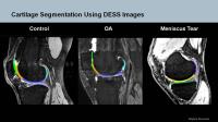
Jedrek Burakiewicz1, Andrew G. Webb1,
Janbernd Kirschner2, Jan J.G.M. Verschuuren3,
Erik H. Niks3, and Hermien E. Kan1
1Gorter Center, Leiden University Medical Center,
Leiden, Netherlands, 2Pediatric
Neurology and Muscle Disorders, University Clinic Freiburg,
Freiburg, Germany, 3Department
of Neurology, Leiden University Medical Center, Leiden,
Netherlands
The interpretation of muscle T2 relaxation times in muscular
dystrophies is complicated by the disease progression, as
both inflammation and increased fat content result in a
longer T2. We measured water-T2 in two muscles of the lower
leg using a tri-exponential fitting of the T2 decay in
patients with DMD and healthy controls. We found a
significantly higher T2-heterogeneity in the soleus muscle
of patients, with no significant difference between the two
groups in average T2 values. T2-heterogeneity should be
taken into consideration when using the water T2 of the
diseased muscle as an outcome measure for therapeutic
interventions.
|
|
2283.
 |
Reliability of fat content measurement of lumbar vertebrae
marrow and lumbar paraspinal muscle using 3D DIXON Fat Fraction
Quantification
Yong Zhang1, Aihong Yu1, Yu Zhang2,
Chao Wang3, Yangyang Duan Mu1, Chenxin
Zhang1, Zhuang Zhou4, Wei Zhao1,
Ling Wang1, and Xiaoguang Cheng1
1radiology, Beijing Jishuitan hospital, Beijing,
China, People's Republic of, 2radiology,
Philips Healthcare, Beijing, China, Beijing, China, People's
Republic of, 3Beijing
Institute of Traumatology and Orthopedics, Beijing, China,
People's Republic of, 4Orthopedics,
The Third Hospital of Hebei Medical University, Beijing,
China, People's Republic of
This study aimed to evaluate the reliability of fat content
measurement of lumbar vertebrae marrow and lumbar paraspinal
muscle using an multi-echo 3D DIXON method. A total of 31
volunteers (15 males and 16 females) were included in this
study and underwent liver mDIXON-quant MR imaging by an
radiologist and this examinations were repeated by another
radiologist within 2 weeks. The radiologists measured fat
content of L3, psoas (PS), erector spinae (ES), and
multifidus (MF) muscles on the central L3 axial MR images on
ISP V7 workstation and after 2 weeks they repeated the same
measurements. Our results showed mean fat content of L3, PS,
ES, MF was 38.19%, 3.52%, 3.48%, 3.53% for males and 32.11%,
3.40%, 7.06%, 7.14% for females. The repeatability and
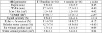
reproducibility of measurement of fat content, T2* and R2*
of L3, PS, ES, MF was high (the intra-observer ICC and
inter-observer ICC all>0.9). Fat content measurement of
lumbar vertebrae marrow and lumbar paraspinal muscle using
mDIXON-quant imaging has high reliability and be potentially
used in clinical practice.
|
|
2284.
 |
Investigating Regional Variations of Acetyl Carnitine In Thigh
and Calf Muscles In Vivo using PRESS Localized Long TE-based MR
Spectroscopy
Rajakumar Nagarajan1, Zohaib Iqbal1,
Manoj K Sarma1, S. Sendhil Velan2, and
M.Albert Thomas1
1Radiological Sciences, University of California
Los Angeles, Los Angeles, CA, United States, 2Laboratory
of Molecular Imaging, Singapore Bioimaging Consortium,
Singapore, Singapore
Skeletal muscle plays a major role in the development of
insulin resistance (IR) and progression to type 2 diabetes.
A recent work has used long TE (350ms) based PRESS
localized spectrum in the vastus lateralis region of thigh
muscle without any exercise to investigate acetylcarnitine,
a compound formed when acetyl-Coenzyme A exceeds use by the
tricarboxylic cycle in the mitochondria. This work focused
on examining regional variations of acetylcarnitine in the
thigh and calf muscles using the long TE MRS. Our
preliminary results show the unequivocal presence of
acetylcarnitine in lean, young healthy thigh muscle regions
and decreased level in one diabetic type 2 patient.
|
|
2285.
 |
Quantitative Off-Resonance-Based Metallosis Assessment Near
Total Hip Replacements: Correlating an Imaging Biomarker with
Histology
Kevin M Koch1, Matthew F Koff2, Parina
Shah2, S S Kaushik1, Andrew Nencka1,
and Hollis G Potter2
1Radiology, Medical College of Wisconsin,
Milwaukee, WI, United States, 2Magnetic
Resonance Imaging, Hospital for Special Surgery, New York,
NY, United States
The failure of hip arthroplasty may be attributed to
metallic or polyethylene debris generated from implant
components. The metallic components, and their associated
debris are composed of cobalt-chromium alloys, which have a
strong paramagnetic magnetic susceptibility relative to
biological materials. Previously, we demonstrated a
mechanism to utilize MRI data to qualitatively highlight
cobalt-chromium debris deposits in
vivo. In the current study, we extend this work to
provide a quantifiable regional metallosis metric. In
addition, this regional quantitative metric is shown to
statistically correlate with local histology metallosis
scores in subjects undergoing total hip revision surgery.
|
|
2286.
 |
Slab Thickness Calibration for Selective 3D-MSI
Kevin M Koch1 and
S S Kaushik1
1Radiology, Medical College of Wisconsin,
Milwaukee, WI, United States
Slab selection is a crucial component of 3D-MSI metal
artifact reduction sequences, due to the need to reduce
phase-encoded fields of view for body imaging applications
in the hip, spine, and shoulder. However, existing
commercial 3D-MSI sequences are prone to signal loss at the
edges of prescribed slabs. Here, we explain the source of
this signal loss and demonstrate a calibration algorithm
that can be used to reduce this slab-boundary signal loss in
3D-MSI. The presented methods are demonstrated on a
calibrated 3D-MSI total hip replacement dataset acquired at
1.5T.
|
|
2287.
 |
Diffusion Tensor Imaging for Peripheral Nerves in the Upper
Extremities using Realtime B0 Correction & Image based
Distortion Correction: A feasibility study
Maggie Mei Kei Fung1, Ek Tsoon Tan2,
David Soon Yiew Sia3, and Darryl Sneag3
1MR Apps & Workflow, GE Healthcare, New York, NY,
United States, 2MR,
GE Global Research Center, Niskayuna, NY, United States, 3MRI
Research Lab, Hospital of Special Surgery, New York, NY,
United States
Diffusion tensor imaging (DTI) can potentially be helpful in
visualizing peripheral nerves and assessing nerve damages.
However, upper extremity DTIs (wrist, elbows & arm) are
susceptible to distortion and fat suppression failure,
especially in arms-down position where the area of interest
is far from iso-center and can have more B0 inhomogeneity.
In this study, we aim to investigate whether a combination
of B0 correction methods can help reduce fat suppression
failure, improve spatial misalignment and thus improve nerve
tracking. We observed consistent fat suppression improvement
at the wrist, but no significant improvement in spatial
accuracy.
|
|
2288.
 |
Development of an Automated Shape and Textural Software Model of
the Paediatric Knee for Estimation of Skeletal Age.
Caron Parsons1,2, Charles Hutchinson1,2,
Emma Helm2, Alexander Kenneth Clarke3,
Asfand Baig Mirza3, Qiang Zhang4, and
Abhir Bhalerao4
1Division of Health Sciences, University of
Warwick, Coventry, United Kingdom, 2Department
of Radiology, University Hospital Coventry & Warwickshire,
Coventry, United Kingdom, 3Warwick
Medical School, Coventry, United Kingdom, 4Department
of Computer Sciences, University of Warwick, Coventry,
United Kingdom
There are multiple methods available for skeletal age
determination in the paediatric endocrine population. Only
two methods, using left hand and wrist x-rays are in
frequent clinical use, however Greulich & Pyle is based on
data collated between 1931 and 1942 and Tanner Whitehouse
uses data from as far back as 1949. We present the initial
results of an automated software model of shape and textural
analysis of the physes of the knee.
|
|
2289.
 |
3D Printed Phantom for Optimization of Trabecular Bone Structure
Imaging
Cem M Deniz1,2, Greg Chang3, and Ryan
Brown1
1Department of Radiology, Center for Advanced
Imaging Innovation and Research (CAI2R) and Bernard and
Irene Schwartz Center for Biomedical Imaging, New York
University School of Medicine, New York, NY, United States, 2The
Sackler Institute of Graduate Biomedical Sciences, New York
University School of Medicine, New York, NY, United States, 3Department
of Radiology, Center for Musculoskeletal Care, New York
University Langone Medical Center, New York, NY, United
States
Phantoms have been used in MRI for sequence optimization and
scanner calibrations. Recent developments in 3D printing
technology have provided tools to manufacture application
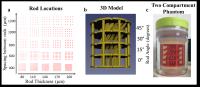
specific phantoms in a fast and reliable way. In this work,
we used 3D printing technology to build a resolution phantom
for optimization of trabecular bone structure imaging. We
used rods with different thickness, orientation and spacing
for capturing the range of possible trabecular bone
structures. Developed phantom was used to investigate the
effect of slice thickness on trabecular bone structure
imaging.
|
|
2290.
 |
High resolution 3D steady-state imaging for peripheral nerves at
7T
Daehyun Yoon1, Sandip Biswal1, Brian
Rutt1, Amelie Lutz1, and Brian
Hargreaves1
1Radiology, Stanford University, Palo Alto, CA,
United States
For the past few decades, MRI has been increasingly used for
identifying peripheral nerve injury, causing chronic and
neuropathic pain. Unfortunately, a substantial number of MRI
examinations fails to find the causative nerve damage,
possibly because it is too subtle or small. Recent
developments of PET-MRI demonstrated improved detection
capability of the nerve damage, but the precise anatomic
characterization of the detected lesion still remains
challenging. We introduce high-resolution 3D steady-state
imaging sequences at 7T that enable examination of
microstructures of peripheral nerves in extremities. We
believe our methods have great potential for improving
diagnosis of various pain syndromes.
|
|
2291.
 |
Diagnostic performance of susceptibility-weighted magnetic
resonance imaging (SWMRI) for the assessment of subacromial spur
formation causing subacromial impingement syndrome (SAIS)
Dominik Nörenberg1,2, Marco Armbruster1,
Yi-Na Bender2, Thula Walter2, Gerd
Diederichs2, Bernd Hamm2, Ben Ockert3,
and Marcus R. Makowski2,4
1Department of Clinical Radiology, Munich
University Hospitals Campus Großhadern, Germany, Munich,
Germany, 2Department
of Radiology, Charité, Berlin, Germany, Berlin, Germany, 3Department
of Trauma and Orthopedic Surgery, Shoulder and Elbow
Service, Munich University Hospitals Campus Großhadern,
German, Munich, Germany, 4King’s
College London, Division of Imaging Sciences and Biomedical
Engineering, London, United Kingdom, London, United Kingdom
Shoulder pain is regarded as the second most common
musculoskeletal disorder in the general population. 44 % of
shoulder pain syndromes are related to subacromial shoulder
impingement (SAIS) due to rotator cuff tear (RCT) and
glenohumeral joint arthritis. Especially subacromial spur
formation is associated with SAIS and RCT. Our study
demonstrates that SWMRI allows for a reliable detection and
precise 3D-localization of subacromial spur formation under
the coracoacromial arch in patients with SAIS and provides
superior evaluation of diamagnetic spur formation compared
to standard shoulder MRI using conventional radiography as a
reference.
|
|
2292.
 |
Metal implant imaging using highly undersampled phase-cycled 3D
bSSFP
Damien Nguyen1,2, Tom Hilbert3,4,5,
Jean-Philippe Thiran5,6, Tobias Kober3,4,5,
and Oliver Bieri1,2
1Radiological Physics, Dep. of Radiology,
University of Basel Hospital, Basel, Switzerland, 2Department
of Biomedical Engineering, University of Basel, Basel,
Switzerland, 3Advanced
Clinical Imaging Technology (HC CMEA SUI DI BM PI), Siemens
Healthcare AG, Lausanne, Switzerland, 4Department
of Radiology, University Hospital (CHUV), Lausanne,
Switzerland, 5LTS5,
École Polytechnique Fédérale de Lausanne, Lausanne,
Switzerland, 6Department
of Radiology, University Hospital Lausanne (CHUV), Lausanne,
Switzerland
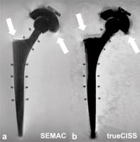
In this study, we explore the possibility of using a highly
undersampled 3D phase-cycled balanced Steady-State
Free-Precession (bSSFP) sequence (trueCISS) to image metal
implants in the body and compare it to the Slice Encoding
for Metal Artifact Correction (SEMAC) method. We show that
the trueCISS approach not only offers qualitatively good
morphological images, but also delivers quantitative maps
that could potentially improve the overall diagnostic
quality and efficiency within a clinically reasonable time.
|
|
2293.
 |
Diagnosis of Chronic Hip Pain After Total Hip Arthroplasty Using
SEMAC-VAT MR Imaging
Yimin Ma1, Panli Zuo2, Mathias Nittka3,
and Xiaoguang Cheng4
1Department of radiology, Department of
radiology, Jishuitan Hospital, Beijing, China, Beijing,
China, China, People's Republic of, 2Siemens
Healthcare, MR Collaborations NE Asia, Beijing, China,
Beijing, China, China, People's Republic of, 3Siemens
Healthcare, Erlangen, Germany, Erlangen, Germany, 4Department
of Radiology, Department of Radiology, Jishuitan Hospital,
Beijing, China, Beijing, China, China, People's Republic of
With the rapid development of medicine technology, total hip
arthroplasty (THA) is now widely used in the treatment of
endstage hip osteoarthritis, severe hip fracture, hip bone
tumor, and so forth. THA can relieve hip pain and improve
the activity of the joints, while it still brings some
unexpected complications, such as periprothesis bone
resorption, periprothesis fractures, and metallic implants
dislocation. Since then, distortion-free MRI near
metal, like SEMAC-VAT MR, has shown its great clinical
potential in diagnosing patients treated with THA.
|
|
2296.
 |
Quantification of Magnetization Transfer parameters in across
different muscle groups
Chun Kit Wong1, Jamie X. M. Ho1, and
Mary Stephenson1,2
1A*STAR-NUS Clinical Imaging Research Centre,
Singapore, Singapore, 2Department
of Medicine, National University of Singapore, Singapore,
Singapore
Quantitative magnetization transfer (qMT) parameters can
potentially be used as biomarker of diseases. In this study,
qMT parameters’ nominal value are determined for selected
muscle groups in healthy human subjects’ forearm, mid-thigh,
and calf. Nominal values of qMT parameters are determined by
taking the mean value across the subjects for each muscle
group. From the results, strong correlations of qMT
parameters between certain muscle groups within the same
individual subjects are observed, suggesting that the qMT
parameters' variation is biological in origin.
|
|
2303.
 |
Assessment of Tibial Nerve and Common Peroneal Nerve in Diabetic
by Diffusion Tensor Imaging: a Feasibility Study
Chao Wu1, Bin Zhao1, Guangbin Wang1,
Shanshan Wang1, and Hongjing Bao1
1Shandong Medical Imaging Research Institute,
Shandong university, Jinan, China, People's Republic of
This study aimed to measure the FA and ADC values by
quantitative DTI at the tibial nerve and common peroneal
nerve and determine whether DTI can be used in the DPN. 25
healthy volunteers and 13 patients with DPN were underwent
MR examinations at 3T including DTI of knee. The FA values
of both tibial nerve and common peroneal nerve in DPN
patients were significantly lower than those in healthy
volunteers. The ADC values in DPN patients were higher than
those in healthy groups. DTI may thus be a reliable method
to added diagnostic value in patients with DPN.
|
|
2297.
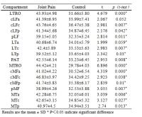 |
Articular Cartilage Assessment Using T1? Mapping in Early
Osteoarthritis Patients with Knee joint Pain
Jin Qu1, Xinwei Lei1, Ying ZHAN1,
Huixia Li1, and Yu Zhang2
1Tianjin First Center Hospital, Tianjin, China,
People's Republic of, 2Philips
Healthcare, Beijin, China, People's Republic of
The purpose of this study was to evaluate articular
cartilage degeneration in healthy subjects and patients with
knee joint-pain as the only clinical manifestation using T1ρ
measurements and to examine the interrelationship between
cartilage abnormalities. Quantitative assessment of
cartilage was performed using T1ρ mapping technique in 5
healthy volunteers and 17 knee joint-pain patients. T1ρ
values were signi?cantly elevated among patients with knee
joint-pain compared to normal controls. Proteoglycan reduced
in patients with knee joint-pain as the only clinical
manifestation. Comparing to routine MR, T1ρ mapping could be
more useful for these patients
|
|