ISMRM 24th Annual Meeting & Exhibition • 07-13 May 2016 • Singapore

Traditional Poster Session: Cancer

2422 -2433 DWI & MRS in Cancer
2434 -2450 Tumour Response to Therapy
2451 -2470 Clinical MRI of Solid Tumour
2471 -2486 Breast Cancer


2422.   
Noise-corrected exponential DWI using multi-acquisition MRI facilitates quantitative evaluation of whole-body skeletal tumour burden in patients with metastatic prostate cancer.
Matthew David Blackledge1, Nina Tunariu1,2, Zaki Ahmed2, Julie Hughes2, Raquel Perez-Lopez1,2, Dow Mu Koh1,2, David J Collins1,2, and Martin O Leach1,2
1CRUK Cancer Imaging Center, Division of Radiotherapy and Imaging, Institute of Cancer Research, London, United Kingdom, 2MRI, Royal Marsden Hospital, London, United Kingdom
We evaluate the potential utility of storing all imaging acquisitions from whole-body diffusion-weighted MRI (WBDWI) studies.  This provides the possibility of using weighted least-squares fitting to obtain maps of ADC uncertainty, invaluable to clinicians wishing to report confidence in ADC estimates.  Furthermore, we describe for the first time a novel post-processing technique that combines ADC and ADC uncertainty information into a single computed image: noise-corrected exponential WBDWI.  We demonstrate that this technique provides excellent contrast between bone metastases and background healthy tissue, but does not suffer from the same T2 shine-through and/or coil sensitivity artefact present in conventional DW-images.


2423.   
Proton MRS monitoring of Atkins-based dietary therapy in patients with glioma
Peter B Barker1,2, Subechhya Pradhan1, Yanqin Lin1,3, Karisa C Schreck4, Doris D.M. Lin1, Jaishri Blakeley4,5, and Roy E Strowd5,6
1Radiology, Johns Hopkins Univ School of Medicine, Baltimore, MD, United States, 2F.M. Kirby Research Center for Functional Brain Imaging, Kennedy Krieger Institute, Baltimore, MD, United States, 3Department of Electronic Science, Xiamen University, Xiamen, China, People's Republic of, 4Neurology, Johns Hopkins Univ School of Medicine, Baltimore, MD, United States, 5Oncology, Johns Hopkins Univ School of Medicine, Baltimore, MD, United States, 6Department of Neurology and Internal Medicine, Wake Forest School of Medicine, Winston Salem, NC, United States
5 patients with high grade glioma were enrolled in an 8-week dietary therapy program based on the modified Atkin’s diet. Tumor and contralateral brain metabolism was monitored before and after therapy using proton MRS. Significant reductions in contralateral NAA were observed post treatment, as well as increases in acetone in both lesion and contralateral brain. MRS may be useful for monitoring the efficacy of dietary therapy in patients with malignant glioma. 


2424.   
MR spectroscopy for 2HG detection in mIDH gliomas: Comparison of sensitivity improvement using different refocusing RF pulses with and without outer volume suppression
Sunitha B Thakur1,2, Ralph Noeske3, Robert Young4, Justin Cross5, and Ingo Mellinghoff6
1Medical Physics, Memorial Sloan Kettering Cancer Center, New York, NY, United States, 2Radiology, Memorial Sloan Kettering Cancer Center, New York, NY, United States, 3Berlin, Germany, 4Radiology, New York, NY, United States, 5Memorial Sloan Kettering Cancer Center, New York, NY, United States, 6New York, NY, United States
Isocitrate dehydrogenase IDH mutations in gliomas have ability to produce R-2-hydroxyglutarate (2HG). Directly measuring 2HG using non-invasive MR spectroscopy is an attractive strategy to accurately predict IDH mutation status and provide useful diagnostic and prognostic information. Quantification of 2HG metabolite may have potential advatages to evaluate treatment response in IDH1/2 targeted inhibitor trails . Here we report the optimization of subecho times on GE 3T scanners using phantoms to detect 2HG with the maximum sensitivity. We also evaluated the effect of different refocusing pulses on 2HG sensitivity with and without outer volume suppression (OVS) pulses. Part of these results were also verified with in-vivo data. 


2425.   
Imaging response to 90-yttrium radioembolization with volumetric ADC DWI in patients with metastatic liver cancer
Russell Rockne1, Syed Rahmanuddin1,2, John J. Park2, and Jinha M. Park2
1Dept. of Information Sciences/ Mathamatical Oncology, Beckman Research Institute City of Hope, Duarte, CA, United States, 2Radiology, Helford Clinical Research Hospital | Beckman Research Institute, Duarte, CA, United States
The purpose of this study is to evaluate changes in tumor volume and mean ADC before and after 90-yttrium (Y90) radioembolization for patients with metastatic liver cancer. Eighteen patients were identified in a retrospective study. Volumetric analysis of lesions before and after therapy showed a consistent increase in ADC (p<0.001), suggesting a decrease in cellularity. Changes in tumor volume measured on post-gadolinium (PG)  T1 MRI were not statistically significant. Additional MRIs following Y90 treatment should be analyzed with clinical outcomes to determine whether early changes in ADC following therapy may serve as an early indicator of response. 


2426.   
Distortion-Free bSSFP-based Diffusion MRI: Preliminary Experience using an MRI-Guided Radiotherapy System
Yu Gao1,2, Yingli Yang3, Novena Rangwala1, and Peng Hu1,2
1Radiological Sciences, University of California, Los Angeles, Los Angeles, CA, United States, 2Physics and Biology in Medicine, University of California, Los Angeles, Los Angeles, CA, United States, 3Radiation Oncology, University of California, Los Angeles, Los Angeles, CA, United States
DWI is a promising imaging biomarker for tumor response evaluation, and the purpose of this work is to develop a distortion-free DW sequence that is reliable for adaptive treatment planning. ADC accuracy of our proposed diffusion-prepared segmented bSSFP-based sequence against the standard spin-echo single-shot echo-planar-imaging was quantitatively validated on a diffusion phantom. Geometric reliability was confirmed on both phantom and in-vivo experiments. Preliminary patient study on a MRI-Guided radiotherapy system showed high geometric accuracy and promising tumor detection capability. All these demonstrate the feasibility of using the DW-SSFP sequence for longitudinal tumor response evaluation and treatment planning. 


2427.   
Application of High B-Value High-Resolution Diffusion-Weighted Imaging in Differentiating Malignant from Benign Thyroid Nodules
Qingjun Wang1, Yong Guo1, Jing Zhang1, Minghua Huang1, Qinglei Shi2, and Tianyi Qian2
1Radiology, Chinese Navy General Hospital, Beijing, China, People's Republic of, 2Scientific Marketing, Siemens Healthcare, Beijing, China, People's Republic of
In this study, we evaluated the advantages of using high b-value high-resolution diffusion-weighted MR imaging (DWI) to differentiate between malignant and benign thyroid nodules. This prospective study included 28 consecutive patients with thyroid nodules (14 malignant nodules in 10 patients and 24 benign nodules in 20 patients). Three b-values including b-values of 0, 800 and 2000 sec/mm2 and a Readout Segmentation Of Long Variable Echo-trains (RESOLVE) imaging technique were used in the high b-value high-resolution DWI. The results showed that the high b-value high-resolution DWI can further increase the diagnostic accuracy for thyroid nodules with the best sensitivity, specificity and area under receiver operating characteristic curve (AUC) than low b-value or single high b-value.


2428.   
Comparison of Capability for Postoperative Recurrence Evaluation in Non-Small Cell Lung Cancer among Whole-Body MRI with and without DWI, MR/PET, PET/CT and Conventional Radiological Examinations
Yoshiharu Ohno1,2, Yuji Kishida2, Shinichiro Seki2, Hisanobu Koyama2, Kota Aoyagi3, Hitoshi Yamagata3, Takeshi Yoshikawa1,2, Masao Yui3, Yoshimori Kassai3, Katsusuke Kyotani4, and Kazuro Sugimura2
1Advanced Biomedical Imaging Research Center, Kobe University Graduate School of Medicine, Kobe, Japan, 2Radiology, Kobe University Graduate School of Medicine, Kobe, Japan, 3Toshiba Medical Systems Corporation, Otawara, Japan, 4Center for Radiology and Radiation Oncology, Kobe University Hospital, Kobe, Japan
    Recurrence assessment is very important for management of postoperative non-small cell lung cancer (NSCLC) patients.  We hypothesized that whole-body PET/MRI and MRI with DWI have a potential for improving recurrence assessment as compared with whole-body PET/CT and conventional radiological examinations in NSCLC patients.  The purpose of this study was to directly compare the diagnostic performance for postoperative lung cancer recurrence assessment among whole-body PET/MRI, MRI with and without DWI, PET/CT and conventional radiological examination in NSCLC patients.


2429.   
Heterogeneity of IDH+ glioma metabolism evaluated by CSI
Benjamin C Rowland1, Min Zhou1, Huijun Liao1, Nils Arvold2, Raymond Y Huang3, and Alexander P Lin1
1Centre for Clinical Spectroscopy, Brigham and Women's Hospital, Boston, MA, United States, 2Radiation Oncology, Brigham and Women's Hospital, Boston, MA, United States, 3Radiology, Brigham and Women's Hospital, Boston, MA, United States
MR spectroscopy is a powerful technique for understanding tumour metabolism. Chemical shift imaging allows spatial variations in spectra to be observed at the cost of signal to noise and acquisition time. In this abstract we examine the heterogeneity of metabolite concentrations in both tumour and healthy tissue, in particular the onco-metabolite 2-hydroxyglutarate, to ascertain if the benefits of CSI outweigh the penalties.


2430.   
Prediction of Medulloblastoma survival using the Water Proton Resonance Frequency
Ben Babourina-Brooks1, Sarah Kohe1, Andrew Peet1, and Nigel Davies2
1University of Birmingham, Birmingham, United Kingdom, 2University Hospitals Birmingham, Birmingham, United Kingdom
The rate of improvement in survival, among children with brain tumours, has decreased in recent years and novel prognostic markers that may contribute to treatment stratification and improved outcomes are required.  Medulloblastoma tumours have a high rate of mortality due to their aggressive nature,  however some do not. MRS can measure absolute water proton resonance frequency (PRF), which is sensitive to temperature and other features of the tumour microenvironment. This could be utilized as a potential prognosis marker, however has not been explored. This study assessed the water PRF as a predictor of survival in a medulloblastoma patients, using MRS at diagnosis. 


2431.   
In-vitro 1H-MRS of living human melanoma cells at 9.4T
Katarzyna Pierzchala1, Nicolas Kunz1, and Rolf Gruetter1
1CIBM, EPFL, Lausanne, Switzerland
There is increasing need of a more human cell model to recapitulate the in-vivo cell-cell interactions, presenting physiological relevance. Magnetic resonance spectroscopy (MRS) has the potential to diagnose many tumors and to characterize their metastatic potential. In this study we present the metabolic profile of highly metastatic human melanoma cells line WM793.We demonstrate the feasibility of characterizing in-vitroliving melanoma cells by 1H-MRS with a total number of 9 metabolites quantified, covering energy markers (Glc, Lac, Ace), amino acids (Glu, Ala) and anti-oxidant (Tau, Asc) and cell membrane precursor (Cho).


2432.   
Characterisation of disease heterogeneity in malignant pleural mesothelioma using mixture modelling of ADC and R2
Lin Cheng1, Matthew D. Blackledge1, David J. Collins1, Nina Tunariu1,2, Neil P. Jerome1, Matthew R. Orton1, Veronica A. Morgan3, Martion O. Leach1, and Dow-Mu Koh1,2
1Division of Radiotherapy and Imaging, Cancer Research UK Cancer Imaging Centre, Institute of Cancer Research, London, United Kingdom, 2Radiology, Royal Marsden Hospital, London, United Kingdom, 3Clinical MRI Unit, Royal Marsden Hospital, London, United Kingdom
Disease heterogeneity in patients with malignant pleural mesothelioma (MPM) makes it challenging to characterise solid disease and assess response following treatment. Computed Diffusion-Weighted MRI (cDWI) provides improved contrast between disease and background tissues, and facilitates total disease segmentation. A mixture modelling of ADC and R2 with semi-automatic segmentation on the cDWI is proposed to assess disease heterogeneity in MPM, with demonstration of its utility on a paired pre/post-treatment dataset. The mixture modelling methodology successfully characterised disease heterogeneity for two MPM patients, and can provide additional quantitative functional disease response characterisation compared with using only a single parameter.


2433.   
Apparent diffusion coefficient ratio as a potential inter-institutional marker for histological grade of bladder cancer
Soichiro Yoshida1, Fumitaka Koga2, Hiroshi Tanaka3, Hiroshi Fukushima2, Yasukazu Nakanishi2, Minato Yokoyama1, Junichiro Ishioka1, Kazutaka Saito1, Yasuhisa Fujii1, and Kazunori Kihara1
1Urology, Tokyo Medical and Dental University Graduate School, Tokyo, Japan, 2Urology, Tokyo Metropolitan Cancer and Infectious diseases Center Komagome Hospital, Tokyo, Japan, 3Radiology, Ochanomizu Surugadai Clinic, Tokyo, Japan
We evaluated standardized apparent diffusion coefficient value (ADC ratio) by taking the ratio of the ADC of the cancer tissue (T-ADC) to that of the gluteus maximus (G-ADC) can be a biomarker for predicting histological grade under two different imaging conditions. Two independent bladder cancer cohorts including 107 and 47 patients were evaluated. There was a significant difference in T-ADC between the cohorts, but no significant difference in the ADC ratio was observed. The ADC ratio significantly correlated with grade, as was the T-ADC. The ADC ratio might serve as an inter-institutional biomarker for predicting histological grade of bladder cancer.

2434.   
GluCEST MRI: A Biomarker for Glutamine Metabolism in Cancer
Rong Zhou1, Puneet Bagga1, Kavindra Nath1, David Mankoff1, Hari Hariharan1, and Ravinder Reddy1
1Radiology, University of Pennsylvania, Philadelphia, PA, United States
We presented the very first evaluation of glutamate chemical exchange saturation transfer (GluCEST) MRI to detect pharmacodynamic effect of small molecule drugs that target cancer glutaminolysis pathway. Conversion of glutamine to glutamate is a rate limiting step along this pathway. Inhibition of this conversion leads to reduction of cellular glutamate concentration that can be detected by GluCEST in vivo and confirmed by ex vivo high resolution 1H MRS of tumor homogenates. 


2439.   
Characterization of HPV positive oropharyngeal tumors using intravoxel incoherent motion DW-MRI before and during radiation therapy
Ramesh Paudyal1, Praveen Venigalla2, Jingao Li2,3, Nadeem Riaz2, Jung Oh Hun1, David Aramburu Nuñez1, Vaois Haztglou4, Yonggang Lu5, Joseph O Deasy1, Nancy Lee2, and Amita Shukla-Dave1
1Medical Physics, Memorial Sloan Kettering Cancer Center, New York, NY, United States, 2Radiation Oncology, Memorial Sloan Kettering Cancer Center, New York, NY, United States, 3Radiation Oncology, Jiangxi Cancer Hospital, Nanchang, China, People's Republic of, 4Radiology, Memorial Sloan Kettering Cancer, New York, NY, United States, 5Radiation Oncology, Washington University, St louis, MO, United States
This study aims to characterize human papillomavirus (HPV) positive (+) oropharyngeal cancer (OPC) using intravoxel incoherent motion (IVIM) DW-MRI performed before and during chemo-radiation therapy. A consensus clustering algorithm based on the hierarchical clustering and Pearson correlation distance was performed using the weekly IVIM DW-MRI metrics of D, f and D*, total tumor volume and delivered radiation dose. It demonstrated the presence of two HPV+ clusters. A trend towards significant increase in D during the pre- and intra-treatment (week 3) IVIM DW-MR data suggested that patients in cluster 2 may benefit with use of less aggressive therapy. 


2440.   
Treatment response assessment of malignant cancer cells to alpha-lipoic acid and a 213Bi-anti-EGFR-MAb with hyperpolarized [1-13C]-pyruvate
Benedikt Feuerecker1, Christian Hundshammer1, Christof Seidl2, Alfred Morgenstern3, Frank Bruchertseifer3, Reingard Senekowitsch-Schmidtke1, and Markus Schwaiger1
1Department of Nuclear Medicine, Technische Universität München, Munich, Germany, 2Department of Obstetrics and Gynaecology, Technische Universität München, Munich, Germany, 3European Commission Joint Research Centre Institute for Transuranium Elements, Karlsruhe, Germany
In the light of up regulation of glycolysis in tumors of the Warburg type, hyperpolarized 13C-labeled metabolic tracers offer new possibilities to probe fast metabolic pathways in real-time. As such, we assessed therapy response of malignant cancer cells to alpha-lipoic acid and a 213Bi-anti-EGFR-MAb with hyperpolarized [1-13C]-pyruvate. Our results point to the fact that treatment of LN18 glioblastoma cells with LPA resulted in decreased proliferation/viability and reduced lactate export. Beyond, we demonstrated that NMR of hyperpolarized [1-13C]-pyruvate proved to be adequate for monitoring the response of bladder carcinoma cells to treatment with a 213Bi-anti-EGFR-MAb as indicated by elevated pyruvate turnover.


2435.   
Use of iron contrast agents to detect brain tumor treatment response based on stimulating the innate immune system
Yang Runze1,2, Susobhan Sarkar2, Daniel J Korchinski1, Ying Wu1, V Wee Yong2,3, and Jeff F. Dunn1,2
1Radiology, University of Calgary, Calgary, AB, Canada, 2Clinical Neuroscience, University of Calgary, Calgary, AB, Canada, 3Oncology, University of Calgary, Calgary, AB, Canada
Glioblastoma Multiforme (GBM) is the most aggressive brain cancer with an abysmal prognosis. It has been shown that monocytes can be activated to suppress GBM stem cells using Amphotericin B (Amp B). We propose that monocytes can be labeled using intravenous injection of ultra-small iron oxide nanoparticles (USPIO), which will allow us to detect a rapid treatment response. We showed that Amp B treated animals significantly decreased T2* compared to vehicles, showing the presence of USPIO within the tumor. This shows that USPIO can be an effective tool to monitor cancer therapies that stimulate innate immunity.


2442.   
MRI biomarkers for pancreatic ductal adenocarcinoma
Navid Farr1, Paolo Provenzano2, Joshua Park3, Sunil Hingorani2, and Donghoon Lee3
1Department of Bioengineering, University of Washington, Seattle, WA, United States, 2Fred Hutchinson Cancer Research Center, Seattle, WA, United States, 3Department of Radiology, University of Washington, Seattle, WA, United States
Pancreatic cancer is a devastating disease with poor prognosis. Pancreatic tumor therapy has been ineffective in part because pancreatic tumors have a dense stroma inhibiting penetration of chemotherapeutic drugs into the tumor. We performed multi-parametric MRI at high resolution to noninvasively assess tumor progression and responses to effective treatment. We used T1 and T2 relaxation, diffusion, magnetization transfer effects along with 3 dimensional volume measurements to characterize the tumors. MR measurements were then compared with histopathological results.


2443.   
Can Diffusion Weighted MRI Assess Early Response of Lymphadenopathy to Induction Chemotherapy in Nasopharyngeal Cancer: A Heterogeneity Analysis Approach
Manijeh Beigi1, Anahita Fathi Kazerooni1, Mojtaba Safari1, Marzieh Alamolhoda2, Ahmad Ameri3, Shiva Moghdam4, Mohsen Shojaee Moghdam5, and Hamidreza Saligheh Rad1
1Medical Physics and Biomedical Engineering, School of Medicine, Quantitative MR Imaging and Spectroscopy Group, Research Center for Cellular and Molecular Imaging, Institute for Advanced Medical Imaging, Tehran, Iran, 2Statistics, Shiraz University of Medical Science, Shiraz, Iran, 3Oncology, Shahid Beheshti University of Medical Science, Tehran, Iran, 4Oncology, Tehran, Iran, 5Payambaran Imaging Center, Tehran, Iran
Induction chemotherapy is an effective way to control subclinical metastasis in locally-advanced nasopharyngeal cancer patients. Diffusion-weighted MRI is a noninvasive imaging technique allowing some degree of tissue characterization by showing and quantifying molecular diffusion. Histogram analysis on ADC map could be carried out to reveal physiological alterations early after IC.  For this purpose, several quantitative metrics from ADC-map were explored to obtain the most accurate feature(s) as potential predictive biomarker for early response of the lymphnode to IC. If the outcome can be predicted at an early stage of the treatment, the patient could be spared from unnecessary treatment toxicity.  


2444.   
Multiparameter MRI Response Assessment in a Phase I Trial of Hypofractionated Stereotactic Irradiation with Pembrolizumab and Bevacizumab in Patients with Recurrent High Grade Gliomas
Olya Stringfield1, John Arrington2, Solmaz Sahebjam3, and Natarajan Raghunand1
1Cancer Imaging & Metabolism, Moffitt Cancer Center, Tampa, FL, United States, 2Radiology, Moffitt Cancer Center, Tampa, FL, United States, 3Neuro-Oncology, Moffitt Cancer Center, Tampa, FL, United States
In this ongoing phase 1 study, we are investigating combined pembrolizumab and bevacizumab therapy with hypofractionated radiotherapy in recurrent glioma. The objective of this work is to develop a non-invasive response assessment measure using multiparameter MRI (mpMRI) scans acquired as part of clinical care which may precede volumetric changes in response to therapy.


2445.   
Integrated Positron Emission Tomography/ Magnetic Resonance Imaging in the Treatment of Cervical Cancer: Preliminary Results
Sharmili Roy1, Dennis Lai-Hong Cheong1, Mary C. Stephenson1, Trina Kok1, Evelyn Laurens1, Joshua D. Schaefferkoetter1, John James Totman1, Vicky Koh2, Johann Tang2, Joseph Ng2, Jeffrey Low2, and Bok Ai Choo2
1A*Star-NUS Clinical Imaging Research Centre, Singapore, Singapore, 2National University Health System, Singapore, Singapore
Response assessment after radical radiation therapy (RT) is typically performed months post treatment completion due to confounding acute RT effects. This study presents feasibility and early results on the ability of PET/MRI in visualizing early tumor changes in the cervix even during RT. This could potentially provide actionable information for treatment modifications prior to the completion of standard of care.  


2446.   
Prostate Cancer: DCE-MRI parameter changes during radiotherapy
Lucy Elizabeth Kershaw1,2, Andrew McPartlin2,3, Ben Taylor4, Ananya Choudhury2,3, and Marcel van Herk2
1CMPE, The Christie NHSFT, Manchester, United Kingdom, 2Manchester Academic Health Sciences Centre, The University of Manchester, Manchester, United Kingdom, 3Oncology, The Christie NHSFT, Manchester, United Kingdom, 4Radiology, The Christie NHSFT, Manchester, United Kingdom
In this pilot work, our aim was to measure plasma flow (Fp) and permeability-surface area product (PS) through the course of RT in prostate tumour and normal tissue to determine whether significant changes could be detected early in treatment.   We detected significant increases in Fp and PS during radiotherapy in this small patient group.


2450.   
Hormonal effect on time-dependent diffusion of the breast fibroglandular tissue
Sungheon Gene Kim1,2, Eric Sigmund1,2, Melanie Moccaldi1,2, Thorsten Feiweier3, and Linda Moy1,2
1Center for Advanced Imaging Innovation and Research, Radiology, New York University School of Medicine, New York, NY, United States, 2Bernard and Irene Schwartz Center for Biomedical Imaging, Radiology, New York University School of Medicine, New York, NY, United States, 3Siemens Healthcare GmbH, Erlangen, Germany
This study is to investigate the potential of the surface-to-volume ratio obtained from multiple diffusion times to measure mammary duct microstructural changes induced by hormonal variation. Seven premenopausal women were scanned twice using a stimulated-echo diffusion sequence; one in the follicular phase of the menstrual cycle and again in the luteal phase. In 6 out of 7 subjects, the surface-to-volume ratio measured with 69 and 173 ms was significantly reduced in the luteal phase compared to the follicular phase. The length scales obtained in our study are consistent with the duct diameters reported in previous ex-vivo studies.


2441.   
Quantification of transverse relaxation time changes in rectal tissue during fixation at ultra-high field MRI
Quincy van Houtum1, Dennis D.W.J. Klomp1, and Marielle M.E.P. Philippens1
1Imaging, UMC Utrecht, Utrecht, Netherlands
In this study we investigate the transverse relaxation effects at 7T during the fixation process in rectal specimens as a preparation for 7T MRI validation of rectal tumor regression with pathology. T2 and T2* were measured every 2 hours during fixation process in six different pig recta. Images allowed mapping of relaxation time at high spatial resolution. The circular muscle showed a decrease of 25% in T2* whilst other tissues remained constant over time for both parameters. A T2* decrease of 25% is seen in the circular muscle of the rectal wall was noticed whilst other tissue-regions remained constant. High resolution and SNR allowed for anatomical delineation and measuring the change of transverse relaxation times.


2436.   
Diffusion weighted magnetic resonance imaging as an early predictor of survival in patients with liver-dominant metastatic colorectal cancer following 90-Yttrium-microsphere radioembolization
Frederic Carsten Schmeel1, Birgit Simon1, Julian Alexander Luetkens1, Frank Träber1, Leonard Christopher Schmeel1, Hans Heinz Schild1, and Dariusch Reza Hadizadeh1
1Department of Radiology, University Hospital Bonn, Bonn, Germany
Imaging-based response assessment to local interventional therapies is essential for further therapy decisions in patients with advanced malignancies.  Therefore, we investigated whether early post-therapeutic changes in diffusion-weighted MRI using quantifications of the apparent diffusion coefficient (ADC) could predict the outcome of patients with liver-dominant metastatic colorectal cancer after radioembolization with 90-Yttrium microspheres (RE). Uni- and multivariate survival analyses were performed comparing various variables with potential impact on overall survival. Our results reveal that an increase in the post-therapeutic minimal ADC remained the strongest and only independent predictor of overall survival shortly after radioembolization.


2437.   
Quality Assurance Methodology for Multicenter Clinical Trials using MRI – Experiences from the NCI National Clinical Trial Network (NCTN) Imaging and Radiation Oncology Core (IROC) Service
Preethi Subramanian1, Jun Zhang2, Shivangi Vora2, Marc Gollub3, Deborah Schrag4, Xiangyu Yang5, Lawrence Schwartz6, and Michael V Knopp7
1Radiology, The Ohio State University, Colu,bus, OH, United States, 2Radiology, The Ohio State University, Columbus, OH, United States, 3Radiology, Memorial Sloan Kettering Cancer Center, New York, NY, United States, 4Dana Farber Cancer Institute, Boston, MA, United States, 5The Ohio State University, Columbus, OH, United States, 6Radiology, Columbia University, New York, NY, United States, 7Radiology - Wright Center of Innovation, The Ohio State University, Columbus, OH, United States
Quality Assurance Methodology for Multicenter Clinical Trials using MRI


2438.   
Early detection of photoimmunotherapy-induced tumor cell death with hyperpolarized [1,4-13C2]fumarate
Shun Kishimoto1, Jeeva Munasinghe2, Marcelino Bernardo1, Hellmut Merkle3, Keita Saito1, James B Mitchell1, Jan Henrik Ardenkjaer-Larsen4, Peter L Choyke1, and Murali Cherukuri1
1NCI, Bethesda, MD, United States, 2NINDS, Bethesda, MD, United States, 3LFMI, Bethesda, MD, United States, 4GE Health Care, brondby, Denmark
Photoimmunotherapy (PIT) is a novel therapy for cancer treatment.  PIT combines a targeted antibody with the photon absorber, IR700, which, after exposure to near infrared (NIR) light induces highly selective tumor necrosis with almost no side effects to normal adjacent tissue. PIT is now in Phase I clinical trials in head and neck cancers.  Although NIR PIT can be highly effective, the size of the lesion does not immediately change and it may take several weeks for the tumor to completely respond anatomically.  Thus, detecting early therapeutic response in the absence of anatomic change is of interest. Here, we demonstrate the effects of NIR PIT on 13C magnetic resonance spectroscopy (MRS) in an animal model (EGFR positive A431 tumor) using 13C labeled hyperpolarized (HP) pyruvate and fumarate. Interestingly, the lactate-to-pyruvate ratio was almost unchanged, while the malate-to-fumarate ratio showed a significant difference in PIT treated tumors. This is explained by the difference of the bio distribution of these tracers.  Hyperpolarized 13C labeled fumarate MRS is a promising method for detecting early PIT mediated cell necrosis.


2447.   
The feasibility of performing intravoxel incoherent motion MRI for esophageal cancer and an initial comparison with dynamic contrast-enhanced MRI
Sophie E. Heethuis1, Lucas Goense1,2, Peter S.N. van Rossum1,2, Irene M. Lips1, Richard van Hillegersberg2, Jelle P. Ruurda2, Marco van Vulpen1, Gert J. Meijer1, Jan J.W. Lagendijk1, and Astrid L.H.M.W. van Lier1
1Department of Radiotherapy, UMC Utrecht, Utrecht, Netherlands, 2Department of Surgery, UMC Utrecht, Utrecht, Netherlands
The aim of this study was to investigate whether intravoxel incoherent motion (IVIM) MRI could be a non-invasive alternative for dynamic contrast-enhanced (DCE) MRI for response prediction in patients with esophageal cancer. The feasibility of IVIM was researched, followed by a first comparison with DCE-MRI. It was found that non-rigid registration improved the fitting of the IVIM parameters significantly. Comparison of IVIM and DCE-MRI suggested an inverse relation between perfusion fraction and area-under-the-concentration curve (AUC).


2448.   
Restriction Spectrum Imaging based tumor cellularity performs better than ADC in patients newly diagnosed with Glioblastoma Multiforme
AnithaPriya Krishnan1, Carrie R. McDonald1, Nikdokht Farid2, Anders M. Dale1,3, and Nathan S. White1,3
1MMIL, Radiology, University of San Diego, La Jolla, CA, United States, 2Division of Neuroradiology, University of San Diego, La Jolla, CA, United States, 3Center for Translational Imaging and Precision Medicine, University of San Diego, La Jolla, CA, United States
Contrast enhancing (CE) volumes are unreliable in the pseudo-progression window post radiotherapy and ADC based estimate of tumor cellularity is significantly affected by edema. Here, we provide preliminary evidence that Restriction Spectrum Imaging (RSI) based tumor cellularity performs better than ADC, supplements structural volumes and is significantly predictive of overall survival (OS). The study also highlights the need for higher b values as the association of ADC (in CE) with outcomes was observed only when high b values were used for ADC estimation and with b=1500s/mm2, ADC in edema ROI was not independent of edema volume (r=0.7)


2449.   
High resolution imaging of ex-vivo humane pancreas specimen at high field MRI
Quincy van Houtum1, Marielle M.E.P. Philippens1, Maarten M.S. van Leeuwen2, Frank F.J. Wessels2, and Dennis D.W.J. Klomp1
1Imaging, UMC Utrecht, Utrecht, Netherlands, 2Radiology, UMC Utrecht, Utrecht, Netherlands
The aim of this study is to demonstrate high resolution MR imaging of ex vivo pancreas-specimen while maintaining the in vivo shape and orientation. A pancreaticoduodenal specimen was positioned inside a 3T MRI while maintaining in vivo shape and orientation using substitutes for anatomical features. A 3D TSE sequence with a spatial resolution of 0.45x0.45x2mm, was used for acquisition.  Images showed contrast between multiple anatomical structures, allowed for discrimination between tumor and healthy tissue and showed an underestimation of tumor size on CT. Image quality holds promise for improved guidance during PA and registration with in vivo MRI.

2451.   
Quantitative Evaluation of the Effect of Bone on Pelvic Lesion Uptake for MR-based Attenuation Correction on an Integrated Time-of-Flight PET/MRI System
Andrew Leynes1, Jaewon Yang1, Dattesh Shanbhag2, Sandeep Kaushik2, Florian Wiesinger3, Youngho Seo1, Thomas Hope1, and Peder Larson1
1University of California San Francisco, San Francisco, CA, United States, 2GE Global Research, Bangalore, India, 3GE Global Research, Munich, Germany
The current clinical standard for extracranial MR-based attenuation correction (MRAC) on hybrid PET/MRI systems is the use of a Dixon-type sequence to generate a continuous-value fat-water map. The exclusion of bone in Dixon MRAC contributes a clinically significant amount of underestimation in bone lesion uptake. Bone information from a zero echo-time (ZTE) MRI pulse sequence is combined with the Dixon MRAC to produce a hybrid ZTE-Dixon MRAC. The work demonstrates, using PET/MR patient data, that the Dixon MRAC (neglecting bone) is underestimating bone lesion uptake by a clinically significant amount (>10%) when compared to the hybrid MRAC (including bone).


2452.   
Multivariate assessment of brain glioma using hybrid IVIM and DK MRI
Ya-Fang Chen1, Hsiang-Kuang? Liang2, and Wen-Chau Wu3,4
1Medical Imaging, National Taiwan University Hospital, Taipei, Taiwan, 2Radiation Oncology, National Taiwan University Hospital, Taipei, Taiwan, 3Graduate Institute of Oncology, National Taiwan University, Taipei, Taiwan, 4Clinical Medicine, National Taiwan University, Taipei, Taiwan
In the present study, we investigated the feasibility of hybrid intravoxel incoherent motion and diffusion kurtosis MR imaging in assessing brain gliomas. Our data showed that combined intravascular fraction (a surrogate measure of cerebral blood volume), diffusion coefficient (a measure of diffusivity), and diffusion kurtosis coefficient (a measure of diffusion heterogeneity) may better demarcate brain gliomas through exploration of multiple pathophysiological aspects.


2453.   
Comparison of HCC Tumor-Size measured in MRI and Histopathology – Does the Sequence matter?
Marco Armbruster1, Dominik Nörenberg1, Katharina Hoffmann2, Joachim Andrassy2, and Harald Kramer1
1Department of Clinical Radiology, Ludwig-Maximilian-University Munich, Munich, Germany, 2Department of Visceral Surgery, Ludwig-Maximilian-University Munich, Munich, Germany
Size measurements of hepatocellular carcinoma lesions play an important role in treatment algorithms of this disease, however little is known about which MRI sequence has the highest accuracy. This study shows, that there is a significant variance in size assessment of different MRI sequences and phases, while the hepatobiliary-phase seems to be best correlated to histopathologic measurements as the standard of reference and delineates HCC lesions most sharply.


2454.   
Fractional Enhancement metric improves SNR and visualisation of quantitative two-point contrast-enhanced MRI in retroperitoneal sarcoma
Matthew David Blackledge1, Christina Messiou1,2, Jessica M Winfield1,2, Dow Mu Koh1,2, David J Collins1,2, and Martin O Leach1,2
1CRUK Cancer Imaging Center, Division of Radiotherapy and Imaging, Institute of Cancer Research, London, United Kingdom, 2MRI, Royal Marsden Hospital, London, United Kingdom
We compare two enhancement fraction parameters that may be used for quantification of two-point contrast-enhanced MRI studies: The relative enhancement and the fractional enhancement. Using computer simulations we show that fractional enhancement is better behaved in the presence of imaging noise, resulting in better SNR for this parameter over a range of intrinsic longitudinal tissue relaxivities and contrast medium concentrations.  Further, in a cohort of 25 patients with retroperitoneal sarcoma, fractional enhancement significantly outperformed the relative enhancement in terms of visual assessment of contrast-to-noise, signal-to-noise, tumour detection, imaging artefacts and within tumour contrast.


2455.   
Assessing Myeloma Focal Lesion Conspicuity on Dixon Images
Timothy James Pengilley Bray1, Saurabh Singh1, Arash Latifoltojar1, Kannan Rajesparan1, Farzana Rahman1, Alan Bainbridge2, Shonit Punwani1, and Margaret A Hall-Craggs1
1Centre for Medical Imaging, University College London, London, United Kingdom, 2Medical Physics, University College London, London, United Kingdom
Dixon imaging is becoming more widely used in multiple myeloma (MM) and can provide both functional and anatomical information. We observed that myeloma lesions seemed more conspicuous on fat only (FO) images than on conventional in-phase T1, and therefore hypothesised that lesion detection rates would be higher on FO images. In this research, we show that reader sensitivity, positive predictive value and confidence are indeed higher on FO images. This may be because myeloma lesions cause a proportionately greater change in fat content than in water content. We suggest that Dixon imaging should be used in preference to T1 imaging alone when performing WB-MRI. 


2456.   
Proton Magnetic Resonance Spectroscopy (1-H MRS) of Sputum and Exhaled Breath Condensate: A Non-invasive Tool for Lung Cancer Screening
Naseer Ahmed1, Tedros Bezabeh2,3, Renelle Myers 4, Omkar B Ijare3, Shantanu Banerji 1, Reem Alomran 1, Zoann Nugent 1, and Zoheir Bshouty 4
1CancerCare Manitoba, Winnipeg, MB, Canada, 2Chemistry, University of Guam, Mangilao, Guam, 3Chemistry, University of Winnipeg, Winnipeg, MB, Canada, 4Health Sciences Centre, Winnipeg, MB, Canada
We undertook this study to determine if 1H Magnetic Resonance Spectroscopy (MRS) can provide an alternate tool for the screening of lung cancer. Metabolic profiles of sputum and exhaled breath condensate (EBC) samples were obtained from 15 patients (cancer, n=8 and control, n=7) using a Bruker Avance 400 MHz NMR spectrometer. Methanol was detected at a significantly lower concentration in the EBC samples (p<0.05). Absence of glucose and lower concentration of glycoprotein (p<0.05) were observed in the sputum samples of the cancer patients. MRS may serve as a screening tool for lung cancer in high-risk patients but this requires validation in a larger study. 


2457.   
Quantifying Pathologies and Improving Tractography in Brain Tumor Using Diffusion Basis Spectrum Imaging
Peng Sun1, Kim J. Griffin1, Hung-Wen Kao2,3, Chien-Yuan Eddy Lin4,5, Ching-Po Lin3,6, and Sheng-Kwei Song1
1Radiology, Washington University in St. Louis, St. Louis, MO, United States, 2Department of Radiology, Tri-Service General Hospital, National Defense Medical Center, Taipei, Taiwan, 3Biomedical Imaging and Radiological Sciences, National Yang-Ming University, Taipei, Taiwan, 4GE Healthcare, Taipei, Taiwan, 5GE Healthcare MR Research China, Beijing, China, People's Republic of, 6Institute of Neuroscience, National Yang-Ming University, Taipei, Taiwan
Patients with brain tumors usually exhibit heterogeneous tissue features. Our results suggest DBSI-derived indices can quantify each individual feature. In addition, DBSI-derived tractography can visualized white matter tracts through confounding tumor and peritumoral edema.


2458.   
Software development for evaluating hepatic heterogeneity and its application in hepatocellular carcinoma
Tae-Hoon Kim1, Chang-Won Jeong1, Jong-Hyun Ryu1, Hong-Young Jun1, Dong-Woon Heo1, Sung-Chan Kang1, and Kwon-Ha Yoon1,2
1Imaging science-based research center, Wonkwang University School of Medicine, Iksan, Korea, Republic of, 2Radiology, Wonkwang University Hospital, Wonkwang University School of Medicine, Iksan, Korea, Republic of
In this paper, we developed the quantification software for evaluating the voxel-based cellular heterogeneity of gadoxetic acid-enhanced magnetic resonance imaging (MRI) in the liver. Our software is clinically applied to accurately quantify and interpret the alterations of liver functions in patients with hepatocellular carcinoma.


2459.   
Statistical Clustering of Parametric Maps from Quantitative Dynamic Contrast Enhanced MRI and an Associated Decision Tree Model for Non-Invasive Tumor Grading of Solid Clear Cell Renal Cell Carcinoma
Yin Xi1, Qing Yuan1, Yue Zhang1, Ananth Madhuranthakam1,2, Jeffrey Cadeddu3, Vitaly Margulis3, James Brugarolas4,5, Payal Kapur3,6, and Ivan Pedrosa1,2
1Radiology, UTSouthwestern Medical Center, Dallas, TX, United States, 2Advanced Imaging Research Center, UTSouthwestern Medical Center, Dallas, TX, United States, 3Urology, UTSouthwestern Medical Center, Dallas, TX, United States, 4Internal Medicine, UTSouthwestern Medical Center, Dallas, TX, United States, 5Developmental Biology, UTSouthwestern Medical Center, Dallas, TX, United States, 6Pathology, UTSouthwestern Medical Center, Dallas, TX, United States
We propose a method that provides a simplified visual representation of tumor vascular heterogeneity in clear cell renal cell carcinoma (ccRCC) based on the combination of multiple parametric maps from quantitative dynamic contrast-enhanced (DCE) MRI analysis. Using this approach we observed an association between the tumor grade and vascular heterogeneity, especially in medium size tumors. A decision tree model was developed to predict high grade and low grade histology in solid ccRCCs, which may help in management decisions by providing additional information about the tumor biology beyond tumor size.


2460.   
Validation of Interstitial Volume Fraction Quantification Performed with Dynamic Contrast-Enhanced Magnetic Resonance Imaging in Skeletal Swine Muscle
Stefan Hindel1, Anika Söhner1, Marc Maaß2, and Lutz Lüdemann1
1University Hospital Essen, Essen, Germany, 2Wesel Protestant Hospital, Wesel, Germany
We assessed the accuracy of interstitial volume fraction v(e) measurements in low-perfused tissue performed using dynamic contrast-enhanced magnetic resonance imaging (DCE-MRI) with a gadolinium-based contrast agent. A 3D gradient echo sequence with k-space-sharing was used to determine v(e) in muscle tissue of twelve pigs. The evaluation was performed with the simple and extended Tofts model and the 2-compartment exchange model using different acquisition durations (ADs). The v(e) values determined by MRI were compared with the histologic analysis of muscle tissue sections. There was good agreement between histology and DCE-MRI modeling but also a strong dependence on AD with the Tofts models.


2462.   
A simplified spin and gradient echo (SAGE) DSC-MRI approach for the simultaneous assessment of brain tumor perfusion, permeability, and cellularity
Ashley M Stokes1, Jack T Skinner2, and C. Chad Quarles1
1Department of Imaging Research, Barrow Neurological Institute, Phoenix, AZ, United States, 2National Comprehensive Cancer Network, Philadelphia, PA, United States
Dynamic susceptibility contrast (DSC-MRI) MRI is routinely used for brain tumor imaging and has shown promise as an early biomarker for treatment response. Conventional DSC-MRI is susceptible to contrast agent leakage effects, reducing the reliability of the resulting blood volume maps. The use of a simplified spin and gradient echo (SAGE) sequence, combined with robust processing strategies for correction of leakage effects, could facilitate more rapid clinical translation and adoption of DSC-MRI for brain tumor imaging. Taken together, the simplified SAGE approach and subsequence leakage correction provides a clinically feasible strategy for the simultaneous assessment tumor perfusion, permeability and cellularity. 


2461.   
Quantification of Blood Volume Fraction Using Dynamic Contrast-Enhanced Magnetic Resonance Imaging in Skeletal Swine Muscle
Stefan Hindel1, Anika Söhner1, Marc Maaß2, and Lutz Lüdemann1
1University Hospital Essen, Essen, Germany, 2Wesel Protestant Hospital, Wesel, Germany
We estimated the blood volume fraction in low perfused tissue using dynamic contrast-enhanced magnetic resonance imaging (DCE-MRI). The blood volume fraction v(b) was measured in hind leg muscle of pigs weighing approx. 60 kg. MRI was performed using a 3D gradient echo sequence with k-space-sharing and either a gadolinium-based (gadoterate meglumine) or an intravascular contrast agent (gadofosveset trisodium). Comparison of the different DCE-MRI methods with histology revealed good agreement between histological findings and the results obtained with the 2-compartment exchange model, bolus deconvolution and equilibrium MRI.


2463.   
Robust and Efficient Pharmacokinetic Parameter Estimation: Application to Prostate DCE-MRI
Soudabeh Kargar1, Eric G Stinson2, Eric A Borisch2, Adam T Froemming2, Akira G Kawashima3, Lance A Mynderse4, Joshua D Trzasko2, and Stephen J Riederer2
1Biomedical Engineering and Physiology, Mayo Graduate School, Rochester, MN, United States, 2Radiology, Mayo Clinic, Rochester, MN, United States, 3Radiology, Mayo Clinic, Scottsdale, AZ, United States, 4Urology, Mayo Clinic, Rochester, MN, United States
The use of MRI for planning targeted biopsy and evaluation of recurrence is becoming more common; in particular, Dynamic Contrast-Enhanced MRI (DCE-MRI) as part of multi-parametric MRI, is used for assessment of tumor angiogenesis and monitoring the effectiveness of therapy. We are interested in accurate estimation of Ktrans and Kep as an indication of change in perfusion patterns in benign and malignant tissue. We developed a robust and efficient numerical optimization technique to find the (nonlinear) least squares estimates of Ktrans and Kep from 3D DCE-MRI. The perfusion maps generated with this technique match the Levenberg Marquardt method and DynaCAD.


2464.   
The Influence of Pre-load Contrast Agent Dosing Schemes on DSC-MRI Data
Natenael B. Semmineh1, Kelly Gardner1, Jerrold L. Boxerman2, and C. Chad Quarles1
1Imaging Research, Barrow Neurological Institute, Phoenix, AZ, United States, 2Diagnostic Imaging, Rhode Island Hospital, Providence, RI, United States
Brain tumor DSC-MRI studies can be confounded by T1 and T2* effects that occur when the contrast agent extravasates. Traditionally a combination of CA pre-loading and leakage correction techniques are used to minimize T1 leakage effects, but currently there is no consensus on the most robust dosing scheme. Using simulations we demonstrate that pre-load dosing schemes significantly alter blood volume estimates. This computational approach is being utilized to identify a CA dosing scheme that minimizes total CA dose and yields robust CBV measures across a range of physical, physiological and pulse sequence parameters.


2465.   
Non-uniform noise correction in dynamic contrast-enhanced MR images reveals superiority of the two compartmental exchange model over the extended Tofts and the adiabatic approximation to the tissue homogeneity models in glioma patients
Georgios Krokos1, Neil Thacker1, Ibrahim Djoukhadar1, David Morris1, Alan Jackson1, and Marie-Claude Asselin1
1Medical and Human Sciences, University of Manchester, Manchester, United Kingdom
Variability in the underlying assumptions and mathematical formulations of the commonly used models for pharmacokinetic analysis of dynamic contrast-enhanced (DCE) MRI data make model selection not straightforward[1].  The variable noise in DCE-MR images affects the precision of parameters such that correcting the chi-square for the non-uniform noise is required for model comparison[2]. After noise correction, the two-compartmental exchange model fitted the data better, particularly in grade IV glioma, compared to the extended Tofts and adiabatic approximation to the tissue homogeneity models. Lengthening the acquisition duration not only provided more precise parameter estimates but also reduced the mathematical correlations between parameters.


2466.   
Cerebral blood flow measurements correlate well in paediatric brain tumour patients using ASL- and DSC-MRI
Jan Novak1,2, Stephanie Withey1,2, Lesley MacPherson3, and Andrew Peet1,2
1Institute of Cancer and Genomic Sciences, University of Birmingham, Birmingham, United Kingdom, 2Oncology, Birmingham Children's Hospital, Birmingham, United Kingdom, 3Radiology, Birmingham Children's Hospital, Birmingham, United Kingdom
Low grade paediatric brain tumours are increasingly being targeted by anti-vasculature therapies.  It is therefore of particular interest to assess perfusion in these pathologies. We assessed paediatric tumour perfusion using both arterial spin labelling (ASL) and dynamic susceptibility contrast (DSC) on a 3T TX Philips Achieva scanner. Blood flow maps were produced for both techniques where good qualitative agreement was found illustrated by strong, significant pixel-by-pixel correlations.  We found that tumour blood flow measured by ASL and DSC significantly correlated (ρ = 0.714, P = 0.047) suggesting ASL can be used instead of DSC for this measurement.


2468.   
Proton-Density Fat Fraction measurement: A Viable Quantitative Biomarker for Differentiating Adrenal Adenomas from Nonadenomas
Xiaoyan Meng1, Xiao Chen1, Yaqi Shen1, Zhen Li1, Xuemei Hu1, Hui Lin2, and Daoyu Hu1
1raidology, Tongji hospital, Tongji medical college, Huazhong university of science and technology, Wuhan,Hubei province, China, People's Republic of, 2GE Healthcare, Wuhan,Hubei, China, People's Republic of
The aim of our study was to compare the accuracy of PDFF measurements and conventional IP/OP images for quantifying the fat content of adrenal gland nodules and for distinguishing adenomas from nonadenomas.Our results showed that PDFF imaging provided almost accuracy compared with IP/OP imaging.PDFF could be a simpler diagnostic tool for discriminating adenomas from nonadenomas, with a high sensitivity and a relatively high specificity, thus avoiding complicated data calculations. This technique is potentially a helpful and a widely applicable method for diagnosing adrenal gland nodules in clinical studies. 


2467.   
Automated segmentation of soft tissue sarcoma into distinct pathological regions using diffusion and T2 relaxation
Shu Xing1, Carolyn R. Freeman2, Sungmi Jung3, and Ives R. Levesque4,5
1Physics, McGill, Montreal, QC, Canada, 2Radiation Oncology, McGill University Health Center, Montreal, Canada, 3Pathology, McGill University Health Center, Montreal, Canada, 4Medical Physics Unit, McGill University, Montreal, QC, Canada, 5Research Institute, McGill University Health Center, Montreal, QC, Canada
In this work, we propose a novel method to automatically distinguish various pathological tissue types within tumors, in particular soft tissue sarcoma. Pathological tissue signatures within the tumor, including high cellularity, high T2 content, or necrosis, can be interpreted from the combination of T2-weighted images, DW-MRI (b=500-1000 s/mm2) and ADC maps. We propose an automated approach that compares the ADC, the T2, and a quantified surrogate for the high-b-value DW-MRI image, between the tumor and a reference tissue, to segment the tumor. Delineating tumor sub-regions is useful in assessing the overall tumor environment and may inform sub-region-targeted radiation dose-painting.


2469.   
Three-time point method of dynamic contrast-enhanced MRI in discrimination of  ACTH-producing and non-functional pituitary adenomas based on whole-tumor quantification
Miaomiao Wang1, Chao Jin1, Jianxin Guo1, Lihong Chen1, Tingting Qu1, Hui Hao1, and Jian Yang1
1Department of Radiology, the First Affiliated Hospital of Xi'an Jiaotong University, Xi'an, China, People's Republic of
Quantitative characterizations of functional vascularity of ACTH-producing and non-functioning pituitary adenomas are critical to identify their differences in clinics. From this, a semi-quantitative method, i.e. three-time point method of DCE-MRI was proposed to detail the dynamic enhanced features and thus distinguish them. The results indicate that based on the enhancement time-signal curve, the volume percentage of the two groups are different in the washout-type curve. Particularly, in wash-out phase of the curve, the descending slope of ACTH-producing is greater than that in non-functioning pituitary adenoma.


2470.   
Amide proton transfer (APT) Imaging in Head and Neck Cancer: preliminary results
Benjamin King Hong Law1, Ann D King1, Kunwar S Bhatia1, Anil T Ahuja1, Brigette B Ma2, David Ka-Wai Yeung2, Yi-Xiang Wang1, and Jing Yuan3
1Department of Imaging and Interventional Radiology, The Chinese University of Hong Kong, Shatin, Hong Kong, 2Department of Clinical Oncology, The Chinese University of Hong Kong, Shatin, Hong Kong, 3Medical Physics and Research Department, Hong Kong Sanatorium & Hospital, Happy Valley, Hong Kong
Amide proton transfer (APT) imaging is a promising functional MRI technique that investigates the chemical exchange processes between free water and mobile amide protons in cancers. It is sensitive to small variations in these amide protons but the potential value of APT imaging in head and neck cancer is unknown. We have shown APT imaging of head and neck cancer is feasible, although the success rate varies with tumour site. No difference was found between the APT parameters of undifferentiated nasopharyngeal carcinoma and head and neck squamous cell carcinoma in this small preliminary study, but larger studies are needed.

2471.   
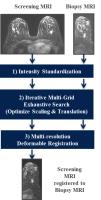
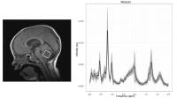
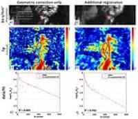
Co-registration of pre-biopsy and biopsy MRIs to facilitate lesion localization for MR-guided breast biopsies
Mirabela Rusu1, Elizabeth A. Morris2, Elizabeth J. Sutton2, and Ileana Hancu1
1GE Global Research, Schenectady, NY, United States, 2Memorial Sloan Kettering Cancer Center, New York, NY, United States
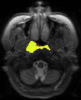
Lesion identification in MR-guided biopsy exams can be hampered by many factors, including large deformations and limited tissue perfusion due to breast compression. Multiple post-contrast scans, image subtraction and maximum intensity projection map generation may be needed to relocate the lesion. This preliminary study suggests that non-rigid registration between the (uncompressed breast) pre-biopsy series and the (compressed breast) biopsy series may facilitate fast and accurate lesion (re)localization, even with limited/absent lesion enhancement. 


2472.   
The test-retest reliability of fat-water ratio MRI derived breast density measurements and automated breast segmentation
Jie Ding1, Patricia A Thompson2, Marilyn T Marron3, Maria Altbach3,4, Denise Roe3,5, Jean-Philippe Galons4, Cynthia A Thomson3, Fang Wang6, Alison Stopeck7, and Chuan Huang1,8,9
1Biomedical Engineering, Stony Brook University, Stony Brook, NY, United States, 2Pathology, Stony Brook Medicine, Stony Brook, NY, United States, 3Cancer Center, University of Arizona, Tucson, AZ, United States,4Medical Imaging, University of Arizona, Tucson, AZ, United States, 5Epidemiology and Biostatistics, University of Arizona, Tucson, AZ, United States, 6Stony Brook Medicine, Stony Brook, NY, United States,7Hematology and Oncology, Stony Brook Medicine, Stony Brook, NY, United States, 8Radiology, Stony Brook Medicine, Stony Brook, NY, United States, 9Psychiatry, Stony Brook Medicine, Stony Brook, NY, United States
It has been shown that breast density (BD) value derived from fat-water-ratio MRI (FWR-MRI) strongly correlates with standard digital mammogram derived BD. The fact that no ionizing radiation is associated with FWR-MRI makes it a lower-risk modality for long term BD monitoring and clinical trials. However, data regarding the individual and group level variability and reliability of this method needs to be established.   Conventional approaches for FWR-MRI derived BD rely on manually drawn regions-of-interest. These processes are cumbersome and prone to measurement bias, which may limit the application of FWR-MRI derived BD. Automated breast segmentation has been proposed to resolve this problem and limited results to date are promising. Additional data including an evaluation of BD reliability from manual versus automated measurements is still needed.   
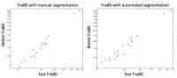
In this study, we evaluate the test-retest reliability of the FWR-MRI derived BD and the quality of data using manual versus automated breast segmentation. Our results demonstrate the high reliability of the FWR-MRI derived BD measure, Fra80, with a typical error of less than 0.02 for both automated and manual breast segmentation. Moreover, our automated breast segmentation protocol yields more reliable Fra80 BD measures compared to the labor-intense manual segmentation method.


2473.   
Dependence of Breast Pharmacokinetic Parameters on pre-contrast T1 and flip angle
Subashini Srinivasan1, Bruce L Daniel1, and Brian A Hargreaves1
1Radiology, Stanford University, Stanford, CA, United States
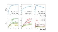
Pharmacokinetic (PK) models have been used to estimate physiological parameters such as permeability and dispersion of the contrast agent and is estimated using the acquired signal, pre-contrast T10, and the acquisition flip angle. In this work, we have determined the dependence of the dispersion models’ and Tofts models’ PK parameters on T10 and B1 maps, as well as the errors introduced by using constant T10 and B1 values in 11 biopsy-proven tumors. Our results show that PK parameters such as kep of Tofts model and kappa of mLDRW dispersion model are less dependent on T10 and B1 and could potentially be used with higher accuracy and precision even when T10 and B1 maps are not acquired.


2474.   
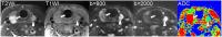
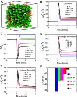
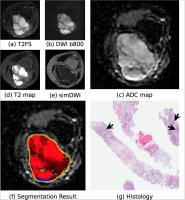
Estimation of breast tumour tissue diffusion parameters from histological images and Monte-Carlo simulations
David Naves Sousa1, Filipa Borlinhas1, and Hugo Alexandre Ferreira1
1Institute of Biophysics and Biomedical Engineering, Faculdade de Ciências da Universidade de Lisboa, Lisboa, Portugal
Diffusion-Weighted Imaging is a MRI technique that is able to distinguish between benign and malignant breast tumours via the Apparent Diffusion Coefficient (ADC). Nevertheless, this parameter provides very limited information regarding tissue microstructure. Here, is presented an approach to estimate the intracellular (Di) and extracellular (De) diffusion coefficients, and cell membrane permeability of tumour tissues which makes use of known ADC values, histological images and Monte-Carlo simulations of diffusion processes. Results show that distinct combinations of (Di, De, P) correlate with tumour type, and that a decreased De was observed in malignant tumours in agreement with known extracellular matrix changes.


2475.   
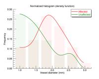
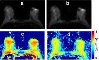
Magnetic resonance lymphangiography in breast cancer related lymphoedema shows differences between affected and unaffected arms
Marco Borri1, Maria A. Schmidt1, Julie C. Hughes1, Erica D. Scurr1, Kristiana D. Gordon2,3, Peter S. Mortimer2,3, Dow-Mu Koh1, and Martin O. Leach1
1CR-UK Cancer Imaging Centre, The Royal Marsden NHS Foundation Trust and The Institute of Cancer Research, London, United Kingdom, 2Cardiac and Vascular Sciences, St. George’s, University of London, London, United Kingdom, 3Skin Unit, The Royal Marsden NHS Foundation Trust, London, United Kingdom
The pathophysiology of breast cancer related lymphoedema (BCRL) is not well understood, one of the main limiting factors being a lack of information on lymphatic collecting vessels. We have recently proposed a novel contrast-enhanced magnetic resonance lymphangiography protocol which allows the identification of lymphatics via the use of associated contrast uptake curves. In this work we have quantified differences between affected and unaffected arms in a cohort of patients with unilateral BCRL. Our analysis did not detect significant differences in vessel counts between the two sides within different sections of the forearm. However, there was a statistically significant difference in vessel diameter between the two arms; lymphatics within the affected arms presented with a larger diameter.


2476.   
Reproducibility of quantitative magnetization transfer imaging of the healthy breast at 3T
Lori R. Arlinghaus1, Richard D. Dortch1,2, Jennifer G. Whisenant2, Hakmook Kang3, and Thomas E. Yankeelov1,2
1Institute of Imaging Science, Vanderbilt University, Nashville, TN, United States, 2Department of Radiology and Radiological Sciences, Vanderbilt University, Nashville, TN, United States, 3Department of Biostatistics, Vanderbilt University, Nashville, TN, United States
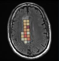
Magnetization transfer (MT) imaging is sensitive to changes in the macromolecular content of tissue and is, therefore, gaining increased attention as a noninvasive approach to probe the complex tumor environment in cancer. The ratio of macromolecular protons to the protons in the free water pool, or pool size ratio (PSR), can be quantified with quantitative MT (qMT) imaging and may be useful for detection of changes in macromolecular content early in the course of treatment. In this study, we explore the repeatability of PSR measurements in healthy breast fibroglandular tissue at 3T to serve as a benchmark for future longitudinal studies of breast cancer treatment.


2477.   
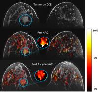
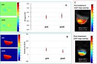
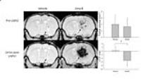
Amide CEST at 7T: A possible biomarker for response to neoadjuvant chemotherapy in breast cancer
Erwin Krikken1, Moritz Zaiss2, Vitaliy Khlebnikov1, Hanneke W.M. van Laarhoven3, Dennis W.J. Klomp1, and Jannie P. Wijnen1
1Radiology, University Medical Center Utrecht, Utrecht, Netherlands, 2Deutsches Krebforschungszentrum, Heidelberg, Germany, 3Medical Oncology, Academic Medical Center Amsterdam, Amsterdam, Netherlands
Neoadjuvant chemotherapy has an important role in the treatment of breast cancer and the need for early detection of treatment response is high. As a non-invasive method able to predict treatment response is lacking, we investigated the feasibility of using amide CEST MRI at 7T as a biomarker. Six patients were included after informed consent was given. The ATP signal was robust and repeatedly detectable in the same patient. Significant differences were seen in amide signal before  and after the first cycle of chemotherapy.


2478.   
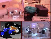
A compact and easy to handle set-up for high quality MR Elastography of the breast.
Jurgen H Runge1, Jules L Nelissen2,3, Larry de Graaf2, Barbara Molenkamp4, Suzan van der Meij4, Klaas Nicolay2, Gustav J Strijkers3, Jaap Stoker1, Anneloes E Bohte1, Aart J Nederveen1, Ondrej Holub5, and Ralph Sinkus5
1Radiology, Academic Medical Center, Amsterdam, Netherlands, 2Biomedical NMR, Eindhoven University of Technology, Eindhoven, Netherlands, 3Preclinical and Translational MRI, Academic Medical Center, Amsterdam, Netherlands, 4Surgery, Academic Medical Center, Amsterdam, Netherlands, 5Biomedical Engineering, King's College London, London, United Kingdom
Distinction between benign and malignant breast lesions remains difficult with conventional (dynamic) contrast-enhanced MRI. MR Elastography (MRE) can distinguish benign and malignant tissues based on their viscoelastic properties but breast MRE has not found widespread use in daily clinical practice, because of the complex equipment required and cumbersome data acquisition. Here we present a compact, easy to handle breast MRE set-up that allows the acquisition of high quality, artefact-free MRE data. This set-up was designed, built and tested at two different institutions in volunteers and a patient.


2479.   
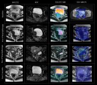
Dual-Parametric MR Imaging with Read-Out Segmented Diffusion-Weighted and High Temporal Resolution Dynamic Contrast-Enhanced Imaging Improves the Differentiation of Malignant and Benign Breast Lesions
Bin Wu1,2, Yanqiong Chen2, Hui Liu3, Xu Yan3, Caixia Fu4, Dan Wang1, Jian Mao2, Dominik Nickel5, Berthold Kiefer5, Yajia Gu2, and Weijun Peng2
1Radiology, Shanghai Proton and Heavy Iron Center, Fudan University Caner Center, Shanghai, China, People's Republic of, 2Radiology, Fudan University Shanghai Cancer Center, Shanghai, China, People's Republic of,3NEA MR Collaboration, Siemens Ltd, Shanghai, China, People's Republic of, 4Siemens Shenzhen Magnetic Resonance Ltd, Shenzhen, China, People's Republic of, 5Siemens Healthcare GmbH, Erlangen, Germany, Forchheim, Germany
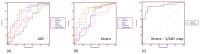
We investigated the clinical value of a dual-parameter classification method in differentiating benign and malignant breast lesions using readout-segmented diffusion-weighted imaging (RS-DWI) and quantitative dynamic contrast-enhanced magnetic resonance imaging (DCE-MRI), and found they correlated with histological results.


2480.   
Proton Density Water Fraction as a Measurement of Breast Fibroglandular Tissue Volume and Concentration
Roberta M Strigel1,2,3, Leah Henze Bancroft2, Diego Hernando1, and Scott B Reeder1,2,3,4,5,6
1Radiology, University of Wisconsin, Madison, WI, United States, 2Medical Physics, University of Wisconsin, Madison, WI, United States, 3Carbone Cancer Center, University of Wisconsin, Madison, WI, United States,4Biomedical Engineering, University of Wisconsin, Madison, WI, United States, 5Emergency Medicine, University of Wisconsin, Madison, WI, United States, 6Medicine, University of Wisconsin, Madison, WI, United States
Elevated breast density confers an increased risk for breast cancer. Accurate and precise measurement of the amount of fibroglandular breast tissue has potential to serve as a quantitative imaging biomarker of risk for the development of breast cancer.  In this work we introduce novel, confounder corrected chemical-shift encoded (CSE)-MRI techniques to measure the proton density water fraction (PDWF). Estimation of PDWF with CSE-MRI addresses potential confounders that negatively impact accuracy, precision, and reproducibility, enabling protocol independent quantification of the volume and concentration of fibroglandular tissue in the breast.


2481.   
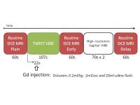
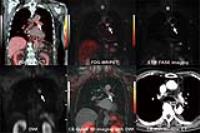
Utility of semi-quantitative analysis of initial enhancement using TWIST-VIBE in the diagnosis of breast lesions
Mariko Goto1, Koji Sakai1, Kayu Takezawa1, Hiroshi Imai2, Elisabeth Weiland3, and Kei Yamada1
1Radiology, Kyoto Prefectural University of Medicine, Kyoto, Japan, 2Siemens Japan K.K., Tokyo, Japan, 3Siemens Healthcare GmbH, Erlangen, Germany
The prototype TWIST-VIBE sequence improves the temporal resolution of breast MRI while preserving spatial resolution. High-temporal resolution TWIST-VIBE was performed during the initial enhancement phase and high-spatial resolution routine DCE MRI in a single session, and whether the additional information of initial enhancement analysis using TWIST-VIBE improved the diagnostic accuracy of breast MRI was evaluated. The combination of BI-RADS and new parameters of initial enhancement (MS and TTE) calculated from TWIST-VIBE has the potential to increase the specificity of breast MRI and may be useful as additional information to determine the need for biopsy.


2482.   
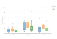
Application of Histogram Analysis of Pharmacokinetic Parameters in Dynamic Contrast-Enhanced MR Imaging of Breast lesions with CAIPIRINHA-Dixon-TWIST-VIBE Technique
Yiqi Hu1, Tao Ai1, and Liming Xia1
1Tongji Hospital, department of radiology, Wuhan, China, People's Republic of
The overlap of pharmacokinetic parameters values exists between benign and malignant lesions. Most previous studies chose mean pharmacokinetic parameters when elevating the state of breast lesions perfusion. However, tumors are heterogeneous that are marked by microenvironmental factors and thus manifests as radiologic heterogeneity. The mean pharmacokinetic parameter values may overlook the subtle but important difference between breast lesions. Thus, the aim of our study is to investigate the feasibility of histogram 
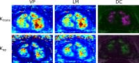
analysis of pharmacokinetic parameters including Ktrans, kep, ve in breast DCE-MRI imaging and determine which metric of each pharmacokinetic parameter may best help differentiate benign from malignant lesions.


2483.   
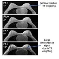
Influence of fat suppression to evaluate T1 values in breast cancer: assessing the reliability of pharmacokinetic parameters
Kayu Takezawa1, Mariko Goto1, Koji Sakai1, Hiroyasu Ikeno1, Katsuhiko Nakatsukasa2, Hiroshi Imai3, and Kei Yamada1
1Radiology, Kyoto Prefectural University of Medicine, Kyoto, Japan, 2Breast Surgery, Kyoto Prefectural University of Medicine, Kyoto, Japan, 3Siemens Japan K.K, Tokyo, Japan
     The influence of fat suppression on T1 values and pharmacokinetic parameters in breast cancer were evaluated using a prototype Dixon-TWIST-VIBE technique. We measured T1 values of breast cancers on both fat suppression and not-fat suppression data sets and we calculated Ktrans values using same ROI that employed on T1 value measurements. Our result suggests that the fat suppression might influence T1 values in breast cancer, and reliability of Ktrans seemed inappropriate as an absolute value. On the other hand, the assessment of intra-patient Ktrans change might be feasible.


2485.   
3D MRI Breast Density Change in Women with Hormonal Positive Breast Cancer Following Adjuvant Hormonal Therapy
Yoon Jung Choi1, Jeon-Hor Chen2,3, Shunshan Li2, Po-Han Chen4, Pei-Yu Liu4, Inyoung Youn1, and Min-Ying Su2
1Department of Radiology, Kangbuk Samsung Hospital, Seoul, Korea, Republic of, 2Center for Functional Onco-Imaging, Department of Radiological Sciences, University of California Irvine, Irvine, CA, United States,3Department of Radiology, Eda Hospital and I-Shou University, Kaohsiung, Taiwan, 4Department of Medical Imaging, China Medical University, Taichung, Taiwan
Hormonal regimens may affect breast tissue with the change of breast volume or composition. This study was to apply a well-established breast and fibroglandular tissue segmentation method to analyze the density changes in patients receiving adjuvant hormonal therapy. The results showed that pre-menopausal women had a higher density reduction, presumably due to their more abundant fibroglandular tissues that can be decreased, but a high variation was observed. The density reduction assessed by 3D MRI may be used as a surrogate marker to correlate with metabolic genotyping, and further used in combination to better predict patient’s prognosis.


2484.   
Rapid T1 and T2 Measurements of Breast Tissue at 3T using Multi-TR, Multi-TE Spectroscopy
Leah C Henze Bancroft1, Roberta M Strigel1,2,3, Gavin Hamilton4, Scott B Reeder1,2,5,6,7, and Diego Hernando2
1Medical Physics, University of Wisconsin-Madison, Madison, WI, United States, 2Radiology, University of Wisconsin-Madison, Madison, WI, United States, 3University of Wisconsin Carbone Cancer Center, University of Wisconsin-Madison, Madison, WI, United States, 4Radiology, University of California, San Diego, San Diego, CA, United States, 5Medicine, University of Wisconsin-Madison, Madison, WI, United States, 6Biomedical Engineering, University of Wisconsin-Madison, Madison, WI, United States, 7Emergency Medicine, University of Wisconsin-Madison, Madison, WI, United States
The highly heterogeneous distribution of fat and fibroglandular tissue in the breast makes obtaining accurate measures of T1 and T2 relaxation times difficult.  Here, a rapid, multi echo, multi TR spectroscopy sequence is used to measure the T1 and T2 relaxation times of fat and fibroglandular tissue in the breast at 3T.  Partial voluming effects are accounted for through accurate measurement of the proton density fat fraction.


2486.   
Does the Initial Enhancement Ratio (IER) Predict which Malignancies are Biologically Significant on a Pre-operative Breast MRI?
Neeti R Bagadiya1, Laura Heacock1, Yiming Gao1, Meghan Jardon1, Samantha Heller1, and Linda Moy1
1Radiology, New York University, New York, NY, United States
Breast MRI allows preoperative identification of patients who may have extensive disease at presentation and allows for appropriate surgical planning and treatment.   Despite the high sensitivity of MRI, the role of preoperative surgical staging of breast cancer patients is controversial. There is concern that the high false positive rates of breast MRI lead to additional biopsy procedures and surgeries [1,2]. Abbreviated breast MRI (AB-MR), defined as the first post-contrast scan, has been proposed as an exam that may have a higher specificity compared to conventional breast MRI  [3,4].  Two recent studies show that AB-MR has a high PPV for and may preferentially selects for biologically significant tumors, thereby reducing overdiagnosis and overtreatment. The concept of a biologically significant breast cancer has not been defined.  We hypothesized that since invasive carcinomas usually demonstrate fast initial uptake of contrast, a threshold of enhancement as determined by initial enhancement ratio (IER) may be associated with the identification of biologically significant breast cancers [5].  We evaluated a cohort of women with known cancer who underwent MRI guided needle localization (MRNL) for a finding that was suspicious for additional disease.  We examined whether there was an association with the IER and the likelihood that it would be detected on AB-MR exam.  Using Dynacad software we retrospectively reviewed the IER of MRI detected synchronous cancers that underwent MRNL.  We found there is a significant correlation between invasive cancers and IER that can aid in the detection of biologically significant synchronous cancers on MRI.
 

The International Society for Magnetic Resonance in Medicine is accredited by the Accreditation Council for
Continuing Medical Education to provide continuing medical education for physicians.