ISMRM 24th Annual Meeting & Exhibition • 07-13 May 2016 • Singapore

Traditional Poster Session: Cardiovascular

2525 -2545 CV Novel Techniques
2546 -2553 Atherosclerosis Imaging
2554 -2581 MR Angiography
2582 -2605 Velocity & Flow Quantification
2606 -2617 Myocardial Perfusion & Function
2618 -2634 Cardiovascular Image Processing


2525.   
A Low cost Cardiac Phantom for Evaluation of Motion and Thermometry
Shivaprasad Ashok Chikop1, Amaresha Shridhar Konar1, Nithin Vajuvalli1, Darshan Shivaram Keelara1, Ashwini Kumnoor1, Sairamesh raghuraman2, Ramesh Venkateshan2, and sairam Geethanath1
1Medical Imaging Research Centre, Dayananda Sagar Institutions, Bangalore, India, 2Wipro GE Healthcare, Bangalore, India
The goal of the work was to build a low cost cardiac phantom that mimics cardiac motion and measure thermometric profile based on applied B1+ fields during MR imaging. The cardiac motion inside the phantom was simulated using the mechanical gear setup. To obtain the thermometric profile of the cardiac phantom 12 probes were inserted into the phantom with required thermal insulation.  The phantom also provides an opportunity to correlate local SAR findings with temperature measurements in the heart phantom. 


2526.   
Interactive cardiac CINE-MRI using an intuitive 3D navigation system
Andrew James Patterson1, Martin John Graves1, and David John Lomas1
1Radiology, University of Cambridge & Addenbrooke’s Hospital, Cambridge, United Kingdom
This work describes the development of a novel approach for prescribing cardiac scan plane locations which will benefit cardiac examinations where the anatomy is abnormal (for example, in congenital anomalies). We have developed a desktop sized constrained surface controller which mimics an ultrasound transducer. This enables retrospective reformatting of a 3D localizer image to determine the desired cardiac view. The cardiac scan plane is then prospectively transferred to a 2D CINE bSSFP sequence to enable interactive assessment of cardiac function. Initial testing has established that our approach allows us to replicate standard cardiac views.


2527.   
Comparing the impact of through-plane motion during the cardiac cycle on steady state signal evolution in cine 2D and 3D balanced steady state free precession
Tyler Joseph Spear1, Tori Stromp1, Steve Leung2, and Moriel Vandsburger1
1Saha Cardiovascular Research Center, University of Kentucky, Lexington, KY, United States, 2Gill Heart Imaging Center, University of Kentucky, Lexington, KY, United States
This project uses cardiac magnetic resonance to compare cine balance steady state free precession signals between 2D and 3D acquisitions in order to better understand how through-plane motion of tissues impacts the change in signal through the cardiac cycle.  As quantitative techniques become more prevalent and refined, it is imperative to be able understand the evolution of these signals to eliminate results due to physical motion of the heart that would skew the results of otherwise diagnostic tests.


2528.   
Evaluation of Cardiac Function in Type-1 Diabetes Using Magnetic Resonance Imaging
El-Sayed H. Ibrahim1, Jadranka Stojanovska1, Scott Swanson1, Claire Duvernoy1, and Rodica Pop-Busui1
1University of Michigan, Ann Arbor, MI, United States
This study evaluates the association between MRI-derived parameteers of cardiac function in Type-1 diabetes (T1DM) at baseline and 3-years follow-up, and compares the results to healthy controls (HC). There were no differences between T1DM and HC at baseline in the measured variables. In T1DM, only left-ventricular (LV) mass-ratio and basal circumferential strain significantly decreased from baseline to follow-up. Mitral early-to-atrial filling-rate (representing diastolic function) increased, and apical torsion decreased from baseline to follow-up, although these differences were not significant. Among the parameters that showed differences between baseline and follow-up, only LV mass-ratio showed significant difference between female and male patients.


2529.   
Development of the phantom to measure the image distortion in the magnetic resonance angiography  and verification of the relationship between geometric positions and the image distortion
Atsushi FUKUYAMA1, Haruo ISODA2, and Shuji KOYAMA2
1Radiological Sciences, Nagoya University Graduate School of Medicine, Nagoya, Japan, 2Brain & Mind Research Center, Nagoya University, Nagoya, Japan
Our objective is to develop a phantom that can evaluate the image distortion along the body axis objectively, and elucidate the relationship between the occurrence and geometrical position of image distortion in MRA. As a result, the phantom that we have developed has made it possible to evaluate image distortion objectively over a wide range of the trunk. We made clear that when separated from the center of the magnetic field in vertical or horizontal directions, image distortion became larger with the increase in distance. 


2530.   
Altered Aortic Geometry in Pediatric Patients with Marfan Syndrome
Roel LF van der Palen1,2, Julio Garcia2, Alex J Barker2, Michael J Rose3, Luciana Young4, Arno AW Roest1, Michael Markl2,5, Joshua D Robinson4,6, and Cynthia K Rigsby3
1Department of Pediatric Cardiology, Willem-Alexander Children and Youth Center, Leiden University Medical Center, Leiden, Netherlands, 2Department of Radiology, Feinberg School of Medicine, Northwestern University, Chicago, IL, United States, 3Department of Medical Imaging, Ann & Robert Lurie Children’s Hospital of Chicago, Chicago, IL, United States, 4Division of Pediatric Cardiology, Ann & Robert Lurie Children’s Hospital of Chicago, Chicago, IL, United States, 5Department of Biomedical Engineering, McCormick School of Engineering, Northwestern University, Chicago, IL, United States, 6Department of Pediatrics, Ann & Robert Lurie Children’s Hospital of Chicago, Chicago, IL, United States
Marfan syndrome (MFS) is a connective tissue disease with high risk of aortic dissection/rupture. Two-thirds of dissections originate in the ascending aorta (AAo), one-third in the descending aorta (DAo). Diameter plays an important role in risk stratification. However, aortic dimension alone may not capture the complex changes in aortic geometry that are often encountered in MFS patients, e.g. elongation and changes in overall shape of the aorta. Aortic 3D geometry was systematically investigated in a cohort of children and adolescents with MFS and was compared to an age appropriate control cohort: altered aortic geometry in pediatric MFS patients was detected.


2531.   
Quantitative Measures of Right Ventricular Shape Abnormalities in ARVC Patients from CMR
Kristin Mcleod1,2, Samuel Wall1,2, Jørg Saberniak2,3, and Kristina Haugaa2,3
1Computational Cardiac Modelling Department, Simula Research Laboratory, Oslo, Norway, 2Centre for Cardiological Innovation, Oslo, Norway, 3Department of Cardiology, Oslo University Hospital, Rikshospitalet, Oslo, Norway
Better understanding of the impact that disease has on ventricular shape remodeling and improved tools for quantifying disease severity are needed in complex diseases such as ARVC. We propose a method to automatically characterise right ventricular shape features in ARVC patients using statistical methods applied to shapes extracted from CMR images in a population of 27 ARVC patients. In addition to characterizing the typical shape features in ARVC, the level of severity of any given shape feature (e.g. dilation) can be measured in a new patient to quantify the degree in which that specific feature is present in that patient.


2532.   
Long-Term Effect of Persistent Microvascular Obstruction on Adverse Post-Infarction Ventricular Remodeling via Prolonged Iron-Driven Inflammatory Process: a Cardiac Magnetic Resonance Study with Ex-vivo Validation
Ivan Cokic1, Avinash Kali1, Hsin-Jung Yang1, Richard Tang1, Frank S Prato2, David Underhill1, and Rohan Dharmakumar1
1Cedars-Sinai Medical Center, Los Angeles, CA, United States, 2Lawson Health Research Institute, London, ON, Canada
Hemorrhagic myocardial infarctions (MI) are associated with persistence of iron depositions in the chronic MI phase. These iron deposits have been shown to lead to perpetual recruitment of new monocytes/macrophages into the infarcted territory. Hemorrhagic MI is accompanied by a persistent microvascular obstruction (PMO) but the converse is not true. In this histologically validated cardiac MRI study we demonstrate that even in the absence of hemorrhage, PMO per se can lead to chronic iron-driven inflammatory response. Our findings are expected and lend improved insight into the mechanisms driving heart failure in infarction with PMO.


2533.   
MRI relaxation parameters predict functional outcome after experimental myocardial infarction
Sebastian Haberkorn1, Christoph Jacoby1, and Ulrich Flögel1
1Molecular Cardiology, Heinrich Heine University, Düsseldorf, Germany
In the present study, we systematically compared myocardial tissue characterization by Gd-based techniques with intrinsic T1/T2 mapping and their correlation with local cardiac function after experimental myocardial infarction (MI). We found that pre-contrast T1 mapping with variable flip angle analysis carried out 1 day after MI predicts the functional outcome after 21 days at least as reliable as LGE. Compared to the latter technique providing plain signal enhancement, the current approach determines quantitative maps with a large dynamic range, which may pave the way for reliable myocardial tissue characterization without any CA.


2534.   
MRI characterisation of iron overload in a humanised mouse model of ß-thalassemia major
Laurence H Jackson1, Evangelia Vlachodimitropoulou Koumoutsea2, Panicos Shangaris3, Thomas M Ryan4, Anna L David3, John Porter2, Daniel J Stuckey1, and Mark F Lythgoe1
1Centre for Advanced Biomedical Imaging, University College London, London, United Kingdom, 2Dept. Haematology, University College London, London, United Kingdom, 3Institute for Women’s Health, University College London, London, United Kingdom, 4Dept. Biochemistry and Molecular Genetics, University of Alabama, Birmingham, AL, United States
β-thalassemia major is a common blood disorder causing the production of abnormal red blood cells (RBCs) leading to severe anaemia. Current treatment of the disease consists of regular blood transfusions with the side effect of iron overload. Due to difficulties in producing a representative animal model of the disease, research into experimental therapy has been limited. A recently developed humanised mouse model of thalassemia has the potential to provide a platform for the development of new treatments. Here we determine whether magnetic resonance imaging (MRI) can be used to assess and quantify biomarkers of disease in-vivo.


2535.   
The Effect of Hypertrophy in CardioCEST Magnetization Transfer Contrast
Scott William Thalman1, Zhengshi Yang2, Ashley Pumphrey2, and Moriel Vandsburger1,2,3
1Department of Biomedical Engineering, University of Kentucky, Lexington, KY, United States, 2Saha Cardiovascular Research Center, University of Kentucky, Lexington, KY, United States, 3Department of Physiology, University of Kentucky, Lexington, KY, United States
Remodeling of the myocardium via hypertrophy and fibrosis increases the risk of adverse cardiac events. Quantitative magnetization transfer weighted imaging has shown promise as a method to identify fibrosis, however the effects of hypertrophy on such measures remains unknown. Using a murine model of chronic Angiotensin-II stimulation characterized by robust hypertrophy with little fibrosis, we demonstrate that despite large increases in myocardial mean cross-sectional area (AngII= 4825μm2±717 vs Saline= 2372μm2±158), no change occurs in the cardioCEST derived measure of magnetization transfer ratio (MTR) (AngII= 30.8%±7.3 vs Saline= 27.2%±8.6). Thus, increases in MTR due to fibrosis are unbiased by concomitant hypertrophy.


2536.   
Metabolic alterations in chemotherapy-induced cardiotoxicity revealed by in vivo hyperpolarized 13C-MRS
Michael S Dodd1, Vicky Ball1, and Damian J Tyler1
1Department of Physiology, Anatomy and Genetics, University of Oxford, Oxford, United Kingdom
Doxorubicin can lead to pre-clinical and clinical heart failure and reduced myocardial energetics. Alterations in metabolism potentially play a role in chemotherapy induced heart failure. The aim of this work was to assess the in vivo metabolic phenotype of doxorubicin treated hearts using hyperpolarized MRS. Doxorubicin cardiotoxicity was induced in Wistar rats and 4 weeks later, CINE-MRI and hyperpolarized [1-13C]pyruvate MRS was performed.  Doxorubicin resulted in significant wall thinning, and reductions in both cardiac function and volume, all indicative of cardiotoxicity. Interestingly, doxorubicin resulted in significant metabolic alterations; with reductions in both PDH flux and 13C label incorporation into alanine.


2537.   
Advanced multi-GPU-based MR simulations (MRISIMUL) on realistic human anatomical models (XCAT)
Christos G. Xanthis1,2, Panagiotis G. Papadimitroulas3, George C. Kagadis3, and Anthony H. Aletras1,4
1Lund Cardiac MR Group, Department of Clinical Physiology, Lund University, Lund, Sweden, 2Department of Computer Science and Biomedical Informatics, University of Thessaly, Lamia, Greece, 3Department of Medical Physics, School of Medicine, University of Patras, Patras, Greece, 4Laboratory of Medical Informatics, School of Medicine, Aristotle University of Thessaloniki, Thessaloniki, Greece
In the past, MR simulations were usually confined to incorporation of simplified anatomical models of limited realism and/or simulations of basic MR physics concepts only. The purpose of this study was the incorporation of the XCAT anatomical phantom in MRISIMUL, a high performance multi-GPU simulation platform. Two different CMR applications were studied: the first for heart morphology evaluation and the second for T1 mapping of the heart. Results showed that the incorporation of realistic human anatomical models in a high performance MR simulation platform may benefit the design and optimization of MR protocols and pulse sequences in the future.


2538.   
In Vivo, High-Frequency 3D Cardiac Magnetic Resonance Elastography:  Feasibility in Normal Volunteers
Arvin Arani1, Kevin J. Glaser1, Shivaram Poigai Arunachalam1, Phillip J. Rossman1, David S. Lake1, Joshua D. Trzasko1, Armando Manduca1, Kiaran P. McGee1, Richard L. Ehman1, and Philip A. Araoz1
1Mayo Clinic, Rochester, MN, United States
Noninvasive stiffness imaging techniques (elastography) can image myocardial tissue biomechanics in vivo.  However, for cardiac magnetic resonance elastography (MRE) techniques the optimal vibration frequency for in vivo experiments is unknown. The purpose of this study was to determine the optimal vibration frequency for cardiac MRE in healthy volunteers. Cardiac MRE displacement fields can be imaged with mean OSS-SNR > 1.6 at frequencies as high as 180Hz, however, mean OSS-SNR values and myocardial coverage was shown to be highest at 140Hz across all subjects. This study motivates future evaluation of high-frequency 3D MRE in patient populations.


2539.   
In-vivo assessment of myocardial stiffness in a pig with induced myocardial infarction using 3D Magnetic Resonance Elastography
Shivaram Poigai Arunachalam1, Arvin Arani1, Francis Baffour1, Joseph Rysavy2, Phillip Rossman1, David Lake3, Kevin Glaser1, Joshua Trzasko1, Armando Manduca3, Kiaran McGee1, Richard Ehman1, and Philip Araoz1
1Radiology, Mayo Clinic, Rochester, MN, United States, 2Surgery, Mayo Clinic, Rochester, MN, United States, 3Physiology and Biomedical Engineering, Mayo Clinic, Rochester, MN, United States
Myocardial stiffness is a novel biomarker with both diagnostic and prognostic potential for a range of cardiac diseases such as myocardial infarction which is known to significantly increase stiffness. Magnetic Resonance Elastography (MRE) is a non-invasive imaging technique that can be applied to the heart for in-vivo myocardial tissue characterization.  The purpose of this pilot study was to assess the feasibility of measuring in-vivo stiffness changes in infarcted tissue and compare with remote (i.e. non-infarcted) myocardium in the same pig using 3D MRE.  Results indicate a 3-fold increase in stiffness of the infarct compared to the normal myocardium. 


2540.   
Dependence of RF Lesion Visibility in Native T1-weighted MRI on Time after RF Ablation
Eugene G. Kholmovski1,2, Ravi Ranjan2, Sathya Vijayakumar1,2, Nathan A. Angel2, and Nassir F. Marrouche2
1UCAIR, Department of Radiology, University of Utah, Salt Lake City, UT, United States, 2CARMA Center, University of Utah, Salt Lake City, UT, United States
Catheter RF ablation is a widely accepted procedure for treatment of cardiac arrhythmias. Recently, native (non-contrast) T1-weighted (T1w) MRI was proposed to characterize RF lesions immediately post-ablation. The main aim of this work was to study how the visibility and volume of RF lesions in native T1w MRI changes with time after ablation. We have found that reduction of T1 relaxation time of RF ablated myocardium is transient. The visibility of RF ablation lesions and volume of the corresponding enhanced regions in native T1w MRI drastically reduce few days after ablation.


2541.   
Assessment of Myocardial B0 over the Cardiac Cycle at 7.0T: Implications for Susceptibility-based CMR Techniques
Teresa Serradas Duarte1, Till Huelnhagen1, and Thoralf Niendorf1,2
1Berlin Ultrahigh Field Facility (B.U.F.F.), Max Delbrück Center for Molecular Medicine in the Helmholtz Association, Berlin, Germany, 2Experimental and Clinical Research Center, a joint cooperation between the Charité Medical Faculty and the Max Delbrück Center in the Helmholtz Associaiton, Berlin, Germany
Magnetic susceptibility-based methods are an emerging technique in CMR for myocardial tissue characterization. Making use of UHF MRI, temporally resolved myocardial T2* mapping was recently demonstrated. Since susceptibility weighted MRI is highly dependent on main magnetic field homogeneity, B0 assessment is crucial for interpretation of results. This pioneering study investigates B0 variation in the heart over the cardiac cycle at 7.0T and its implications for myocardial T2* mapping in a cohort of healthy volunteers. Results show that septal macroscopic field inhomogeneities are minor regarding their effects on T2*. This provides encouragement for temporally resolved susceptibility-based CMR at UHF.


2542.   
Accelerated Multi-contrast Late Enhancement with Isotropic Spatial Resolution Using Compressed Sensing with Edge Preservation for Improved Characterization of Infarct Heterogeneity: Initial Clinical Experience
Li Zhang1,2, Mihaela Pop1,2, and Graham A Wright1,2
1Department of Medical Biophysics, University of Toronto, Toronto, ON, Canada, 2Sunnybrook Health Sciences Centre, Toronto, ON, Canada
Accurate characterization of infarct heterogeneity depends on high spatial resolution imaging1. This study was performed to evaluate the use of an accelerated multi-contrast late enhancement acquisition (MCLE)2with COmpressed Sensing with Edge Preservation (COSEP) for isotropic spatial resolution imaging to improve characterization of infarct heterogeneity in the clinical setting. We have shown that an isotropic resolution of 2.2mm could be achieved using an accelerated three-dimensional (3D) MCLE acquisition within a single breath-hold in a patient study and the COSEP reconstruction provides improved characterization of infarct heterogeneity compared to an alternative compressed sensing method.


2543.   
Multi-parametric Cardiac Magnetic Resonance for Prediction of Cardiac Complications in Thalassemia Intermedia: a Prospective Multicenter Study
Antonella Meloni1, Nicola Giunta2, Pietro Giuliano2, Maria Giovanna Neri1, Stefania Renne3, Antonino Vallone4, Massimiliano Missere5, Emanuele Grassedonio6, Vincenzo Positano1, Daniele De Marchi1, and Alessia Pepe1
1Fondazione G. Monasterio CNR-Regione Toscana, Pisa, Italy, 2"ARNAS" Civico, Palermo, Italy, 3P.O. “Giovanni Paolo II”, Lamezia Terme (CZ), Italy, 4Az. Osp. "Garibaldi" Presidio Ospedaliero Nesima, Catania, Italy, 5P.O. “Giovanni Paolo II”, Campobasso, Italy, 6Policlinico "Paolo Giaccone", Palermo, Italy
We prospectively assessed the predictive value of MRI for cardiac complications in talassemia intermedia (TI). All our patients underwent MRI examination assessing multi-parametric findings (heart and liver iron, myocardial fibrosis, atrial areas, and biventricular function).

Righ ventricular (RV) hypertrophy identified patients at high risk for arrhythmias and pulmonary hypertension. Both RV hypertrophy and fibrosis detected by LGE were independent predictive factor for cardiac complications.  So, a widespread program using MRI exploiting its multi-parametric potential, including the measurement of RV mass,  can have considerable power for opening the prognosis of TI patients by the early identification and treatment of patients at risk for cardiac complications.


2544.   
Microvascular obstruction and hemorrhage influence T1 and T2 relaxation parameters in the detection of edema in acute myocardial infarction
Nilesh R. Ghugre1,2,3, Venkat Ramanan1, Jing Yang1, Idan Roifman3, Mohammad I Zia3, Bradley H Strauss3, Kim A Connelly4, and Graham A Wright1,2,3
1Physical Sciences Platform, Sunnybrook Research Institute, Toronto, ON, Canada, 2Department of Medical Biophysics, University of Toronto, Toronto, ON, Canada, 3Schulich Heart Research Program, Sunnybrook Health Sciences Centre, Toronto, ON, Canada, 4Cardiology, St. Michaels Hospital, Toronto, ON, Canada
T1 and T2 relaxation parameters have been instrumental in quantifying both edema and hemorrhage in acute myocardial infarction (AMI). Unlike T2, the combined effect of edema and hemorrhage on T1 has not been well described.  Our study assessed and compared the impact of microvascular obstruction (MVO) and hemorrhage on the sensitivity of T1 and T2 in the quantification of edema. Our study indicated that the capacity of T1 to detect edema is affected by the counteracting influence from hemorrhage. Furthermore, T2 may be more sensitive to edema than T1 in AMI, particularly in the presence of MVO and hemorrhage.


2545.   
Motion Averaged MR-Based Attenuation Correction for Coronary 18F-Fluoride Hybrid PET/MR
Philip M Robson1, Marc R Dweck1, Nicolas A Karakatsanis1, Maria Giovanna Trivieri2, Ronan Abgral1, Johanna Contreras2, Umesh Gidwani2, Jagat P Narula2, Valentin Fuster2, Jason C Kovacic2, and Zahi A Fayad1
1Translational and Molecular Imaging Institute, Icahn School of Medicine at Mount Sinai, New York, NY, United States, 2Department of Cardiology, Icahn School of Medicine at Mount Sinai, New York, NY, United States
Cardiac and coronary imaging using hybrid PET/MR is gaining increasing interest.  PET image reconstruction requires knowledge of the PET-photon attenuation of the object in order to produce accurate images of PET tracer activity.  The standard approach for MR-based attenuation correction is breath-hold volumetric imaging to freeze motion of the chest and abdomen.  However, for imaging the heart, alignment of anatomy during PET data collection and attenuation measurement is crucial.  In this work, we propose mapping attenuation using a free-breathing golden-angle radial gradient echo sequence and compare the PET images produced with this novel approach and the standard breath-hold approach.

2546.   
Prospective Acceleration and CS Reconstruction for 3D Isotropic High-Resolution Carotid Imaging
Bram F Coolen1, Abdallah G Motaal1, Wouter V Potters1, Maarten J Versluis2, Gustav J Strijkers3, and Aart J Nederveen1
1Department of Radiology, Academic Medical Center, Amsterdam, Netherlands, 2Philips Healthcare, Benelux, Netherlands, 3Department of Biomedical Engineering and Physics, Academic Medical Center, Amsterdam, Netherlands
We present a prospectively accelerated 3D black-blood sequence in conjunction with CS reconstruction and show the feasibility of 3D high-resolution carotid vessel wall imaging. Results show that acceleration in combination with CS reconstruction increased SNR efficiency and resulted in improved vessel wall visibility as compared to the full data acquisition. More importantly, high-resolution imaging carotid imaging (0.4-0.5 mm) was shown feasible within a clinically feasible imaging time of 4 min.  


2547.   
Measurement of Plaque Burden using 3D SNAP vessel wall MRI
Niranjan Balu1, Jie Sun1, Daniel S Hippe1, Huijun Chen2, and Chun Yuan1
1Radiology, University of Washington, Seattle, WA, United States, 2Tsinghua University, Beijing, China, People's Republic of
We have developed a single sequence (SNAP) for identification of high-risk features such as stenosis, intraplaque hemorrhage and juxtaluminal calcification. However measurement of plaque burden, an early marker for disease, required a separate black-blood scan. In this work, we demonstrate that plaque burden can be measured using an alternate reconstruction (SNAP2). In patients with atherosclerosis, we compare vessel wall area measurements on SNAP2 to established black-blood MRI measurements and show that our method provides results comparable to established plaque MRI methods.


2548.   
Artery Imaging with Flexible Contrast in a Single Scan using 3D Golden Angle Radial Sampling: Feasibility Demonstration in Carotid Arteries
Haikun Qi1, Shuo Chen1, Xinlei Pan1, and Huijun Chen1
1Center for Biomedical Imaging Research, School of Medicine, Tsinghua University, Beijing, China, People's Republic of
Multi-contrast imaging is needed for comprehensive characterization of atherosclerosis, which often involves a series of separate scans. But the mis-registration between scans due to motion and the long scan time limits its application. So acquisition of multi-contrast images in one scan is preferable. In this study, we developed an inversion recovery prepared 3D golden angle radial sampling sequence enabling flexible posteriori selection of TI time and frame duration for arbitrary-contrast reconstruction. The proposed method may be a one-stop solution for 3D large coverage plaque imaging by providing inherently co-registered multi-contrast images in a single scan within short scan time.


2549.   
Combined High Resolution, Four Dimensional, CE-MRA and DCE-MRI of Carotid Atherosclerotic Plaque
Jianmin Yuan1, Andrew J Patterson2, Ammara Usman1, Gregory C Makris1, Zhongzhao Teng1, Jonathan H Gillard1, and Martin J Graves1,2
1Department of Radiology, University of Cambridge, Cambridge, United Kingdom, 2Department of Radiology, Cambridge University Hospitals NHS Foundation Trust, Cambridge, United Kingdom
Carotid neovascularization and lumen surface morphology both play important roles in the progression of atherosclerotic plaque. This study uses a high-spatial and temporal resolution dynamic 3D contrast-enhanced (CE) imaging technique to simultaneously acquire and explore the relationship between the two factors. Results demonstrate that carotid luminal stenosis and appearance of ulceration are correlated with the plaque neovascularization. The study shows the ability to perform high-resolution carotid plaque morphology and functional assessment within a reasonable scanning time.


2550.   
Accumulation of Atherosclerotic Plaque in Mouse Aortic Valves Determined by MRI
Hanne Hakkarainen1, Jaana Rysä2, Anna-Kaisa Ruotsalainen1, Anna-Liisa Levonen1, and Timo Liimatainen1,3
1A.I. Virtanen Institute for Molecular Sciences, University of Eastern Finland, Kuopio, Finland, 2School of Pharmacy, University of Eastern Finland, Kuopio, Finland, 3Imaging Center, Kuopio University Hospital, Kuopio, Finland
High resolution cine-MRI was applied for examining aortic valve function and atherosclerotic plaque accumulation in the valves in mice on a high fat diet. The end systolic images of the aortic valves of these mice showed clear signs of atherosclerotic fat accumulation and dysfunction of the valves. This implies that the cine-MRI method provides a noninvasive tool for monitoring atherosclerotic plaques in the mouse aortic valves.  


2551.   
Feasibility of 3D multi-sequence PET/MRI of carotid atherosclerosis
Rik PM Moonen1, Stefan Vöö2, Jan Bucerius2, Joachim Wildberger1, and Eline Kooi1
1Department of Radiology, Maastricht University Medical Center +, Maastricht, Netherlands, 2Department of Nuclear Medicine, Maastricht University Medical Center +, Maastricht, Netherlands
Noninvasive imaging can improve risk stratification in carotid atherosclerosis. Multi-sequence MRI allows visualization of plaque burden and components, while PET can be used to study inflammation. Combining the two modalities with hybrid PET/MRI in a one-stop shop approach may improve assessment of vulnerable plaque.

The goal of this work was to optimize a 3D multi-sequence carotic PET/MRI protocol including 3D MPRAGE, 3D SPACE pre- and post-contrast, and UTE, using simulations and optimization in healthy volunteers.

Feasibility of the protocol was demonstrated in a patient suffering from carotid atherosclerosis (>70% stenosis).


2552.   
Quantitative Susceptibility Mapping of Atherosclerosis in Carotid Arteries
Chaoyue Wang1, Saifeng Liu2, Sagar Buch2, Hyun Seok Choi3, Eo-Jin Hwang3, Zhaoyang Fan4, and E. Mark Haacke1,2,5,6
1School of Biomedical Engineering, McMaster University, Hamilton, ON, Canada, 2The MRI Institute for Biomedical Research, Waterloo, ON, Canada, 3Department of Radiology, St. Mary’s Hospital, Seoul, Korea, Republic of, 4Department of Radiology, Cedars Sinai Hospital, Los Angeles, CA, United States, 5Department of Radiology, Wayne State University, Detroit, MI, United States, 6Biomedical Engineering, Northeastern University, Shenyang, China, People's Republic of
Due to complex structures in neck, such as the presence of bone and air, existing methods for neck quantitative susceptibility mapping (QSM) have several limitations. The purpose of this study was to find a reliable method for data collection in order to use QSM to detect carotid plaque and recognize vulnerable features. Therefor, we proposed a multi-echo SWI approach for data collection and regional 2D polynomial fitting method for data processing. Preliminary results show this method is able to image the vessel wall clearly and recognize calcified atherosclerosis and thrombosis by their susceptibility value. 


2553.   
Black- and Gray- Blood Dual Contrast Magnetic Resonance Imaging  Based on Compressed Sensing
Hanjing Kong1, Bo Li1, Huarui Du1, Jue Zhang1, and Li Dong2
1Peking university, Beijing, China, People's Republic of, 2Anzhen Hospital, Beijing, China, People's Republic of
Multi-contrast weighted imaging methods with a single scan provide efficient means for viewing and characterizing the atherosclerotic plaques. We extend previous studies by establishing a new strategy (CS-BLGDuC) and further reducing scan time with compressed sensing . 16 subjects including 6 healthy volunteers and 10 patients with carotid stenosis were carried out.The scan time of CS-BLGDuC were dramatically reduced compared with conventional 3D TOF and QIR. Superficial vascular calcification was clearly showed in gray blood images. It is feasible for the CS-BLGDuC strategy to depict superficial vascular calcifications and improve scan efficiency without decreasing image quality.

2554.   
Non-contrast-enhanced MRA using Velocity-sensitised, Acceleration-sensitised and Combined Sensitisation with Fast-Spin-Echo Readout
Andrew Nicholas Priest1, Ian G Murphy1, and David John Lomas1
1Radiology, Addenbrookes Hospital and Cambridge University, Cambridge, United Kingdom
This work develops an NCE-MRA technique based on the flow-sensitised dephasing (FSD) method but using a fast-spin echo (FSE) readout to avoid off-resonance artifacts and allow a larger field of view. The flow-preparation module may be velocity-sensitised, acceleration-sensitised or mixed velocity-and-acceleration-sensitised. The FSE readout allows these approaches to be combined with additional flow sensitisation by ‘adaptive refocus’ which reduces the refocusing flip angle in systole. These approaches performed well in healthy volunteers, in particular the velocity-sensitisation  and mixed-sensitisation approaches. Adding adaptive refocus increased venous contamination and thus reduced the arterial image quality. Future work will evaluate these methods in patients.


2555.   
Peripheral MR Angiography using velocity-sensitive gradient-echo technique
Dongchan Kim1, Hyunseok Seo1, Jaejin Cho1, Kinam Kwon1, and HyunWook Park1
1KAIST, Daejeon, Korea, Republic of
Recently quiescent-interval single-shot (QISS) non-contrast MR angiography (NC-MRA) technique was developed to obtain high-contrast peripheral angiographic images in a reasonable imaging time. QISS enhances contrast between the artery and unwanted signal by saturating signals from background, fat and vein. Thus, QISS needs multiple RF pulses for background saturation, which is limited in perfect saturation due to the inhomogeneity of main magnetic field. In this work, we propose a new MRA technique, which could generate peripheral angiogram without saturation pulses and an external ECG trigger using the velocity-sensitive gradient-echo (GRE) sequence.


2556.   
Single center experience with a prototype self-navigated 3D SSFP whole heart sequence in assessing coronary artery origin AUTHORS (LAST NAME
Arni Nutting1, Amos Varga-Szemes2, Shahryar Chowdhury1, Davide Piccini3, and Anthony Hlavacek1
1Pediatrics, Medical University of South Carolina, Charleston, SC, United States, 2Radiology, Medical University of South Carolina, Charleston, SC, United States, 3Lausanne, Switzerland
We performed a retrospective review of studies obtained using a prototype, self navigated, free breathing 3D SSFP sequence. Scans were reviewed for diagnostic sensitivity of coronary artery origin, diagnostic quality, and were graded for the severity of respiratory or cardiac motion or blood pool inhomogeneity. A diagnostic scan was obtained in 80.7% of cases. Blood pool inhomogeneity was common but very rarely affected diagnosis. The greatest factor affecting diagnostic ability was cardiac motion. We concluded that self-navigated 3D sequences can provide excellent sensitivity in diagnosing coronary origins with significant time savings compared to diaphragm navigated sequences.


2557.   
Selective Arterial Spin Labeling in Conjunction with Phase-encoded Information for the Simultaneous Visualization of Morphology, Flow Direction and Velocity of Individual Arteries in the Cerebrovascular System
Thomas Lindner1, Naomi Larsen1, Olav Jansen1, and Michael Helle2
1Clinic for Radiology and Neuroradiology, University Hospital Schleswig-Holstein, Kiel, Germany, 2Philips GmbH Innovative Technologies, Research Laboratories, Hamburg, Germany
In the presented study, selective Arterial Spin Labeling and Phase encoded information are obtained in a single acquisition, thereby allowing for artery-selective velocity and flow imaging. From the data, several image contrasts can be derived. First, anatomical images are available. From these, static artery-selective angiograms are obtained, which are processed as binary image masks and applied to the phase encoded images. Thereby, vessel-selective Phase encoded angiograms are created. Furthermore, it is possible to calculate venous-only images by subtraction of the arterial information derived from the ASL measurements from the phase-encoded images.


2558.   
3T Coronary MRI in Patients Treated with Bioresorbable Vascular Scaffolds for the Assessment of Vascular Patency and Blood Flow Velocity Quantification
Simon Reiss1, Axel Joachim Krafft1,2,3, Marius Menza1, Lisa Caroline Besch4, Timo Heidt4, Christoph Bode4, Constantin von zur Mühlen4, and Michael Bock1
1Dept. of Radiology - Medical Physics, University Medical Center Freiburg, Freiburg, Germany, 2German Cancer Consortium (DKTK), University Medical Center Freiburg, Heidelberg, Germany, 3German Cancer Research Center (DKFZ), Heidelberg, Germany, 4Department of Cardiology and Angiology I, University Heart Center, Freiburg, Germany
The implantation of bioresorbable Vascular Scaffolds (BVS) is a rapidly evolving technique in the treatment of coronary artery disease. BVS consist of a polylactate-based backbone which is completely dissolved after 2-3 years. Due to the organic material, coronary MRI can be used as a non-invasive technique for post-interventional imaging of arteries treated with BVS, which is not possible for metal stents. In this study, we evaluated the use of 3T MRI in assessing luminal patency after implantation of the BVS, as well as flow measurements for myocardial blood flow quantification as a possible indicator of post-interventional angina.


2559.   
A simulation study and application of UTE-?R2-?R2* combined MR whole brain angiogram using dual contrast superparamagnetic iron oxide nanoparticles.
HoeSu Jung1, SeokHa Jin1, DongKyu Lee1, SoHyun Han1, and HyungJoon Cho1
1Ulsan National Institute of Science and Technology, Ulsan, Korea, Republic of
  Transverse-relaxation-based ΔR2- and ΔR2*- micro MRAs are being investigated for imaging cerebral vasculature in rodent brains with increased sensitivity for intracortical arterioles and venules, in conjunction with exogenous blood pool contrast agents. In this study, we simulated extravascular signal decay behaviors of ΔR2, and ΔR2* values for multiple cylindrical models with varying diameters to quantitatively assess both sensitivity and size overestimation issues in micro MRA. The benefits of following synergistic combination of ΔR2, and ΔR2* angiograms along with the UTE-derived positive angiogram were investigated, and corresponding UTE-ΔR2-ΔR2* combined angiogram was applied to normal and C6 glioma tumor model for the verifications.


2560.   
Optimization of high resolution contrast-enhanced magnetic resonance angiography via modulation transfer function analysis
Toshimasa James Clark1, Gregory J Wilson2, and Jeffrey H Maki2
1Department of Radiology, University of Colorado Denver, Aurora, CO, United States, 2Department of Radiology, University of Washington, Seattle, WA, United States
CE-MRA spatial resolution is determined by a complex interaction between acquired voxel size, gadolinium injection rate, contrast volume, cardiac output, magnetic field strength, and acquisition duration. R1 relaxivity non-linearity and R2* signal degradation inherent in SPGR acquisitions play important roles in observed signal intensity during bolus passage. Through generation of modulation transfer functions spanning the gamut of parameter values likely to be seen in clinical practice we derive injection rates that produce optimal image contrast and resolution in our model.


2561.   
Flip-Angle and dose optimization in Ferumoxytol-enhanced MRA: Preliminary results compared with Gadolinium-enhanced MRA.
Tilman Schubert1,2, Utaroh Motosugi3, Samir Sharma4, Sonja Kinner1, Shane Wells1, Diego Hernando1, and Scott Reeder1
1Radiology, University of Wisconsin Madison, Madison, WI, United States, 2Clinic for Radiology and Nuclear Medicine, Basel University Hospital, Basel, Switzerland, 3Department of Radiology, University of Yamanashi, Yamanashi, Japan, 4Medical Physics, University of Wisconsin Madison, Madison, WI, United States
Ferumoxytol has gained interest as a positive MR-contrast agent in patients with renal failure. However, limited data exist regarding the optimal/minimal dose for MRA applications and optimal scanning parameters. Therefore, this study evaluated image quality with different doses of ferumoxytol with gadolinium as reference. Furthermore, flip angle optimization was performed in the steady state. Relative SNR was found to be significantly higher for gadolinium- compared to ferumoxytol-enhanced MRA during first pass. However, this did not lead to qualitative penalty for ferumoxytol-enhanced MRA. Flip angle-optimization indicated that adapting the flip angle to dose may help to achieve optimal results. 


2562.   
Sensitivity of cardiac magnetic resonance imaging to fluid shifts induced by an external leg compression device
Salvatore Saporito1, Ingeborg H.F. Herold 1,2, Silviu Dovancescu3, Jacques A. den Boer1, Ronald M. Aarts1,3, Arthur R. Bouwman2, Harrie C.M. van den Bosch4, Hendrikus H.M. Korsten 1,2, Hans C. van Assen1, and Massimo Mischi1
1Department of Electrical Engineering, Eindhoven University of technology, Eindhoven, Netherlands, 2Department of Anesthesiology and Intensive Care, Catharina Hospital Eindhoven, Eindhoven, Netherlands, 3Philips Research, Eindhoven, Netherlands, 4Department of Radiology, Catharina Hospital Eindhoven, Eindhoven, Netherlands
The assessment of thoracic fluid status is crucial for diagnosis, management, stratification, and follow-up of heart failure patients. Indicator dilution theoretical framework allows absolute volume estimation; magnetic resonance contrast agents have been proposed as indicators, with the advantage of a non-invasive detection. In this pilot study, we investigated the changes in intra-thoracic blood volume (ITBV) measured by cardiac magnetic resonance during fluid shifts induced by a pneumatic leg compression device. Preliminary results on 8 healthy volunteers suggest the sensitivity of the proposed measurement technique; a significant increase in ITBV after the leg compression was observed.


2563.   
Exploring Other Vascular Dimensions:  Comparison of 3-dimensional and 2-dimensional Vessel Wall Imaging Techniques for the Assessment of Large Artery Vasculopathies
Mahmud Mossa-Basha1, Matthew Alexander2, Jeffrey H. Maki1, Wendy Cohen1, Daniel S Hippe1, Chun Yuan1, Hannu Huhdanpa1, and Tobias Saam3
1Radiology, University of Washington, Seattle, WA, United States, 2Radiology, University of California San Francisco, San Francisco, CA, United States, 3Radiology, Ludwig-Maximilians-Universität München, Munich, Germany
Prompt diagnosis of large artery vasculopathies is important to avoid significant morbidities that can arise from delayed diagnosis.  This is difficult however, as patients frequently present with nonspecific sign and symptoms and luminal imaging techniques are limited in the detection of non-stenotic disease.  Vessel wall imaging is an emerging technique for vasculopathy evaluation throughout the body.  We compare quantitative and qualitative measures of 2D and 3D vessel wall imaging techniques in patients with suspected large artery vasculopathy.


2564.   
Ascending Aortic Nomograms derived from Contrast Enhanced Magnetic Resonance Angiography: Preliminary Values for Sex, Body Surface Area and Age Related Variability
Lara Mrak1, Christopher J. François1, Sonja Kinner1,2, and Mark L. Schiebler1
1Radiology, UW-Madison, Madison, WI, United States, 2University Hospital Essen, Essen, Germany
The size of the ascending aorta is of critical importance for patient survival as it is directly related to the likelihood of aortic dissection and risk of death.  The traditional imaging method that has been used for this measurement has been computed tomography of the chest. With many more children and young adults now being imaged with contrast enhanced magnetic resonance angiography examinations (CE-MRA) , it is useful to know what the range of variability is for the normal aorta on these exams. We present here age and sex specific values for nomograms of the ascending aorta  derived from non-gated CE-MRA examinations that are indexed to body surface area.


2565.   
Quadruple inversion-recovery high-resolution contrast-enhanced MR for vulnerable carotid plaque imaging
Zhefeng Yu1, Jianzhong Sun1, Yong Zhang2, and Minming Zhang1
1Radiology, Second Affiliated Hospital of Zhejiang University School of Medicine, Hangzhou, China, People's Republic of, 2MR Research China, GE Healthcare, Beijing, China, People's Republic of
This preliminary study found 25 carotid plaques in eighteen patients with 40-99% stenosis degree. Both readers gave their scores of enhanced QIR T1 weighted images (p>0.05). SNRs were evaluated as 109±21 and 113±18 (p>0.05) before and after the contrast enhancement. And CNRs were 98±19 and 82±22 (p>0.05) for the same comparison. Gd-enhanced QIR for carotid plaque imaging can achieve high quality due to good inhibition of blood flow.


2566.   
PETRA quiet MRA: Improved Robustness with 3D Elastic Cross-Registration
Yutaka Natsuaki1, Robert Grimm2, Xiaoming Bi1, David Grodzki2, Peter Schmitt2, and Gerhard Laub1
1Siemens Healthcare, Los Angeles, CA, United States, 2Siemens Healthcare, Erlangen, Germany
Previously, Ultra-Short TE sequences and their subtraction-based non-contrast MR angiography (e.g. PETRA qMRA) have shown promising results in intracranial applications, in particular with tortuous carotid arteries that has been problematic for the current clinical gold standard 3D Time Of Flight (TOF).  As with any subtraction based MRA techniques, PETRA qMRA is also sensitive to motion occurred in subsequent acquisitions.  The current work demonstrates that the proposed 3D elastic cross-registration can solve this limitation for the PETRA qMRA, and it takes to the next level in motion robustness and in the highest attainable isotropic resolution.  


2567.   
Utility of Non-contrast-enhanced MR Angiography for the Aneurysmal follow-up and Prediction of Endoleaks status post Endovascular Aortic Repair
Hiroshi Kawada1, Satoshi Goshima1, Kota Sakurai2, Yoshifumi Noda1, Kimihiro Kajita3, Nobuyuki Kawai1, Hiromi Ohno1, and Masayuki Matsuo1
1Radiology, Gifu university hospital, Gifu, Japan, 2Radiology, China Kosei Hospital, Seki, Japan, 3Radiology Service, Gifu university hospital, Gifu, Japan
We found that standard measurements showed strong correlation between CTA and MRA (r = 0.92- 0.99) and perfect agreement between two observers (ICC = 0.97 - 0.99). We also demonstrated four MRA features which are feasible for the detection of endoleaks. The “mottled high intensity” and “creeping high intensity with low band rim” were significant positive findings (P < 0.001), and “no signal black spot” and “layered high intensity area” were also significant negative findings (P < 0.031). The observation of these findings showed feasible diagnostic performance for detecting endoleaks which also showed reliable reproducibility among blinded observers.


2568.   
The Diagnostic Value of QISS MRA in Lower-Extremity Arteriosclerosis: A Comparison with CT Angiography
Gang Wu1, Tianjing Zhang2, Peter Schmitt3, and Xiaoming Li4
1Tongji Hospital, Huazhong university of science and technology, Wuhan, China, People's Republic of, 2Shanghai, China, People's Republic of, 3Erlangen, Germany, 4Wuhan, China, People's Republic of
The Diagnostic Value of QISS MRA in Lower-Extremity Arteriosclerosis: A Comparison with CT Angiography

one abstract

four figures


2569.   
Non-Contrast Enhanced MR Angiography of Lower Extremities: Initial Experience of Using motion-sensitized driven equilibrium (MSDE or iMSDE) Prepared balance Steady-State Free Precession (bSSFP) in Patients with Diabetes
Shuiqing Yang1, Lee Jiang2, Yingjie Mei3, Queenie Chan4, Allan Jin2, Feng Huang2, and Guihua Jiang1
1Medical Image Center, Guangdong NO.2 Provincial People's Hospital, Guangzhou, China, People's Republic of, 2Philips Healthcare (Suzhou), Suzhou, China, People's Republic of, 3Philips Healthcare, Guangzhou, China, People's Republic of, 4Philips Healthcare, HongKong, China, People's Republic of
Arterial stenosis of the lower extremities is the most common cause of amputation in diabetic patients. Contrast-enhanced MR angiographyis images suffer from venous contamination and is reported potential renal toxicity. Our NCE-MRA showed a high percentage of stenosis segments and provides higher image quality than the CE-MRA. The NCE-MRA technique is capable of clear depiction and accurate detection of the whole lower extremity arterial tree in patients with diabetes.


2570.   
Assessment of Reproducibility of Noncontrast Renal 3D MRA Using Time-Spatial Labeling Inversion Pulse with Respiratory Triggering(Time-SLIP Renal MRA)in Clinical Setting
Yuki Ohmoto-Sekine1, Junji Takahashi2, Takashi Yoshida2, Makiko Ishihara3, Hiroshi Tsuji1, Yasuji Arase1, and Mitsue Miyazaki4
1Health Management Center, Toranomon Hospital, Tokyo, Japan, 2Toranomon Hospital, Tokyo, Japan, 3Imaging Cenetr, Toranomon Hospital, Tokyo, Japan, 4Toshiba Medical Resarch Institute, Chicago, IL, United States
Noncontrast renal 3D MRA using time-spatial labeling inversion pulse (Time-SLIP) with respiratory triggering (Time-SLIP renal MRA) is proven to be valuable for non-invasive assessment of renal arteries. However, to our knowledge, there are no studies demonstrating the reproducibility of Time-SLIP renal MRA. Thus, we examined the reproducibility of Time-SLIP renal MRA and the result showed acceptable inter-scan agreement between the original and repeated scans.


2571.   
Koosh ball whole-heart coronary magnetic resonance angiography with zoom imaging without a navigator
Yuki Furukawa1, Takashige Yoshida1, Kouhei Yuda1, Takumi Koyano1, Masami Yoneyama2, Nobuo Kawauchi1, Tetsurou Shirai3, and Atushi Senoo4
1Radiology, Tokyo Metropolitan Police Hospital, Nakano-ku, Japan, 2Phlips Electronics Japan, Minato-ku, Japan, 3Cardiovascular Medicine, Tokyo Metropolitan Police Hospital, Nakano-ku, Japan, 4Division of Radiological Sciences, Tokyo Metropolitan University, Arakawa-ku, Japan
We applied a 3D isotropic radial (koosh ball) trajectory WHC-MRA as zoom imaging.  Navigation was not conducted; however, imaging was completed using high resolution. Zoom-KB WHC-MRA demonstrated good delineation of both the myocardial wall and coronary arteries. This was obtained without using a phrenic motion correction algorithm. When Zoom-KB WHC-MRA was compared with the reference standard, there was no significant difference (P > 0.05). The Zoom-KB enabled high isotropic spatial resolution with a short scan time. Motion artifact was controlled by equalizing noise. This was a simple, very efficient robust sequence.


2572.   
Evaluation of time-resolved non-contrast 4-D dMRA technique using both Cartesian and 3D stack-of-stars golden-angle radial samplings in cerebrovascular malformations: A preliminary study
Lirong Yan1, Songlin Yu1, Samantha Ma1, Yeang Chng2, Salamon Noriko2, Nader Pouratian2, and Danny JJ Wang1
1Neurology, University of California Los Angeles, Los Angeles, CA, United States, 2Radiology, University of California Los Angeles, Los Angeles, CA, United States
A non-contrast 4-D MRA using stack-of-star golden angle acquisition (Radial-TrueSTAR) has been recently introduced, which can accelerate the imaging speed up to 3 fold, compared to conventional Cartesian acquisition. Here we evaluated the clinical utility of Cartesian- and Radial-TrueSTAR in cerebral arteriovenous malformation patients by comparison with time-of-flight and DSA. Our preliminary data demonstrates that Radial-TrueSTAR provides shorter scan time while preserving similar image qualities compared to Cartesian-TrueSTAR. Compared to TOF, the heterogeneity within the nidus can be observed using both Cartesian- and Radial-TrueSTAR. Radial-TrueSTAR may become a promising approach with reduced scan time and patient comfort in clinical applications.


2573.   
Breath-hold coronary MR angiography:  comparison of flow-dependent QISS with flow-independent T2-prepared 2D balanced steady-state free precession
Robert R. Edelman1,2, Marcos Paulo Botelho1, Amit Pursnani1, Shivraman Giri3, and Ioannis Koktzoglou1,4
1Radiology, NorthShore University HealthSystem, Evanston, IL, United States, 2Radiology, Feinberg School of Medicine, Northwestern Univesity, Chicago, IL, United States, 3Siemens Healthcare, Chicago, IL, United States, 4Radiology, University of Chicago Pritzker School of Medicine, Chicago, IL, United States
Radial quiescent-interval slice-selective (QISS) is flow-dependent due to the application of an in-plane FOCI inversion pulse, whereas radial 2D T2-prepared bSSFP is essentially flow-independent.  Both techniques show promise as efficient breath-hold alternatives to standard-of-care free-breathing 3D techniques for coronary MR angiography.  Comparison of radial QISS with T2-prepared radial bSSFP was performed in healthy subjects and patients with coronary artery disease (CAD).  Although the appearance of the coronary arteries in healthy subjects was similar using flow-dependent QISS and flow-independent T2-prepared 2D bSSFP, aside from increased pericardial fluid signal and streak artifact with the latter technique, the appearance diverged in severe CAD. 


2574.   
Feasibility of non-gated Single 20-second Breath-Hold non-contrast-enhanced MR angiography of renal artery with 3D bSSFP: Comparison with Respiratory Gated time-spatial labeling inversion pulse (Time-SLIP)
AKIYOSHI YAMAMOTO1, KATSUMI NAKAMURA1,2, HIROKI MATOBA1, YUJI SHINTANI1, DAIJI UCHIYAMA1, SEIGO YOSHIDA1, and MITSUE MIYAZAKI3
1Radiolgy, Tobata Kyoritsu Hospital, Kitakyushu, Japan, 2Nexus Image Lab, Japan, Kitakyushu, Japan, 3Toshiba Medical Research Institute USA, Vernon Hills, IL, United States
A non-gated single 20-sec breath-hold non-contrast-enhanced 3D-MRA technique using bSSFP was developed and the feasibility of the proposed method for the visualization of renal artery was compared using respiratory gated non-contrast-enhanced time-SLIP 3D-MRA with several minutes of scan time. The single breath-hold bSSFP-MRA technique was optimized in flip angle and the number of segmentations to obtain excellent renal blood signal with maintaining about 20 sec breath-holding. The proposed bSSFP-MRA technique gave equal to time-SLIP MRA in the visualization of renal artery.


2575.   
Variable TR and Variable Refocusing Flip Angle Fresh Blood Imaging for Non-Contrast MRA at 3T: A 7-Minute Iliac to Calf Station Run-Offs Scan
Xiangzhi Zhou1 and Mitsue Miyazaki1
1Toshiba Medical Research Institute USA, Vernon Hills, IL, United States
Fresh Blood Imaging (FBI) scan time can be greatly reduced using variable TR (vTR) technique for imaging the peripheral artery without contrast. To reduce the SAR in FSE acquisition so that the shortest TR can be enabled for the slice encodings at the kz edge, variable refocusing flip angle (vFA) is proposed in addition to vFA for FBI in this study. The optimized FBI sequence with vTR and vFA was applied on volunteers to achieve a 7-minutes 3 station run-off scan to cover from iliac to calf.


2576.   
Structural and functional evaluation (SaFE)-MRI of peripheral artery disease (PAD) using 3D double-echo steady-state and time-resolved velocimetry
Michael Langham1, Benoit Desjardins1, Erin Englund1, Emile R Mohler, III2, Thomas Floyd3, Jamal J Derakshan1, and Felix W Wehrli1
1Department of Radiology, University of Pennsylvania, Philadelphia, PA, United States, 2Department of Medicine, University of Pennsylvania, Philadelphia, PA, United States, 3Department of Anesthesiology, SUNY Stony Brook, Stony Brook, NY, United States
An alternative unenhanced structural and functional evaluation (SaFE-MRI) MRI is proposed and described for assessing peripheral artery disease (PAD). The SaFE-MRI protocol interleaves 3D double-echo steady-state (DESS) acquisition and velocity mapping at each station. In SaFE-MRI velocity maps in all major conduit arteries (from abdominal aorta to runoff arteries) serve as a “road map” for locating hemodynamically significant stenoses and gray- and black-blood images are used to grade stenoses and detect vascular calcium. Initial results in patients with PAD agree with the finding from CTA and CE-MRA examinations as well as pressure volume recordings. 


2577.   
Aortic Functional Measurement with Accelerated Non-Contrast-Enhanced 3D Aortic CINE MRI
Yan Wang1, Liang Ge2, Hsin-Wei Shen3, Evan Kao3, Chengcheng Zhu1, David Saloner4, and Jing Liu1
1Radiology, UCSF, San Francisco, CA, United States, 2Surgery, UCSF, San Francisco, CA, United States, 3Radiology and Biomedical Imaging, UCSF, San Francisco, CA, United States, 4Radiology and Biomedical Imaging, UCSF/VA, San Francisco, CA, United States
3D aortic CINE imaging provides precise functional measurements compared to the 2D imaging techniques. We proposed a highly accelerated free-breathing self-gated 3D MRI method to image the aorta through the entire cardiac cycle. Automatic segmentation based on Level-Set Methods was developed to efficiently segment the aorta lumen with the acquired 3D aortic CINE data. The calculated aortic measurements have potentials for many applications such as aortic aneurysm evaluation and aortic stiffness measurement.


2578.   
Imaging Lenticulostriate Arteries at 3 Tesla using Flow-Sensitive Black-Blood Acquisition with Adaptive Flow-SensitiveGradient
Lyu Li1, Zechen Zhou1, Bida Zhang2, Bin Xie2, Feng Huang3, Chun Yuan4, and Hua Guo1
1Center for Biomedical Imaging Research, Department of Biomedical Engineering, School of Medicine, Tsinghua University, Beijing, China, People's Republic of, 2Healthcare Department, Philips Research China, Shanghai, China, People's Republic of, 3Philips Healthcare (Suzhou) Co., Ltd., Suzhou, China, People's Republic of, 4Department of Radiology, University of Washington, Seattle, WA, United States
Occlusion of lenticulostriate arteries (LSAs) were reported to lead to lacunar infarction. Currently, the major technique to image LSAs is digital subtraction angiography (DSA) for its high resolution and good definition of small vessels. For MRI, qualified images of LSAs can only be acquired from 7T scanners in recent studies. In this abstract, we optimized a technique independent of in-flow effect called flow-sensitive black-blood (FSBB). With this optimized method, qualified images of LSAs can be achieved on a 3T scanner, which makes it more practical for imaging LSAs using MRI in clinical applications.


2579.   
Non-invasive vascular resistance assessment of the main branch of the cerebral venous tree using 3D phase-contrast MR angiography
Sidy Fall1, Gwenael Page2, Jean-Marc Constans3, and Olivier Baledent1,4
1BioFlowImage, University of Picardy Jules Verne, AMIENS, France, 2BioFlowImage, University of Picardy Jules Verne, Amiens, France, 3Radiology Department, University Hospital of Picardy, Amiens, France, 4Image Processing Department, University Hospital of Picardy, Amiens, France
The venous drainage system may be influenced by the blood outflow resistance. Precise quantitative measurements of intracranial venous resistance are few. Phase-contrast (PC) MR angiography was used to quantify vascular resistance (VR) of the global cerebral venous system and to explore the correlations between the estimated resistances and the venous outflows measured by cine phase contrast (PC) MRI. Twenty six healthy volunteers participated in this study. The sum of resistances of the transverse sinus, sigmoid sinus and jugular vein correlated with the blood flows measured at C2C3 level: left R2 =0.39 and right R2 = 0.40 .


2580.   
Towards Plaque and Thrombus Imaging with Hybrid of Opposite-Contrast (HOP) MR Angiography at 7T
Sören Johst1, Karsten H Wrede1,2, Harald H Quick1,3, and Mark E Ladd1,4
1Erwin L. Hahn Institute for Magnetic Resonance Imaging, University Duisburg-Essen, Essen, Germany, 2Department of Neurosurgery, University Hospital Essen, Essen, Germany, 3High Field and Hybrid MR Imaging, University Hospital Essen, Essen, Germany, 4Medical Physics in Radiology, German Cancer Research Center (dkfz), Heidelberg, Germany
Imaging of intracranial plaques and thrombi is important, as they play a role e.g. in rupture risk of cerebral aneurysms. 7T provides the potential to increase spatial resolution due to increased SNR. A time-of-flight MR angiography (TOF MRA) sequence was extended to a hybrid of opposite-contrast MRA sequence providing two contrasts, bright and black blood. Inverting and subtracting both contrasts makes thrombi/plaques visible. Sufficient flow dephasing moments for black-blood contrast were determined for three resolutions, image post-processing was implemented, and contrast evaluated. This approach potentially enables patient studies with higher resolution than previously achievable while maintaining reasonable acquisition times.


2581.   
Accelerated Visualization of Individual Intracranial Arteries with Reduced Number of Control Acquisitions in Super-selective Arterial Spin Labeling
Thomas Lindner1, Naomi Larsen1, Olav Jansen1, and Michael Helle2
1Clinic for Radiology and Neuroradiology, University Hospital Schleswig-Holstein, Kiel, Germany, 2Philips GmbH Innovative Technologies, Research Laboratories, Hamburg, Germany
In this study, different approaches for obtaining selective Arterial Spin Labeling (ASL) angiograms are presented. Conventionally, the label image of each artery has to be matched with a separate control image. In this study, the number of control acquisitions is reduced, thus, making it possible to reduce acquisition times considerably. In one approach, a shared control condition is used for three selectively labeled arteries to obtain the final images (“cycled super-selective ASL”). This means that only one control image is used for subsequent data processing with three images of different arteries. In the second approach, no control image is required at all and the angiography information can be obtained from the label images only (“self-control ASL”). Image quality appeared similar in all approaches. Compared to super-selective ASL, image acquisition times are reduced in the presented approaches.

2582.   
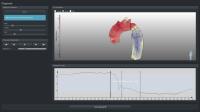
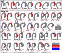
Unattended Processing of 4D Flow MRI in the Aorta: Assessment of Aortic Dimension, Blood Flow, and Demographics in 782 Subjects
Julio Garcia1, Alex J. Barker1, Susanne Schnell1, Jeremy D. Collins1, James C. Carr1, and Michael Markl1,2
1Radiology, Northwestern University, Chicago, IL, United States, 2Biomedical Engineering, Northwestern University, Evanston, IL, United States
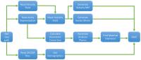
The processing of time-resolved 3D phase-contrast MRI with three-directional velocity encoding (4D flow MRI) cases can be highly time consuming given the large multi-dimensional datasets (3D+time of the cardiac cycle+3-directional blood flow velocities). However, the fully automated processing of cases in large databases is still challenging. The purpose of this study was to introduce an automated workflow allowing the unattended retrospective processing of aortic 4D flow MRI data from a large database of subjects.
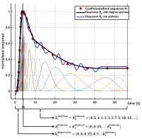

2583.   
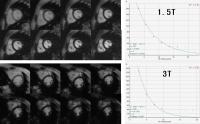
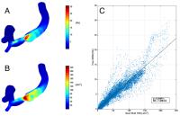
Variability of flow parameters when subjected to changes of MR acquisitions parameters in 4D flow MRI using a realistic thoracic aortic phantom.
Cristian Montalba1, Jesus Urbina1,2, Julio Sotelo1,3, Marcelo Andia1,4, Cristian Tejos1,3, Pablo Irrarazaval1,3, Israel Valverde5,6, and Sergio Uribe1,4
1Biomedical Imaging Center, Pontificia Universidad Católica de Chile, Santiago, Chile, 2School of Medicine, Pontificia Universidad Católica de Chile, Santiago, Chile, 3Electrical Engineering, Pontificia Universidad Católica de Chile, Santiago, Chile, 4Radiology Department, Pontificia Universidad Católica de Chile, Santiago, Chile, 5Institute of Biomedicine of Seville, Universidad De Sevilla, Seville, Spain, 6Cardiology Unit, Hospital Virgen del Rocio, Universidad de Sevilla, Seville, Spain
4D flow is a MRI technique characterized by long scanning times. Because of that, it is difficult to study the variability of flow parameters when subjected to changes of the MR parameters. The purpose of this work is to study the variability of different flow parameters due to changes of spatial and temporal resolutions in 4D flow acquisitions through controlled experiments using a realistic normal adult thoracic aortic phantom. We conclude that changing the spatial and temporal resolutions in the 4D flow imaging greatly affects different flow parameters with induced errors of up to 23.9%.


2584.   
Background Phase Correction for Quantitative Phase-Contrast MRI
Rizwan Ahmad1, Ning Jin2, and Orlando P Simonetti3
1Electrical and Computer Engineering, The Ohio State University, Columbus, OH, United States, 2Siemens Healthcare, Columbus, OH, United States, 3Radiology and Internal Medicine, The Ohio State University, Columbus, OH, United States
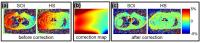
Virtually every phase-contrast MRI (PC-MRI) measurement is contaminated with background phase (BP) from eddy currents and concomitant gradient terms. A widely reported method to correct BP relies on a polynomial fitting of the static pixels within regions of static tissue. This method requires sufficient static tissue in close proximity to the region of interest—a requirement that cannot be met for imaging of the heart or great vessels. In this work, we propose a BP correction method that leverages information from multiple slices collected under identical conditions but with different table positions.


2585.   
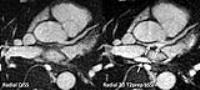
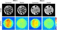
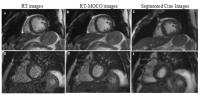
Does Respiratory Motion Influence Tissue Phase Mapping Velocities?
Jan Paul1, Stefan Wundrak1, Peter Bernhardt1, Wolfgang Rottbauer1, and Volker Rasche1
1Internal Medicine II, University Hospital of Ulm, Ulm, Germany
Cartesian Tissue Phase Mapping (TPM) usually necessitates respiratory navigators or other means of motion selection/correction to avoid ghosting artifacts. In radial MRI, however, motion artifacts result in image blurring rather than ghosting, which might allow using all respiratory states for reconstruction. The aim of this study is to investigate the influence of respiratory motion on velocities obtained from radial Tissue Phase Mapping MRI. Only small biases towards reduced velocity peaks were found in ungated compared to motion-compensated reconstructions. Overall velocity agreement of ungated data was very high compared to gated reconstructions.


2586.   
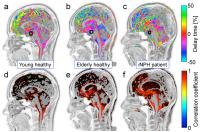
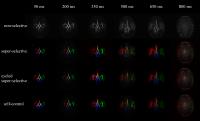
Characterization of Pulsatile Cerebrospinal Fluid Motion Among Young, Elderly and Idiopathic Normal Pressure Hydrocephalus By Correlation Mapping Technique
Satoshi Yatsushiro1, Saeko Sunohara2, Naokazu Hayashi3, Akihiro Hirayama3, Mitsunori Matsumae3, Afnizanfaizal Bin Abdullah4, and Kagayaki Kuroda2
1Course of Science and Technology, School of Science and Technology, Tokai University, Hiratsuka, Kanagawa, Japan, 2Course of Electrical and Electronic Engineering, Graduate School of Engineering, Tokai University, Hiratsuka, Kanagawa, Japan, 3Department of Neurosurgery, Tokai University School of Medicine, Isehara, Kanagawa, Japan, 4Department of Software Engineering, Faculty of Computing, Universiti Teknologi Malaysia, Johor Bahru, Malaysia
                Correlation mapping technique composed of delay time and correlation coefficient mapping to characterize propagation properties of cerebrospinal fluid (CSF) motion was applied to young, elderly healthy and idiopathic normal pressure hydrocephalus (iNPH) patient groups for classification. Brightness of the color of maximum correlation map was adjusted according to the amplitude of the CSF velocity waveform for assisting clinicians to understand the propagation properties intuitively. The groups were classified by quantifying the standard deviation of the correlation distributing in the intracranial CSF space. The technique was expected to classify diseases related to CSF dynamics such as iNPH.


2587.   
Temporal Dynamics and Sampling Rate Effects for Background Phase Estimates in 4D Flow MRI
Michael Loecher1, Peng Hu1, and Daniel B Ennis1,2
1Department of Radiological Sciences, University of California, Los Angeles, CA, United States, 2Biomedical Physics, University of California, Los Angeles, CA, United States
4D Flow phase contrast MRI acquisitions inherently require a measure of background phase to remove phase contributions from non-velocity based components.  The temporal dynamics of this background phase are not well understood.  Consequently, the background phase may be measured too infrequently or too often for accurate and/or time efficient measurements. The purpose of this work was: 1) to measure the temporal dynamics of the background phase with high temporal resolution; and 2) to demonstrate methods of selecting time optimal background phase sampling strategies that improve the measurement efficiency of 4D Flow acquisitions.


2588.   
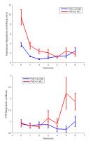
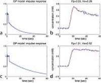
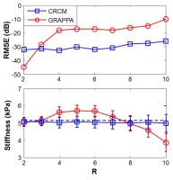
Velocity Transfer Function from Phase Contrast MRI -  A Non-Invasive Method for Assessing Pulmonary Arterial Stiffness and Impedance
Himanshu Gupta1,2, Ankur Gupta1, and Thomas S Denney3,4
1Department of Medicine, Division of Cardiovascular Disease, University of Alabama at Birmingham, Birmingham, AL, United States, 2VA Medical Center, Birmingham, AL, United States, 3Auburn University MRI Research Center, Auburn University, Auburn, AL, United States, 4Electrical and Computer Engineering, Auburn University, Auburn, AL, United States
Pulmonary arterial (PA) impedance accounts for pulsatile blood flow through elastic pulmonary arteries as compared to static pulmonary vascular resistance. Increased PA impedance is an early physiological manifestation of PA remodeling. Currently, PA impedance can only be detected invasively, is expensive and cumbersome to calculate and not done in routine clinical practice. Non-invasive assessment of PA impedance can provide insights in evaluation of patients with normal PA pressures or mild pulmonary hypertension such as in patients with chronic obstructive lung disease. We propose a novel non-invasive parameter, the velocity transfer function (VTF), which is related to PA stiffness and impedance.


2589.   
3D quantification of Vorticity, Helicity, Kinetic Energy and Energy loss in the Left Ventricle from 4D flow data using a finite element method
Julio Sotelo1,2,3, Jesús Urbina1,4, Bram Ruijsink5, David Nordsletten5, Israel Valverde6,7, Cristian Tejos1,2,8, Pablo Irarrazaval1,2,8, Marcelo Andia1,4,8, Daniel E Hurtado3,8, and Sergio Uribe1,4,8
1Biomedical Imaging Center, Pontificia Universidad Catolica de Chile, Santiago, Chile, 2Electrical Engineering Department, Pontificia Universidad Catolica de Chile, Santiago, Chile, 3Structural and Geotechnical Engineering Departement, Pontificia Universidad Catolica de Chile, Santiago, Chile, 4Radiology Department, School of Medicine, Pontificia Universidad Catolica de Chile, Santiago, Chile, 5Biomedical Engineering Department, King's College London, London, United Kingdom, 6Pediatric Cardiology Unit, Hospital Virgen del Rocio, Seville, Spain, 7Cardiovascular Pathology Unit, Institute of Biomedicine of Seville (IBIS), Hospital Virgen del Rocio, Seville, Spain, 8Biological and Medical Engineering Institute, Schools of Engineering, Medicine and Biological Sciences, Pontificia Universidad Catolica de Chile, Santiago, Chile
A quantitative characterization of vortex flow as turbulence and energy may offer a novel index of left ventricle (LV) dysfunction not available in conventional indexes. In this work we propose a novel method based on finite element interpolations to obtain a 3D quantitative maps of vorticity, helicity density, kinetic energy, and energy loss derived from 4D-flow data sets of the LV.? This new method may offer a novel index of LV dysfunction, permitting identify the vortex ring and the magnitude of turbulence values not available in conventional indexes. In future work we pretend validate clinically our method with patient data. 


2590.   
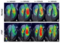
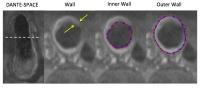
Evaluate Right Ventricular Energy Propagation for Patients With Repaired Tetralogy of Fallot by Using Phase-Contrast MRI
Meng-Chu Chang1, Ming-Ting Wu2, Marius Menza3, Mao-Yuan Su4, Hung-Chieh Huang2, and Hsu-Hsia Peng1
1Department of Biomedical Engineering and Environmental Sciences, National Tsing Hua University, Hsinchu, Taiwan, 2Department of Radiology, Kaohsiung Veterans General Hospital, Kaohsiung, Taiwan, 3Medical Physics, Department of Radiology, University Hospital Freiburg, Freiburg, Germany, 4Department of Medical Imaging, National Taiwan University Hospital, Taipei, Taiwan
The association between right ventricle (RV) volume or pressure overloading pathology and intraventricular flow of repaired tetralogy of Fallot (rTOF) patient is still unclear. Therefore, we evaluated RV input- and output kinetic energy and intraventricular flow patterns for rTOF patients to speculate the energy propagation by using phase-contrast MRI. During systole, rTOF patients presented higher RV output KE. Moreover, in rTOF patients, the blood flow filled into RV with a high velocity, accompanying several local vortices. In conclusion, higher output KE and the visualization of intraventricular vectors helped to comprehend the energy propagation in RV.


2591.   
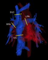
Kinetic Energy Distributions in Fontan Circulation - Evaluation of Respiration Effects
Alejandro Roldán-Alzate1,2, Eric Schrauben3,4, Oliver Wieben2,3, and Christopher J Francois2
1Mechanical Engineering, University of Wisconsin - Madison, Madison, WI, United States, 2Radiology, University of Wisconsin - Madison, Madison, WI, United States, 3Medical Physics, University of Wisconsin - Madison, Madison, WI, United States, 4Centre for Advanced MRI, Auckland, New Zealand
The purpose of this study was to evaluate changes in blood flow and kinetic energy distribution between inspiration and expiration in TCPC patients for assessing efficiency of the system using 4D flow MRI. Six TCPC patients were imaged using a PC-VIPR scheme that allows for double gating to the ECG and respiratory cycles providing flow data for separate respiratory phases. Results exhibit greater respiratory-induced flow changes within a single subject than previous work has shown in the same analysis performed on healthy controls, suggesting that respiration plays a larger role in regulating flow in these patients.


2592.   
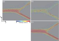
Finite-Element Computational Fluid Dynamics Simulations Constrained by Phase-Contrast MRI Data
Giordanno B. F. Borges1, Ivan R. Siqueira2, Joao L. A. Carvalho3, Jon-Fredrik Nielsen4, and Vinicius C. Rispoli5
1Department of Mathematics, University of Brasilia, Brasilia, Brazil, 2Department of Mechanical Engineering, Pontifical Catholic University of Rio de Janeiro, Rio de Janeiro, Brazil, 3Department of Electrical Engineering, University of Brasilia, Brasilia, Brazil, 4Biomedical Engineering, University of Michigan, Ann Arbor, MI, United States, 5UnB Gama College, University of Brasilia, Brasilia, Brazil
Phase-contrast MRI (PC-MRI) data has been vastly used as boundary conditions in computational fluid dynamics (CFD) simulations. Recently, many authors also used measured flow data to enforce CFD solutions, based on the finite volume method (FVM). On the other hand, the finite element method (FEM) has notable advantages over FVM, such as higher order accuracy and more flexibility dealing with complex geometries. This work presents a finite-element implementation of a MRI-constrained CFD solver. This hybrid solver can be used to regularize PC-MRI data, providing solutions closer to the PC-MRI measurements than pure CFD. Feasibility of this approach is demonstrated using a modified 2D discretization of the Navier-Stokes and continuity equations, using FEM. In this demonstration, two velocity components were taken from a 4D PC-MRI dataset, and used to constrain the CFD solution over a 2D domain.


2593.   
Energy loss and turbulent formations reveal the pressure loss in coarctation flows: A novel 4D Flow MRI-Based quantification method using a finite element approach
Julio Sotelo1,2,3, Jesús Urbina1,4, Cristian Montalba1, Israel Valverde5,6, Cristian Tejos1,2,7, Pablo Irarrazaval1,2,7, Marcelo Andia1,4,7, Daniel E Hurtado3,7, and Sergio Uribe1,4,7
1Biomedical Imaging Center, Pontificia Universidad Catolica de Chile, Santiago, Chile, 2Electrical Engineering Department, Pontificia Universidad Catolica de Chile, Santiago, Chile, 3Structural and Geotechnical Engineering Departement, Pontificia Universidad Catolica de Chile, Santiago, Chile, 4Radiology Department, School of Medicine, Pontificia Universidad Catolica de Chile, Santiago, Chile, 5Pediatric Cardiology Unit, Hospital Virgen del Rocio, Seville, Spain, 6Cardiovascular Pathology Unit, Institute of Biomedicine of Seville (IBIS), Hospital Virgen del Rocio, Seville, Spain, 7Biological and Medical Engineering Institute, Schools of Engineering, Medicine and Biological Sciences, Pontificia Universidad Catolica de Chile, Santiago, Chile
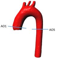
Aortic coarctation (CoA) cause an irreversible pressure loss post-CoA given by the energy dissipation, increasing the ventricular workload. Turbulent flows through CoA generate an irreversible damage in the surrounding tissue for mechanical stresses. We implement a finite elements method to obtain 3D maps of energy loss, kinetic energy, vorticity and helicity from 4D flow data. We performed an in-vitro study that related the pressure gradient, pulse wave velocity and elastic modulus with the energy loss and vorticity and helicity parameters. Concluding that our method may allow assessing the severity of the CoA and the identification of the regions affected.


2594.   
Validation of "WaVelocity" Image Analysis Toolbox for Cardiac Magnetic Resonance Pulse Wave Velocity Measurements
Danilo Babin1, Daniel Devos2, and Patrick Segers3
1TELIN, Ghent University, Ghent, Belgium, 2Ghent University Hospital, Ghent, Belgium, 3ibiTech-bioMMeda, Ghent University, Ghent, Belgium
The purpose is to validate our cardiovascular image analysis toolbox "WaVelocity" for measuring pulse wave velocity (PWV) from cardiac magnetic resonance images against PWV measurements of in-place pressure catheter. The validation was performed using two phantoms: a straight latex tube and an aortic phantom with two different water flow rates. Phase-Contrast para-sagittal image sequences in multiple planes were processed with our image analysis software. Ground truth PWV values were calculated from pressure curves measured by pullback of the catheter. The results show sufficiently high correspondence between calculated MR and catheter PWV to plan for clinical use.


2595.   
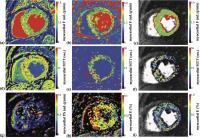
Accelerated Phase-Contrast MRI using Hybrid One- and Two-sided Flow-Encodings Only (HOTFEO)
Da Wang1,2, Jiaxin Shao1, Daniel B Ennis1,2, and Peng Hu1,2
1Radiology, University of California, Los Angeles, Los Angeles, CA, United States, 2Biomedical Physics, University of California, Los Angeles, Los Angeles, CA, United States
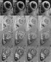
In conventional 4D-flow phase-contrast MRI, each cardiac phase acquires the flow-compensated and three-directional flow-encoded echoes, which often limits the achievable temporal-resolution and temporal-footprint. This can result in under-estimation of maximum peak velocity. Herein, we propose a fast 4D-flow strategy that eliminates the flow-compensated acquisition using hybrid one- and two-sided flow encoding only (HOTFEO). The flow-compensated background phase is derived from three-directional flow-encoded data based on a velocity direction constraint that assumes the velocity direction, not the magnitude, changes very little between two cardiac phases. HOTFEO provides accurate blood flow and velocity measurements compared with conventional 4D-flow technique.


2596.   
MRI flow quantification of Head and Neck arteries
Jérémie Bettoni1, Gwenaël Pagé2, Stéphanie Dakpé1, Jean-Marc Constans3, Sylvie Testelin1, Bernard Devauchelle1, and Olivier Balédent2,4
1Maxillo-Faciale surgery, Amiens Hospital, Amiens, France, 2BioFlow Image, University of Picardie Jules Verne, Amiens, France, 3Diagnostic Radiology, Amiens Hospital, Amiens, France, 4Department of Image Processing, Amiens Hospital, Amiens, France
The aim of this study is to create the first physiological database of the blood flow quantification in the external carotid tree in order to help the surgeon in facial reconstruction by free flap. An original protocol association with 32 head coils channel and microscopic coil is created and 2D PC-MRI are performed on arteries from head and neck area. Blood flow average for each artery is 17 mL/min in superior thyroid artery, 6.5 mL/min in lingual artery, 30.5 mL/min in facial artery, 23.5 mL/min in internal maxillary artery, 21.5 mL/min in superficial temporal artery.


2597.   
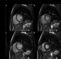
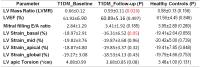
Monitoring the Disease Progression and Aortic Hemodynamics of Pediatric Bicuspid Aortic Valve Patients Using Longitudinal 4D Flow MRI
Michael Rose1, Emilie Bollache2, Kelly Jarvis2,3, Alex Barker2, Susanne Schnell2, Bradley Allen2, Joshua Robinson4,5, Michael Markl2,3, and Cynthia Rigsby1,2
1Medical Imaging, Ann & Robert H. Lurie Children's Hospital of Chicago, Chicago, IL, United States, 2Radiology, Northwestern University, Chicago, IL, United States, 3Biomedical Engineering, Northwestern University, Chicago, IL, United States, 4Pediatrics, Northwestern University, Chicago, IL, United States, 5Pediatric Cardiology, Ann & Robert H. Lurie Children's Hospital of Chicago, Chicago, IL, United States
Over the course of two 4D flow MRI studies (mean duration between studies: 19 months), 12 pediatric BAV patients were evaluated for any changes in aortic hemodynamics. Hemodynamics were characterized via visual grading of flow patterns, peak systolic velocity and regional mean wall shear stress. There were no significant changes in visual grading scores, peak systolic velocities or mean wall shear stress values between baseline and follow up studies suggesting little BAV disease progression during this time.


2598.   
Perioperative assessment of aortic tissue at risk for dysfunction in patients undergoing valve and/or aortic replacement using 4D flow MRI
Emilie Bollache1, Paul W.M. Fedak2,3, Pim van Ooij1, David Guzzardi2, S. Chris Malaisrie3, Alex Hong1, Patrick M. McCarthy3, James Carr1, Jeremy Collins1, Michael Markl1,4, and Alex J. Barker1
1Department of Radiology, Northwestern University, Chicago, IL, United States, 2Department of Cardiac Sciences, University of Calgary, Calgary, AB, Canada, 3Division of Surgery-Cardiac Surgery, Northwestern University, Chicago, IL, United States, 4Department of Biomedical Engineering, Northwestern University, Chicago, IL, United States
The effect of the type of aortic surgery in patients with aortopathy is not well known. We studied 23 patients who underwent 4D flow MRI both before and after aortic valve (AVR) and/or ascending aortic (AAR) replacement, from which we estimated the pre- and post-surgical area of aortic ‘at-risk’ tissue, with an elevated wall shear stress. After surgery, in most AVR patients, at-risk tissue area was decreased while in most AAR patients, it was increased. This pilot study suggests the usefulness of 4D flow MRI to provide longitudinal aortic hemodynamic follow-up after surgery, which should be confirmed in larger populations.


2599.   
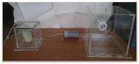
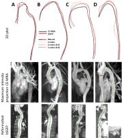
A realistic aortic phantom with a kinking of the aorta: one to one replica of a patient and comparison using PC-MRI and cardiac catheterization
Jesús Urbina1,2, Julio Sotelo1,3, Cristian Montalba1, Tomás Fernández1, Felipe Valenzuela1,3, Cristián Tejos1,3, Pablo Irarrázaval1,3, Marcelo Andia1,4, Israel Valverde5,6, and Sergio Uribe1,4
1Biomedical Imaging Center, Pontificia Universidad Católica de Chile, Santiago, Chile, 2School of Medicine, Pontificia Universidad Católica de Chile, Santiago, Chile, 3Electrical Engineering Department, Pontificia Universidad Católica de Chile, Santiago, Chile, 4Radiology Department, Pontificia Universidad Católica de Chile, Santiago, Chile, 5Pediatric Cardiology Unit, Hospital Virgen del Rocio, Seville, Spain, 6Institute of Biomedicine of Seville, Universidad de Sevilla, Seville, Spain
The aim of this work was to generate a one to one replica of the aorta of a patient with a kinking and to compare the hemodynamic parameters with the ones obtained from patient's PC-MRI and cardiac catheterization. A silicone model was built from CE-MRA data and connected to a MRI compatible pulsatile pump setup. PC-MRI and catheterization data were obtained in the phantom. Most hemodynamic parameters were similar between the patient and the phantom.


2600.   
Turbulent wall shear stress assessment using 4D flow MRI
Magnus Ziegler1,2, Jonas Lantz1,2, Tino Ebbers1,2, and Petter Dyverfeldt1,2
1Division of Cardiovascular Medicine, Department of Medical and Health Sciences, Linköping University, Linköping, Sweden, 2Centre for Medical Image Science and Visualization (CMIV), Linköping University, Linköping, Sweden
Chaotic velocity fluctuations caused by turbulent blood flow create fluctuations in the shear stress acting on the vascular wall. This turbulent wall shear stress can cause vascular remodeling and increased endothelial cell turnover. This work explores the use of MR-estimated turbulent kinetic energy (TKE) for mapping the turbulent wall shear stress. Time-resolved velocity data for non-pulsatile flow was obtained using computational fluid dynamics in two patient-derived geometries and used to simulate PC-MRI measurements. Near-wall TKE was estimated using a novel sampling method and was found to correlate strongly to turbulent wall shear stress, opening new avenues for analysis.


2601.   
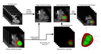
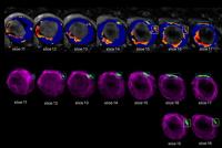
Superquadric Glyphs for Visualizing Myocardial Motion in 3D
Teodora Chitiboi1,2, Mathias Neugebauer1, Susanne Schnell3, Michael Markl3, Lars Linsen2, and Anja Hennemuth1
1Fraunhofer MEVIS, Bremen, Germany, 2Jacobs University, Bremen, Germany, 3Department of Radiology, Northwestern University, Chicago, IL, United States
Various cardiac diseases can be diagnosed by analyzing myocardial motion. The local myocardial velocity can be efficiently computed using tissue phase mapping MRI. While radial, longitudinal, and rotational myocardial velocities are relevant biomarkers, it is challenging to find a single 3D representation that gives a global overview of these three motion directions for the entire cardiac muscle. We designed a visual encoding that maps the three velocities to glyph shapes according to a barycentric space formed by 3D superquadric glyphs. The glyphs show the aggregated myocardial motion information for each AHA segment and are displayed in a 3D layout.


2602.   
4D Flow MRI for the Evaluation of Vasodilation in Patients with Sickle Cell Disease
Lena Václavu1, Bart J Biemond2, John C Wood3, Henk Mutsaerts4, Charles BLM Majoie1, Ed van Bavel5, Aart J Nederveen1, and Pim van Ooij1
1Radiology, Academic Medical Center, Amsterdam, Netherlands, 2Internal Medicine, Academic Medical Center, Amsterdam, Netherlands, 3Cardiology, Children's Hospital Los Angeles, Los Angeles, CA, United States,4Sunnybrook Research Institute, Toronto, ON, Canada, 5Biomedical Engineering and Physics, Academic Medical Center, Amsterdam, Netherlands
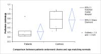
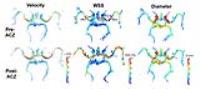
We employed 4D Flow MRI in patients with Sickle Cell Disease. 4D Flow MRI is a non-invasive technique allowing blood flow velocity measurements and estimation of WSS. We investigated dynamic changes in velocity, WSS and vessel diameter in the anterior circulation of the Circle of Willis (CoW) in response to a vasodilator (acetazolamide [ACZ]).  We found that velocity and WSS  increased in the CoW after administration of the vasodilator ACZ, as measured with 4D flow MRI. The change in velocity after administration of ACZ was larger in controls than in patients.


2603.   
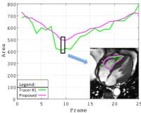
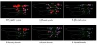
Fixed-Volume Particle Trace Emission for the Analysis of Left Atrial Blood Flow using 4D Flow MRI
Stephen Gaeta1, Petter Dyverfeldt2,3, Jonatan Eriksson2,3, Carl-Johan Carlhäll2,3, Tino Ebbers2,3, and Ann F Bolger4
1Department of Medicine, Duke University, Durham, NC, United States, 2Department of Medical and Health Sciences, Linköping University, Linköping, Sweden, 3Center for Medical Image Science and Visualisation (CMIV), Linköping University, Linköping, Sweden, 4Department of Cardiology, University of California San Francisco, San Francisco, CA, United States
The aim of this study was to develop a novel fixed-volume approach for particle tracing and employ this to develop quantitative analysis of 4D blood flow characteristics in the left atrium (LA). The proposed fixed volume approach for emission of particle traces permits sampling of LA blood volumes and intuitive visualizations where each trace represents the same volume. Using fixed-volume particle traces, LA flow can be separated into different components based on the transit of blood through the LA. Quantitative analysis of functionally distinct subsets of LA flow may provide new perspectives on LA function in health and disease. 


2604.   
Breath-Hold Real-Time Phase Contrast MRI using Radial k-space Sampling and Compressed Sensing
Hassan Haji-Valizadeh1, Elwin Bassett2, Genesh Adluru3, Edward VR DiBella 3, and Daniel Kim3
1Bioengineering, University of Utah, Salt Lake City, UT, United States, 2Physics, University of Utah, Salt Lake City, UT, United States, 3Radiology,UCAIR, University of Utah, Salt Lake City, UT, United States
Phase contrast (PC) MRI is a useful tool for assessing hemodynamic, but suffers from low data acquisition efficiency. In this study we compared real-time PC MRI between Cartesian and Radial undersampling trajectories. Our results show that both real-time MRI pulse sequences yield velocity measurements that agree well with those produced by reference breath-hold PC MRI pulse sequence. Compared with real-time MRI with Cartesian sampling, Radial sampling produced images with fewer artifacts.  This study demonstrates feasibility of real-time PC MRI using radial k-space sampling and constrained reconstruction. 


2605.   
Simultaneous 3D velocity and temperature mapping in fluid flow using MRI
Waltraud B. Buchenberg1, Florian Wassermann2, Sven Grundmann3, Jürgen Hennig1, and Bernd Jung4
1Radiology - Medical Physics, University Medical Center Freiburg, Freiburg, Germany, 2Center of Smart Interfaces, Technische Universität Darmstadt, Darmstadt, Germany, 3Institute of Fluid Mechanics, University of Rostock, Rostock, Germany, 4Interventional and Pediatric Radiology, University Hospital, Institute of Diagnostic, Bern, Switzerland
Since MR thermometry and MR velocimetry allow non-invasive measurements of temperature fields and velocity fields, they are widely applied to address medical questions; however, they are also suited to investigate 3D fluid flow and heat transfer phenomena in technical devices. This work investigates velocity fields and temperature distributions in a countercurrent double pipe heat exchanger. 3D velocity and temperature measurements were performed consecutively. A combination of forced convection (external pump providing laminar flow) and free convection (heating) using MRI can add valuable new insights into heat transfer processes.

2606.   
Measurement of extracellular volume fraction and blood flow from dynamic contrast enhanced myocardial perfusion images applied in non-ischemic cardiomyopathy
Yoon-Chul Kim1, Sung Mok Kim1, Sung-Ji Park1, and Yeon Hyeon Choe1
1Samsung Medical Center, Sungkyunkwan Univ. School of Medicine, Seoul, Korea, Republic of
Recently there have been several studies of estimation of extracellular volume (ECV) fraction from dynamic contrast enhanced (DCE) myocardial perfusion images using a distributed parameter (DP) model. We apply the DP model to DCE perfusion images acquired from a standard stress perfusion protocol, and demonstrate ECV measurements in aortic stenosis (AS) patients (n=13) and hypertrophic cardiomyopathy (HCM) patients (n=11).  Preliminary results from sector-wise analysis indicate 1) higher mean ECV values in HCM patients than normal volunteers and AS patients and 2) lower mean blood flow values in AS and HCM patients than those in normal volunteers. 


2607.   
Myocardial Perfusion using radial MS-CAIPIRINHA
Tobias Wech1, Michael Braun1,2, Daniel Stäb3, Peter Speier4, Henning Neubauer1, Walter Kullmann2, Thorsten A. Bley1, and Herbert Köstler1
1Department of Diagnostical and Interventional Radiology, University of Würzburg, Würzburg, Germany, 2Institute of Medical Engineering, University of Applied Sciences Würzburg-Schweinfurt, Schweinfurt, Germany,3The Centre for Advanced Imaging, The University of Queensland, Brisbane, Australia, 4Siemens Healthcare, Erlangen, Germany
The benefit of using radial instead of Cartesian readouts for MS-CAIPIRINHA accelerated myocardial perfusion measurements was explored. Conjugate gradient SENSE was used to determine fully sampled data for the two slices acquired simultaneously. A phantom study was performed first, which revealed lower g-factors for radial MS-CAIPIRINHA in comparison to a corresponding Cartesian acquisition. Finally, saturation-recovery-prepared and ECG-triggered radial MS-CAIPIRINHA was applied for perfusion investigations of the myocardium in a healthy volunteer.


2608.   
Comparison of myocardial perfusion and permeability estimates from dynamic contrast-enhanced MRI with three quantitative analysis methods
Wang Jing1, Yudong Zhang2, Yang Fan3, HaiBin Shi2, and Xiaoyan Liu1
1Center for Medical Device Evaluation, CFDA, Beijing, China, People's Republic of, 2Department of Radiology, the First Affiliated Hospital with Nanjing Medical University Nanjing, Nanjing, China, People's Republic of,3GE Healthcare, MR Research China, Beijing, China, People's Republic of
This study compares three methods for quantitatively analyzing dynamic contrast-enhanced MRI datasets for the extraction of myocardial perfusion, permeability, and other hemodynamic parameters. The two-compartment exchange model and the adiabatic approximation to the tissue homogeneity models are compared with reference to model-free deconvolution. The goal was to determine if these three models could reliably estimate hemodynamic parameters and be able to distinguish between normal patients and those with hypertrophic cardiomyopathy. Results demonstrate that all three methods yield consistent estimates for perfusion, blood volume, and mean transit times, and that these parameters were significantly different for the hypertrophic heart.


2609.   
Measuring microvascular flow characteristics with myocardial DCE-MRI perfusion data using a model-independent, multi-resolution spline approach in patients at stress
Karl P Kunze1, Christoph Rischpler1, Markus Schwaiger1, and Stephan G Nekolla1
1Department of Nuclear Medicine, Klinikum rechts der Isar der TU München, Munich, Germany
This abstract proposes a new B-spline based approach for model-independent deconvolution of myocardial DCE-MRI perfusion data and the reconstruction of the vascular transit time distribution function. It allows the model-independent quantification of vascular mean transit time and vascular transit time heterogeneity, whose relationship is of importance in understanding the implications of different ischemic microvascular disease patterns. The presented algorithm was tested in simulations and showed stability for the range of perfusion parameters expected under stress conditions. 12 DCE-MRI patient datasets from adenosine stress examinations were analyzed, showing a steady increase of heterogeneity with mean transit time.


2610.   
Semi-quantitative stress perfusion CMR for assessing complications in the transplant heart
Madeline Schwid1, Hannah Recht1, Kai Lin1, Jeremy Collins1, Michael Markl1, Dan Lee1, and James Carr1
1Northwestern University, Chicago, IL, United States
This study evaluated semiquantitative measures of ischemia using noninvasive stress perfusion CMR for the detection and quantification of CAV using invasive coronary angiography as the standard reference on a cohort of chronic heart transplant patients. Based on angiogram results, patients were divided into groups by disease severity. The upslope and myocardial perfusion reserve was measured per segment for each patient based on the AHA 16 segment model. These values were then correlated to the angiogram results and averaged by segment to create bullseye plots for each group. As disease severity increases, both myocardial perfusion reserve and stress upslope decrease. 


2611.   
Comparison of Cardiac Perfusion and Cardiac Global Functional Parameters between Healthy Volunteers and Cardiac Transplant Patients
Travis DeSa1, Jeremy D Collins1, James C Carr1, Kai Lin1, and Michael Markl1
1Radiology, Northwestern University Feinberg School of Medicine, Chicago, IL, United States
Myocardial perfusion using MRI has become a promising non-invasive method to evaluate myocardial ischemia in CVDs. However, assessment of CMR's effectiveness in differentiating regional myocardial perfusion in patients with CVD and comparison of regional myocardial perfusion indices to cardiac global functional parameters has not been thoroughly studied. We evaluated 12 volunteers and 9 cardiac transplant patients and found significant differences in segmental and average CMR perfusion, but little correlation between perfusion and global functional parameters. This weak correlation indicates that myocardial perfusion may be more sensitive to detect early changes in LV dysfunction before the onset of global functional impairment.


2612.   
Anaplerotic Flux Into Citric Acid Cycle 4-Carbon Intermediates is Phenotypically Increased in a Murine Model of Heart Failure
Aslan Turer1, Thomas Gillette1, Shawn Burgess2, Craig Malloy2, and Matthew Merritt3
1Cardiology, UT Southwestern Medical Center, Dallas, TX, United States, 2AIRC, UT Southwestern Medical Center, Dallas, TX, United States, 3Biochemistry and Molecular Biology, University of Florida, Gainesville, FL, United States
Heart failure (HF) was studied using a murine model of aortic constriction. Hearts were perfused to steady state using [1,6-13C2]glucose, [1,3-13C2]acetoacetate, and [U-13C]fatty acids. Substrate selection for acetyl-CoA production was measured using isotopomer analysis by carbon-13 NMR. A standard model which includes oxidative flux as well as pyruvate anaplerosis (YPC) via pyruvate carboxylase or the malic enzyme was evaluated. Inconsistencies in the fits led to proposal of a more complicated model that also includes anaplerosis through the succinyl-CoA pathway (Ys), leading to significantly better fits. We hypothesize that induction of the Ys anaplerotic pathway is phenotypic of HF.  


2613.   
Feasibility Study: Free-Breathing 3-D CINE Imaging with Respiratory Gating Based on Pilot Tone Navigation
Jens Wetzl1,2, Lea Schroeder1, Christoph Forman3, Felix Lugauer1, Robert Rehner4, Matthias Fenchel3, Andreas Maier1,2, Joachim Hornegger1,2, and Peter Speier3
1Pattern Recognition Lab, Department of Computer Science, Friedrich-Alexander-Universität Erlangen-Nürnberg, Erlangen, Germany, 2Erlangen Graduate School in Advanced Optical Technologies, Friedrich-Alexander-Universität Erlangen-Nürnberg, Erlangen, Germany, 3Magnetic Resonance, Product Definition and Innovation, Siemens Healthcare GmbH, Erlangen, Germany, 4Magnetic Resonance, Research and Development, Hardware, Siemens Healthcare GmbH, Erlangen, Germany
Respiratory monitoring during continuous, free-breathing acquisitions is challenging. Using self-navigation, a respiratory signal can be derived from the imaging data, but  requires frequent sampling of the k-space center. Pilot Tone navigation promises continuous respiratory monitoring independent of the imaging sequence. In this feasibility study, we compared both strategies for free-breathing 3-D CINE imaging. We found good agreement between both respiratory signals, and an excellent match in both reconstructed images and computed ventricular function parameters. Pilot Tone navigation can thus be considered an alternative to self-navigation, with the benefit of working with arbitrary imaging sequences.


2614.   
Pro-B-Type Natriuretic Peptide is associated with regional left ventricular function and dyssynchrony measured by MRI: The Multi-Ethnic Study of Atherosclerosis (MESA)
Amir Ali Rahsepar1, Mohammadali Habibi2, Cheeling Chan3, Nadine kawel2, Kiang Liu3, Joao Lima2, and James Carr1
1Radiology, Northwestern University, Chicago, IL, United States, 2Cardiology, Johns Hopkins University, Baltimore, MD, United States, 3Preventive medicine, Northwestern University, Chicago, IL, United States
In this large cross-sectional study, we found that higher levels of Pro-B-Type Natriuretic Peptide is associated with regional left ventricular function and dyssynchrony measured by MRI in asymptomatic patients participated in The Multi-Ethnic Study of Atherosclerosis (MESA) study.


2615.   
Effect of exercise on skeletal muscle and cardiac function in mouse models of Duchenne muscular dystrophy
Bauke Kogelman1, Margriet Hulsker2, Christa Tanganyika-de Winter2, Ralf Werring2, Annemieke Aartsma-Rus2, Maaike van Putten2, and Louise van der Weerd1,2
1Department of Radiology, Leiden University Medical Center, Leiden, Netherlands, 2Department of Human Genetics, Leiden University Medical Center, Leiden, Netherlands
Duchenne muscular dystrophy is affecting skeletal and cardiac muscle tissue, due to non-functional dystrophin protein. Potential therapies restore dystrophin expression in skeletal muscle, while cardiac muscle is more difficult to target. To elucidate whether exercise is beneficial or disadvantageous for (cardio)myopathy, we subjected several mouse models, mdx (0% dystrophin), mdx-Xist?hs (varying dystrophin levels), Bl10-WT and Xist-WT wild type mice (100% dystrophin) to low intensity or no exercise. Results showed that low dystrophin levels improve skeletal muscle and cardiac function and suggest that low intensity exercise is beneficial for skeletal and cardiac muscle function in both dystrophic and wild type mice. 


2616.   
Validation of a novel technique to extract cine cardiac cycle without respiratory motion from real-time free-breathing images with unsupervised motion correction
Amir Ali Rahsepar1, Haris Saybasili 2, Ahmadreza Ghasemiesfe 1, Bruce Spottiswoode 2, Ann Ragin1, Jeremy Collins 1, and James Carr1
1Radiology, Northwestern University, Chicago, IL, United States, 2Siemens Medical Solutions, Siemens Healthcare, Chicago, IL, United States
In this study, we presented a novel technique to extract cine cardiac cycle without respiratory motion from real-time free-breathing images with unsupervised motion correction.


2617.   
Impaired myocardial resting perfusion in patients undergo chemo therapy, a quantitative MRI perfusion study
Yi Wang1
1St. Francis Hospital, Albertson, NY, United States
We tried to develop and validate an early detection technique of chemo therapy cardiotoxicity in the cancer treatment using cardiovascular magnetic resonance imaging. We hypothesized myocardial blood flow (MBF), more sensitive technique than EF or DHE, provides information on the altered physiology and function conditions of the heart. Chemo treatment induces changes in myocardial perfusion at rest enough to be detected with model based quantitation, with sequential imaging on the same subject. It provides an earlier indication of cardiotoxicity.

2618.   
4D-PC MRI Pressure Mapping for Therapy Planning of Coarctation of the Aorta
Anja Hennemuth1, Christian Schumann1, Mathias Neugebauer1, Hanieh Mirzaee1, Sarah Nordmeyer2, Marcus Kelm2, Leonid Goubergrits2, and Titus Kühne2
1Fraunhofer MEVIS, Bremen, Germany, 2Department of Congenital Heart Disease / Pediatric Cardiology, German Heart Center Berlin, Berlin, Germany
The induced pressure gradient due to the vessel narrowing associated with coarctation of the aorta is a crucial parameter for treatment planning. Previous studies have shown that pressure differences derived from 4D PC MRI correlate well with conventional pressure catheter measurements. The purpose of the presented work was to investigate how MRI-based pressure maps can be employed in treatment planning of coarctation of the aorta. To this end, a combined 3D maximum value projection, which highlights relative value changes with respect to a reference point, and a curve diagram showing the pressure course along the centerline are provided for interactive exploration. Two cardiologists retrospectively explored 5 datasets of patients with treated coarctation of the aorta. The pressure gradient derived from the 4D PC MRI measurement corresponded well with the interventional measurements. Furthermore, additional relevant information could be derived regarding the location of the critical vessel sections. These findings show the potential of 4D PC MRI pressure mapping as a useful non-invasive tool for treatment planning of coarctation of the aorta.


2619.   
Automatic coronary centerline tracking from coronary MRI
Arna van Engelen1, Markus Henningsson1, and Rene Botnar1
1Department of Biomedical Engineering, Division of Imaging Sciences and Biomedical Engineering, King's College London, London, United Kingdom
The aim of this study was to perform automatic coronary artery centerline tracking on coronary MRI data, in a clinically relevant population. Our method consisted of computation of a vesselness filter followed by fast marching. Parameters were optimized on three subjects, and performance was evaluated on 27 other subjects. Centrelines were traced between a start and end point, and when needed with one additional point in between. Tracking was successful in 92% (RCA), 88% (LAD) and 65% (LCX), with a median distance from manual annotation of 1.0mm. These results show that automatic centerline tracking on coronary MRI data is possible.


2620.   
Accelerated MR Elastography for Quantitative Measurement of Myocardial Stiffness
Rizwan Ahmad1, Samuel Schroeder2, Richard D White3, and Arunark Kolipaka3
1Electrical and Computer Engineering, The Ohio State University, Columbus, OH, United States, 2Mechanical Engineering, The Ohio State University, Columbus, OH, United States, 3Radiology, The Ohio State University, Columbus, OH, United States
Alterations in myocardial stiffness have been linked to wide-ranging cardiovascular conditions. Magnetic resonance elastography (MRE) is a noninvasive, quantitative technique to estimate stiffness of soft tissue.1 Long scan times, however, limit the clinical utility of MRE, especially for cardiac imaging. We propose a data processing technique that not only exploits sparsity in the MRE images but also imposes a constant magnitude constraint to achieve accurate stiffness values from highly undersampled data. The method is validated using a physical phantom of known stiffness value and is then applied to a healthy volunteer to assess myocardial stiffness. 


2621.   
Patient Factors and Electrode Placement Factors that Cause 3T-CMR to Poorly Synchronize with Waveforms of Vectorcardiography – Examination of Factors Worsening R/T-wave Amplitude Ratio –
KEI FUKUZAWA1, Rieko Ishimura2, Takashi Yoshida1, Chiharu Yoshihara1, Junji Takahashi1, and Masakatsu Tano1
1Radiology, Toranomon Hospital, Tokyo, Japan, 2Cardiovascular center, Toranomon Hospital, Tokyo, Japan
The lower R/T ratio inside a bore are obtained in patients with bathycardia, a condition in which the heart axis is close to the body axis. In addition, the short distance between the apex of the heart and the apex electrode and closeness of lead II and the heart axis are factors of a worse R/T ratio. These two points should be improved to obtain waveforms with a high R/T ratio.


2622.   
Feasibility, reproducibility, and reliability for cardiac iron quantification at 3T in comparison with 1.5T
Long Li Ling1, peng peng1, and Huang Zhong Kui1
1The First Affiliated Hospital of Guangxi Medical University, Nanning, China, People's Republic of
To demonstrate that assessment of cardiac iron burden by T2* imaging is feasible, reproducible, and reliable at 3T. With 3T MRI scanners increasingly used in clinical settings, this study may lay a foundation for further MRI development of quantitative markers for noninvasive cardiac iron overloaded assessment at higher fields. Though cardiac T2* measurement at 3T seemed not to offer any greater advantages than 1.5T at present, it is more useful for some hospitals having only 3T scanner.


2623.   
Whole heart coverage in a single breath hold using compressed sensing combined with parallel imaging.
Jinho Park1, Panki Kim2, Young Joong Yang1, Jong-Hyun Yoon1, Byoung Wook Choi2, and Chang-Beom Ahn1
1Kwangwoon University, Seoul, Korea, Republic of, 2Yonsei University, Seoul, Korea, Republic of
For patients with impaired breath-hold capacity, highly accelerated cardiac cine MRI may be useful. The purpose of this study is to perform functional MR imaging of the whole heart in a single breath-hold using an under-sampled data acquisition in combination with compressed sensing with parallel imaging.  The proposed technique is applied for in-vivo functional cardiac imaging of the whole heart in a single breath-hold.  Applied compression factors are 4 of ITSC and 2 of SENSE. The whole heart coverage in a single breath-hold using ITSCxSENSE can be used to achieve adequate spatial resolution (1.8 mm × 1.8 mm) and relatively high temporal resolution (31 ms), with good image quality and functional movement.  It takes 23s for the reconstruction of single slice CINE images (22 cardiac phases). The ITSCxSENSE may be useful for patients with reduced breath-hold capacity.


2624.   
Electrode Placement Positions for Vectorcardiography to Control T waves and Obtain High R waves in 3T CMR
Kei Fukuzawa1, Rieko Ishimura2, Takashi Yoshida1, Junji Takahashi1, Chiharu Yoshihara1, and Masakatsu Tano1
1Radiology, Toranomon Hospital, Tokyo, Japan, 2Cardiovascular center, Toranomon Hospital, Tokyo, Japan
To obtain the highest R/T ratio, for good ECG synchronization, The R/T ratio increased as the angle of lead II axis became further away from the body axis and as the apex electrode and the apex of the heart became a little further apart than the basic placement position. The basic placement position was the lowest in the order of R/T ratios among the five kinds of electrode positions (p < 0.01).


2625.   
Fully Automatic Vessel Wall Contour Detection and Wall Thickness Assessment in Common Carotid and Descending Aorta
Shan Gao1, Ronald van't Klooster1, Anne Brandts2, Stijntje D. Roes2, Reza Alizadeh Dehnavi3, Albert de Roos2, Jos J.M. Westenberg1, and Rob J. van der Geest1
1Division of Image Processing, Department of Radiology, Leiden University Medical Center, Leiden, Netherlands, 2Department of Radiology, Leiden University Medical Center, Leiden, Netherlands, 3Department of Cardiology, Leiden University Medical Center, Leiden, Netherlands
Atherosclerosis is characterized by thickening of the arterial wall. To monitor disease progression and regression, vessel wall segmentation is required for wall thickness assessment.  In this study, we developed a fully automatic method to detect the vessel wall boundaries and quantify the wall thickness for both the common carotid artery and the descending aorta in MR images. The results demonstrated that our method has good agreement with manual segmentation, excellent scan-rescan reproducibility and capability of detecting vessel wall thickening in hypertensive patients. Therefore, the presented method is promising for application in future cohort studies.


2626.   
Segmentation of Abdominal Aortic Aneurysms Wall and Intraluminal Thrombus using 3D Black Blood MRI with Registration based Geometric Active Contour Model
Yan Wang1, Florent Seguro1, Farshid Faraji1, Chengcheng Zhu1, Henrik Haraldsson1, Michael Hope1, Jing Liu1, and David Saloner2
1Radiology, UCSF, San Francisco, CA, United States, 2Radiology, UCSF/VA, San Francisco, CA, United States
An abdominal aortic aneurysm (AAA) may rupture if left untreated. Automatic segmentation methods allow quick and reproducible AAA morphology quantification, which is favorable for the diagnosis of AAA disease. This study developed a novel registration based geometric active contour model to segment vessel wall of AAA from 3D black blood MRI. This method was initially tested on six patients. The proposed method agreed well with manual segmentation with an average Dice value of 91.22%, demonstrating good segmentation accuracy.  These methods can be potentially used for AAA evaluation in clinical setting.


2627.   
Aortic centreline tracking for PWV measurements in multiple MRI sequences
Arna van Engelen1, Torben Schneider2, Hubrecht de Bliek3, Miguel Silva Vieira4, Isma Rafiq4, Tarique Hussain4, Rene Botnar1, and Jordi Alastruey1
1Department of Biomedical Engineering, Division of Imaging Sciences and Biomedical Engineering, King's College London, London, United Kingdom, 2Philips Healthcare, Guidford, United Kingdom, 3Philips Healthcare, HSDP Clinical Platforms, Best, Netherlands, 4Department of Cardiovascular Imaging, Division of Imaging Sciences and Biomedical Engineering, King's College London, London, United Kingdom
Accurate 3D length measurements through the aorta are required for Pulse Wave Velocity (PWV) measurements. We evaluate automatic centreline tracking, requiring only a start and end point, on three different types of MR data (balanced-SSFP, contrast-enhanced and black-blood MRI), in 12 elderly subjects and 10 patients post-coarctation repair. Our algorithm uses vesselness filtering, fast marching and centreline refinement. Length differences between manual and automatic centrelines are generally below 1cm, with corresponding PWV differences well below 0.5m/s. This shows that with minimal user interaction, accurate PWV measurements can be performed using automatic centreline tracking, on commonly used types of MR data.


2628.   
Segmentation of the Right Ventricle in 4-Chamber Cine Cardiac MR Images
Jose A. Rosado-Toro1, Ryan Avery2, Maria I. Altbach3, Aiden Abidov4, and Jeffrey J. Rodriguez1
1Electrical and Computer Engineering, University of Arizona, Tucson, AZ, United States, 2Radiology, University of Arizona, Tucson, AZ, United States, 3Medical Imaging, University of Arizona, Tucson, AZ, United States,4Medicine, University of Arizona, Tucson, AZ, United States
We present a semi-automated algorithm for right ventricle segmentation in 4-chamber cardiac MR images.  The algorithm takes post-processor landmarks in the end-diastolic and end-systolic frames and generates a segmentation of the right ventricle throughout the cardiac cycle.  For the 175 images we analyzed (i.e., 7 patients with 25 frames per patient), the Dice metric was within human variability.


2629.   
Automatic Ventricular Function Measurement with Free-breathing Self-Gated 4D Whole-Heart Cardiac MRI
Yuhua Chen1,2, Jianing Pang2, David Neiman2,3, Yibin Xie2, Christopher T. Nguyen2, Zhengwei Zhou2, and Debiao Li2
1Computer and Information Science, University of Pennsylvania, Philadelphia, PA, United States, 2Biomedical Imaging Research Institute, Cedars-Sinai Medical Center, Los Angeles, CA, United States, 3Electrical Engineering, University of Wisconsin, Madison, WI, United States
Measuring left ventricle (LV) function using MRI currently involves a highly user-dependent and labor-intensive workflow, which includes manual segmentation of 2D cine images acquired during patient breath-hold at multiple short axis locations. In this work, we propose a fully automated LV segmentation method based on a recently developed free-breathing, self-gated 4D whole-heart imaging technique and multi-atlas label fusion, which enables streamlined, “push-button” LV function assessment. We performed cross validation study on five healthy subjects where the proposed method was shown to offer consistent results with manual labelling.


2630.   
Automatic Classification and 3D Visualisation of Abdominal Aortic Aneurysms to Predict Aneurysm Expansion and Rupture
Yolanda Georgia Koutraki1,2, Rachael O. Forsythe2, Chengjia Wang1,3, Olivia Mcbride2, Jennifer Robson2, Tom J. MacGillivray1, Calum D. Gray1, David E. Newby1,2, and Scott I. Semple1,2
1Clinical Research Imaging Centre, University of Edinburgh, Edinburgh, United Kingdom, 2Centre for Cardiovascular Science, University of Edinburgh, Edinburgh, United Kingdom, 3Toshiba Medical Visualization System-Europe, Edinburgh, United Kingdom
The measurement of the diameter of abdominal aortic aneurysms (AAA) as a criterion for repair has been proved to be imperfect, thus new methods are required.  Uptake of Ultrasmall Superparamagnetic Particles of Iron Oxide (USPIO) in AAA has been shown to correlate with aneurysm growth-rate. We previously suggested the use of an automatic AAA classification technique in order to replace manual processing. We have now improved our algorithm to include 3D data analysis and visualisation, multivariate analysis of metrics, batch processing and a Graphical User Interface. We are improving growth prediction with full reproducibility, 40 times faster than before.


2631.   
Quantitative Assessment of Functional Variability with Real-time MRI
Markus Huellebrand1, Mathias Neugebauer1, Michael Steinmetz2, Jens Frahm3, and Anja Hennemuth1
1Fraunhofer MEVIS, Bremen, Germany, 2Universitätsmedizin Göttingen, Göttingen, Germany, 3Biomedizinische NMR Forschungs GmbH am Max-Planck-Institut für Biophysikalische Chemie, Göttingen, Germany
New real-time MRI imaging techniques enable the acquisition of serial images with a temporal resolution of up to 20 ms. These new imaging sequences provide cardiac parameters such as cardiac function and blood flow over multiple heart cycles and their variation over time. To quantify these parameters new analysis methods are required. Our proposed solution combines automatic image processing with interactive exploration techniques in a web application setup. The solution has been successfully tested with data from arrhythmic patients as well as volunteers performing Valsalva maneuver and physical exercise tests.


2632.   
Left Ventricle Rotational Motion From Polar Tagging MRI Using Monogenic Signal Method
Shahriar Shalikar1, Elham Mohammadi1, and Abbas Nasiraei Moghaddam1
1Biomedical Engineering, Amirkabir University of Technology (Tehran Polytechnic), Tehran, Iran
The correlation between left ventricle (LV) rotation and cardiovascular diseases, encourages the studying of regional parameters such as regional rotation in cardiac evaluation. Radial Tagged MRI (tMRI) is a promising technique for rotational motion assessment. However, a robust and reproducible method is required to quantify displacement from tMRI. Monogenic signal method and its performance on radial tMRI is investigated in this study for the first time. The proposed method, due to possibility of dense displacement vector estimation, provides an opportunity to accurately analyze the LV twist and its effect on the cardiac function. Results  showed a very robust pattern for transmural changes of the rotation across the wall, calculated as -0.033 degrees per millimeter. 


2633.   
Dual Active-Shape Modeling for Efficient Right Ventricular Segmentation from MRI Images
Hossam El-Rewaidy1, El-Sayed H. Ibrahim2, and Ahmed Fahmy1
1Nile University, Cairo, Egypt, 2University of Michigan, Ann Arbor, MI, United States
Active-shape modeling (ASM) has potential for segmenting the right ventricle (RV) from MRI images. Nevertheless, the complexity of the RV shape does not allow for concisely capturing all possible shape variations. In this work, we propose a new ASM framework in which the RV contour is split into two simpler segments, septal and free-wall, whose shape variations are independently modeled using two ASM models. Further, the RV contours are aligned using the Bookstein coordinate-transformation. The results from a dataset of 10 patients show that the proposed framework can efficiently model complex RV shape variation with high accuracy in few iterations.


2634.   
Automatic ellipsoidal ROI placement for heart localization
R Reeve Ingle1, Kangrong Zhu2, N Okai Addy1, Ken O Johnson1, Michelle M Nystrom1, William R Overall1, Galen D Reed1, Bob S Hu1,3, and Juan M Santos1
1HeartVista, Inc., Menlo Park, CA, United States, 2Electrical Engineering, Stanford University, Stanford, CA, United States, 3Cardiology, Palo Alto Medical Foundation, Palo Alto, CA, United States
A 3D heart localization algorithm is proposed to automatically determine the placement and orientation of a 3D ellipsoidal ROI for optimal heart coverage. A fast, multi-slice scout scan is used to acquire a time series of cardiac-triggered images covering the heart. These images are used by the algorithm to automatically compute a cardiac motion map for the proposed ellipsoid fitting optimization. In eight subject scans, the algorithm is shown to yield good ellipsoidal placement and orientation that is in good conformance with the shape and position of the heart.
 

The International Society for Magnetic Resonance in Medicine is accredited by the Accreditation Council for
Continuing Medical Education to provide continuing medical education for physicians.