10:30
|
|
Advanced Techniques for MRA - Permission Withheld
Gabriele Krombach
Contrast enhanced and non-contrast enhanced MR
angiography represent the two main methods for
delineation of vessels. In contrast enhanced MRA,
classically spatial resolution and temporal resolution
have to be balanced against each other. View sharing and
central read out of k-space have been introduced for
subsecond acquisition of high resolution dynamics. This
technique has a broad spectrum of clinical applications.
In non-contrast MRA the classical approaches
time-of-flight and phase contrast angiography suffered
from long acquisition time and were prone to flow
artifacts in regions with non-laminar flow. Application
of balanced steady state free precession with flow
sensitive dephasing allows for selective delineation of
arteries with high signal intensity and high spatial
resolution without flow related artefacts. This
technique has already been demonstrated to be of high
clinical impact in many vessel territories including the
upper and lower extremities.
|
11:00
|
0886.
 |
Feasibility of Time-Resolved Subtractionless Contrast
Enhanced Dixon MRA of the lower legs on 1.5T 
Marc Kouwenhoven1, Silke Hey1,
Christine Nabuurs1, Alan Huang1,
Adri Duijndam1, Elwin de Weerdt1,
Holger Eggers2, Niels Blanken3,
and Tim Leiner3
1Philips, Best, Netherlands, 2Philips
Research, Hamburg, Germany, 3Radiology
Dept., University Medical Center, Utrecht, Netherlands
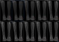
In this work, the feasibility is explored for
subtractionless first-pass time-resolved contrast
enhanced MRA of the lower legs on 1.5T using Dixon,
viewsharing and parallel imaging with high acceleration
factors. Results in seven consecutive patients are
analyzed and compared with the conventional subtraction
method. It is demonstrated that with the
subtractionless method, bulk motion artifacts are
eliminated, and SNR is significantly increased.
|
11:15
|
0887.
 |
Clinical Performance of a Non-contrast MR Angiography
Protocol in the Pre-Transplant Evaluation of the Liver
Vasculature
Jeremy Collins1, Eric Keller2,
Edouard Semaan3, Riad Salem2,
Maria Carr2, Michael Markl2, and
James C Carr2
1Radiology, Northwestern University, Chicago,
IL, United States, 2Northwestern
University, Chicago, IL, United States, 3Chicago,
IL, United States
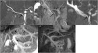
Assessment of the hepatic vasculature is critical as
part of the pre-liver transplant evaluation. The
prevalence of renal insufficiency and concerns regarding
gadolinium administration in this cohort has created a
need for a non-contrast alternative for vascular
assessment. We evaluated the clinical performance of a
non-contrast MRA (NCMRA) protocol at 3T in the
assessment of the hepatic vasculature in patients with
cirrhosis, with contrast-enhanced MRA (CEMRA) as the
reference standard. The NCMRA protocol was diagnostic in
94% of subjects, identifying all relevant variant
anatomy. Clinically available NCMRA techniques when
combined into a comprehensive protocol enable assessment
of the hepatic vasculature.
|
11:30
|
|
Advanced Techniques for Flow Imaging
Michael Hope1
1UCSF
We will focus on the emerging applications of
multidimensional MR flow imaging (4D Flow). The
techniques and hemodynamic biomarkers that we will
discuss can be applied broadly throughout the
cardiovascular system. Two
key issues must
be addressed when considering these applications: 1)
clear advantages over ultrasound/echocardiography and 2)
matching advanced imaging capabilities with clinical
questions that change the management of patients with
cardiovascular disease. The goal is to provide a unique
understanding of how abnormal flow promotes or
exacerbates disease. This understanding, in turn, could
allow patients to be risk-stratified based on flow,
guide medical therapy, and identify new pathways to
target with drug therapy and patients that may benefit
from early intervention. The
outline of the talk is
1) review of two current clinical applications for MRI
flow imaging and 2) discussion of four emerging
applications for 4D Flow.
|
12:00
|
0888.
 |
Impact of Bicuspid Aortic Valve Fusion Phenotype and Valve
Stenosis on Aortic 3D Hemodynamics: New Insights from a
Large Cohort 4D Flow MRI Study in 312 subjects
Alex J Barker1, Pim van Ooij2,
Emilie Bollache1, David Guzzardi3,
S. Chris Malaisrie4, Patrick M McCarthy4,
Jeremy D Collins1, James Carr1,
Paul WM Fedak3, and Michael Markl1,5
1Radiology, Northwestern Univeristy, Chicago,
IL, United States, 2Academic
Medical Center, Amsterdam, Netherlands, 3University
of Calgary, Calgary, AB, Canada, 4Cardiac
Surgery, Northwestern Univeristy, Chicago, IL, United
States, 5Bioengineering,
Northwestern University, Chicago, IL, United States
Bicuspid aortic valve (BAV) morphology will alter
transvalvular blood flow patterns and vessel wall shear
stress (WSS). These hemodynamic changes have been
associated with the regional expression of BAV
aortopathy. However, the presence of aortic stenosis can
confound the regional expression of WSS. The purpose of
this study was to use aortic WSS atlases to understand
the role of aortic valve morphology and stenosis on the
expression of WSS in the ascending aorta of a large
control and BAV patient cohort (n=312).
|
12:15
|
0889.
 |
MRI assessment of aortic flow in patients with pulmonary
hypertension in response to exercise
Jacob Macdonald1, Omid Forouzan2,
Naomi Chesler2, Christopher Francois3,
and Oliver Wieben1,3
1Medical Physics, University of Wisconsin -
Madison, Madison, WI, United States, 2Biomedical
Engineering, University of Wisconsin - Madison, Madison,
WI, United States, 3Radiology,
University of Wisconsin - Madison, Madison, WI, United
States
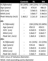
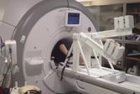
Cardiopulmonary exercise testing is gaining increased
recognition as a useful tool for assessing pulmonary
hypertension (PH). Using an MRI-compatible exercise
device that allows subjects to exercise in the bore of
the magnet, we investigated the effects of exercise
stress on blood flow in the ascending aorta in healthy
controls and patients with PH. The measurements we
obtained demonstrated a decreased exercise capacity in
PH subjects and in older controls. Some parameters, such
as cardiac output, demonstrated statistically
significant changes between rest and stress, while
others were unclear due to the relatively low exercise
power tolerated by the PH patients.
|
12:30
|
|
Adjournment & Meet the
Teachers |
|